CELLS OF IMMUNE SYSTEM

BY: SAI MANOGNA (MSIWM014)

The significant majority of cells involved in the immunity of mammals are derived from the bone-marrow precursors (Left half of the figure) and circulate in the blood, entering if possible and leaving the tissues. In the adult bone marrow at a frequency of 1 in 100,000 cells, an intermittent stem cell survives and maintains the potential to differentiate into all blood cell forms.

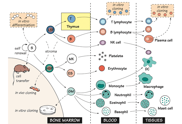
Haemopoiesis has been studied either by injecting a group of cells into the recipient mice of genetically marked marrow cells and examining the progeny they give rise to (Invivo cloning) or by cultivating the bone marrow precursors in the presence of sufficient growth factors (Invitro cloning).

The proliferation and differentiation of all these cells is regulated by the soluble membrane bound growth factors that usually generate the stroma of bone marrow and each other. Such signals turn-on the particular growth factors inside the cell, DNA binding molecules that serve as master switches, establish the subsequent genetic program, resulting in the production of various types of cells.

Bone Marrow :

Unlike any other tissues or organs, the hematopoietic system continues to regenerate itself. In adults hematopoietic cell production occurs primarily in the bone marrow. In the fetal hematopoiesis occurs first in the yolk sac and then in the liver until the bone grows.

Stroma : In haemopoiesis, epithelial and endothelial cells that provide protection and secrete growth factors.

Lymphoid stem cell : These cells are assumed to be capable of differentiating into  B lymphocytes and T lymphocytes. Recent research findings indicate that the distinction might actually be more complex between lymphoid and myeloid stem cells.

Haemopoietic stem cell: Spleen nodule precursors are presumably capable of differentiating into all but lymphoid pathways i.e, granulocyte, erythroid, monocyte, megakaryocyte, also referred to as CFU-GEMM. CFU-GEMM refers to the colony forming unit that generated myeloid cells. GEMM refers to Granulocyte, Erythroid, Monocyte, Megakaryocyte.

Erythroid Stem cell : These erythroid stem cells generate erythrocytes. In response to hypoxia, erythropoietin, a glycoprotein hormone produced in the kidney, hastens the differentiation of red cell precursors, and thus adapts red cell development to the demand for its oxygen carrying capacity which is an example of NEGATIVE FEEDBACK.

Granulocyte–Monocyte : This is the common precursor. The growth or colony stimulating factors influence the relative proportion of these two types of cells.

Cloning : By isolating the single cells and allowing them to differentiate several times, and then studying what cell types can be found in the progeny. Thus the ability of individual stem cells to give rise to one or more types of hematopoietic cells has been explored. This approach is referred to as cloning. A clone is a group of daughter cells originating from the single parent cell. These evidence  indicate that a single stem cell can give rise to all the fully differentiated cells of an adult hematopoietic system under specified conditions.

Neutrophils : These are the short-lived phagocytic cells whose granules contain voluminous bactericidal substances, is the most prevalent leukocyte in the human blood. The first cells to exit the blood and to enter the sites of infection or inflammation are neutrophils.

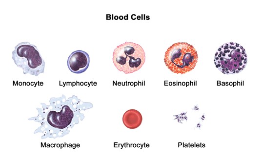
Eosinophils : Eosinophils are the leucocytes with large refractile granules, containing a number of proteins that are highly basic or cationic proteins possibly essential for the killing of large parasites, including worms.

Basophils : These are the leukocytes with large basophilic granules containing heparin and vasoactive compounds which are essential in inflammatory response. The three types of cells which are neutrophils, eosinophils and basophils are collectively known as GRANULOCYTES.

Megakaryocyte : These are the parent cells of the blood platelets.

Platelets : These are the small cells which are responsible for sealing the damaged blood vessels (Which is known as haemostasis) but these cells are also the source of several inflammatory mediators.

Monocyte : The mononuclear phagocytic system consists of monocytes. Monocytes circulate in the bloodstream for about 8 hours and then migrate into the tissues. When these migrate into the tissues, they differentiate into specific tissue known as macrophages. Additionally monocytes are drawn to inflammation sites, replenishing a reservoir for macrophages and possibly dendritic cells as well.

Macrophages : Macrophages are strewed throughout the body. In specific tissues some take up residence, becoming fixed macrophages, while others remain motile called as free or wandering macrophages. These free macrophages migrate across the tissues by amoeboid movement.

Macrophage like cells serve various functions in different tissues and these are named according to the site of location;

Kupffer cells in Liver

Osteoclasts in Bone

Alveolar macrophages in Lungs

Mesangial cells in Kidney

Intestinal macrophages in Gut

Microglial cells in Brain

Histiocytes in Connective tissue.

Dendritic Cells : Dendritic cells are present in all body tissues (eg; Skin cells of Langerhans) where they pickup the antigen and then migrate through the lymphatics or the blood to the T-cell areas of lymph nodes or spleen. Their main function is to activate T-cell immunity, but they might also be involved in the induction of tolerance as well. The plasmacytoid DC is the name derived from their morphological resemblance to the plasma cells. The second subset of these cells are the principal producers of type I interferons, which is an essential group of viral proteins. Experimentally, while these cells are derived from the myeloid cells, the developmental lineage of bone-marrow dendritic cells is still the subject of debate.

Natural Killer Cells (NK Cells) : NK cells have been shown to play a significant role in host defence against both tumor cells and cells infected with certain, but not all the viruses. The membrane molecules and receptors that differentiate T and B cell lines do not express these cells. These cells make up 5%-10% of lymphocytes in the human peripheral blood.

T and B Lymphocytes : These cells are main cellular components of adaptive immunity. T-lymphocytes are derived from the thymus, while B-lymphocytes are derived from the bone marrow of birds or bursa derived. The precursor for antibody forming cells is B-lymphocytes. The liver may play the part of bursa in the liver.

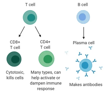
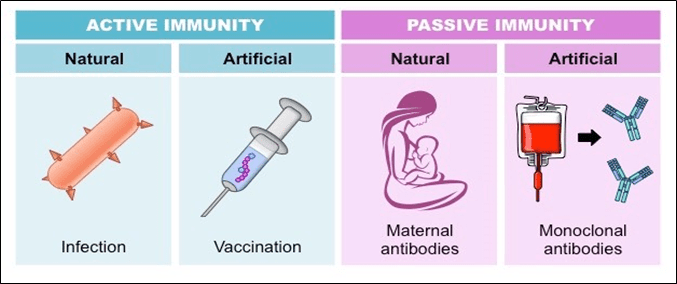
Plasma Cell : Plasma cells are rarely seen in blood. These cells are present if an antibody is formed in the spleen, lymph node etc; Plasma cells do not differentiate and cannot be retained invitro for extended periods. B lymphocytes that generate particular antibodies may however be fused with a tumor cell to generate an immortal hybrid clone or hybridoma that continues to secrete a predetermined range of antibodies. As unique instruments in many branches of biology, such monoclonal antibodies have proven to be of immense benefit and some are now being widely used for the treatment of autoimmune disorders or cancers.

Mast cells : Mast cells are the large tissue cells that are derived from the circulating basophils. To start an inflammatory response, that causes several types of allergy, these mast cells are rapidly activated by tissue damage.

Growth factors : The molecules that regulate hematopoietic cell proliferation and differentiation are also often involved in the regulation of immune responses, such as interleukins or cytokines. In these some of them were first discovered by hematologists and are referred to as Colony stimulating Factors (CSF), but there is no particular meaning to the various names, and one ; IL-3 is sometimes referred to as multi-CSF. In clinical practice growth factors are used to improve specific blood cell subsets and erythropoietin was one of the first proteins developed by recombinant technology in the modern century.

HIV AND IT’S LIFE CYCLE

BY: ABHISHEKA G (MSIWM013)          

INTRODUCTION:

HIV (Human immunodeficiency virus) is a kind of virus that belongs to the retrovirus family and causes AIDS (Acquired immunodeficiency syndrome) in humans. It destroys the immune system of the individual by making the patient vulnerable to different other infections including neurological disorders. The disease of AIDS was first observed in 1981 in the united states. It is an epidemic disease present throughout the world. This disease can be preventable, but not curable.

TYPES OF HIV: There are two types of HIV viruses are present based on the origin of the virus.

  1. HIV-1: It is isolated in America, Europe, and Central Africa. It is more virulent and spread among individuals very rapidly
  2. HIV-2: It is isolated in West Africa, it is less virulent and does not spread rapidly as HIV-1

STRUCTURE OF HIV VIRUS:

  1. It is spherical in nature and measures about 90-120nm in diameter and it is composed of two copies of positive single-stranded RNA enclosed by a conical capsid composed of viral protein P24. The capsid also contains the enzymes which are necessary for viral replication such as Reverse transcriptase (P55/66), Integrase(p32), and protease(p10).
  2. The RNA genome of HIV consists of nine different genes that contain the information needed to make the structural proteins for new virus particles.
  3.  The matrix is composed of the viral protein P17 which surrounds the capsid ensuring the integrity of the virion particles. And the matrix is surrounded by 2 layers of phospholipids which are embedded by 70 copies of viral glycoprotein and lipoprotein which consist of two units namely anchoring transmembrane pedicles (Gp41) and surface projecting knob like spikes (Gp120).

MODE OF TRANSMISSION OF HIV VIRUS:  HIV is transmitted in many different ways.

1.Sexual contact: Unprotected sexual relationship with an infected individual.

2. From mother to child: The virus of HIV is transmitted from mother to child through the uterus, during delivery, and also during breastmilk.

3. Through needles: Through the injections during injecting drugs and needle prick injury which are already used by the infected individuals.

4. Blood transfusion and organ transplantation: The virus is transmitted through transfusion of blood and transplantation of organs from infected individuals to normal individuals.

LIFE CYCLE OF HIV VIRUS:

There are 7 different steps in the life cycle of the HIV virus.

  1. Attachment to the host/Binding to the target cell: The specific site for the HIV virus to bind are CD4 receptors and other co-receptors(CCR5/CXCR4) which are enveloped by the glycoprotein Gp120. Viral binding to host cell triggers fusion of the viral and cell membrane of the host cell, which is mediated by gp41 and allows the virus core into the cytoplasm of the host cell.
  2.  Reverse transcription of its RNA: HIV consists of single-stranded RNA. The single-stranded RNA is converted into DNA. This process is carried out by the enzyme Reverse transcriptase.
  3. Integration into the host genome: The viral double-stranded DNA is integrated into the genome of the infected host cell through the action of the viral integrase enzyme which causing a latent infection.
  4. Transcription of viral proteins: The transcription of viral proteins is mediated by Viral reverse transcriptase. The infected DNA of the host cell makes proviral RNA and protease enzyme cuts or cleaves the polypeptides into functional HIV proteins and the virion assembles.
  5. Assembly of a new viral particle: Viral strands and other proviral products combine to form a package and head for the cell membrane.
  6. Release of immature virions: Immature virus finds a suitable position to push or come out of the host cell by taking a piece of the membrane with it.
  7. Maturation of the virion: The new free virus matures and gets ready to infect another host cell.

SYMPTOMS OF THE DISEASE:

Diarrhea, Fatigue or weakness, fever, headache, joint pain, night sweats, rashes on the body, swollen glands, sudden weight loss, yeast infections in the body, and sexual organs that occur frequently and last for a long time.

LABORATORY DIAGNOSIS OF HIV: HIV is mainly detected by  different clinical tests:

  1. ELISA (Enzyme-linked immunosorbent Assay/ Enzyme Immunoassay): This test detects and measures the antibodies in the blood sample along with the presence of antibodies related to any infections.
  2. Western Blot: This test is used to detect the specific proteins called HIV antibodies present in the blood sample. If ELISA gives positive for HIV, then the western blot technique is used to confirm the positive result of ELISA and gives 99.9% accurate results.

AVAILABLE ANTIRETROVIRAL DRUGS TO HIV:

  1. Nucleoside reverse transcriptase: Zidovudine and Stavudine
  2. Non-nucleoside reverse transcriptase inhibitor: Efavirenz and Nevirapine.
  3. Protease inhibitor: Atazanavir and Darunavir.

PREVENTION OF HIV VIRUS:

  1. Avoid multiple sexual contacts.
  2. Using new and sterile needles for injection.
  3. Care should be taken and testing should be done during blood transfusion and pregnancy.
  4. Biomedical waste should be properly disposed of from hospitals and households.
  5.  Proper sex education should be provided to people.


THE COMPLEMENT SYSTEM

BY: RAHUL ANDHARIA (MSIWM001)

Introduction:

  • It is a part of immune system which consists of series of proteins that interacts with one another in an organized manner to eliminate pathogens.
  • Complement pathway has a role to play in removal of damaged cells and pathogens by helping antibodies and phagocytic cells in this process.
  • Proteins taking part in complement system are known as complement proteins and works together as biological cascade (sequence of reactions, in which each being the catalyst for the other).
  • Mostly, complements are soluble proteins and glyco-proteins produced by special cells called hepatocytes.
  • These complements circulate in the body in form of Zymogens (inactive forms) and more than 20 such types have been recognized in the serum.
  • When an antibody binds to an antigen, it triggers complement pathway. It can also be triggered by few components of innate immunity. This system can also work in acquired immunity.
  • During inflammation, these complements gets activated and reaches infected area of the intestinal tissue through dilated blood vessels, which are activated by proteolytic cleavage and exposes active site of the complements.

History:

  • Complements were identified as heat-sensitive components in the blood in the year 1895 by Jules Bordet.
  • Complement levels will be low during recurrent microbial infections, auto-immune diseases. (Lupus disease).

Overview and components of Compliment pathways:

  • C letter is used to denote complement proteins along with numbers like C1, C2, C3 and so forth. Some can also be denoted by B and D and some are represented by names like homologous restriction factor.
  • Initial step varies in different complement pathways, however, all pathways forms enzyme complexes.
  • C3 is converted into C3a and C3b by C3 convertase. C5 gets cleaved by C5 convertase into C5a and C5b. C3 convertase is bound by C3b to form C5 convertase.
  • Initiation of late components of complement system to form Membrane attack complex (MAC) and to kill pathogen is done by C5 convertase generated by various complement pathways.
  • These are some of the common steps in different complement system pathways.

Types of Complement Pathways:

  1. Classical Pathway:
  2. Initial step in the classical pathway is the formation of Antigen-Antibody complex. When antibody binds to antigen, a conformational change is induced in the FC (fragment crystallization) portion of the antibody. This FC region exposes binding site for C1 protein.
  3. C1 is composed of c1q and two molecules of c1r and c1s each. C1q remains bounded to the FC portion. C4 and C2 are cleaved with the help of proteases, c1s and c1r.
  4. C1 bounded to the immune complex, calls for another protein C4 which gets cleaved into two; C4a and C4b. C4b is activated and attaches to target surface near to C1q whereas C4a goes away(eliminated).
  5. C4b than cleaves C2 into C2a and C2b. C4b2a complex is formed upon C2a binding to C4b, whereas C2b goes away (eliminated).
  6. C3 complex is then activated by C4b2a. This complex is also called as C3 convertase complex, as C3 is converted to an active form by separating C3a and C3b.
  7. To cleave large number of C3 molecules, one molecule of C4b2a is enough. C3b can bind to both, to the microbial surface or to the convertase itself.
  8. C4bC2aC3b complex is formed upon C3b binding to C3 convertase. C5 gets activated by C5 convertase into C5a and C5b. C5b gets stabilized by binding to C6 while C5a is eliminated. C5bc6 complex is formed, which than binds to C7.
  9. Complex called C5BC6C7 formed, binds to phospho-lipid bilayer of the cell membrane and further binds to C8.
  10. At the end all this C5b678, results in activation of C9 which forms a macromolecular structure called Membrane attack complex. (MAC).   
  11. Due to the formation of membrane attack complex, hole is produced in the bacterium resulting in leaking of cellular contents and unwanted substances can get in. As a result of this cell looses its osmotic stability resulting in lysis by influx of water and loss of electrolytes.
  12. The system (MAC complex formation) is found to be more effective in gram negative than in gram positive bacteria as MAC complex can be easily formed in thin peptidoglycan layers of gram negative bacterial cell walls rather than thick layered gram positive cell walls.
  13. There are few exceptions where, some of the c3b molecules do not associate with C4b2a. Instead these molecules are known to coat microbial cell surfaces and immune complexes and works their as Opsonins (an antibody or cell that binds to foreign antigen and exposes them to phagocytosis process). This process of opsonin formation is called as Opsonisation.

2. Alternate Pathway:

  • In this pathway there is no formation of antigen-antibody complex.
  • In this pathway, the complement system is initiated by Cell surface Constituents, which are foreign to the host. Example- Lipopolysaccharide.
  • During inflammation, bacteria enter host body and reaches to the site where C3 is directly bounded to antigens surface, and becomes active.
  • C3 contains thioester bonds which undergo hydrolysis to give C3a and C3b.C3b now binds to surface of the foreign particle and then binds to Factor B.
  • This factor B exposes site which serves as enzymatic substrate for serum protein D. As a result of this, factor D cleaves B into Ba and Bb. This results in formation of C3 convertase (C3bBb).
  • C5 convertase gets formed by C3 convertase and C5 then forms MAC complex, similar to the Classic pathway.

3. Mannose Binding Lectin Pathway (MBL):

  • Without Antibody and Endotoxin, complement pathway can be activated with the help of MBL pathway. When circulating lectin binds to Mannose residues on carbohydrates surface of Micro-organisms, this MBL pathway gets activated.
  • Salmonella, Listeria and Neisseria strains can induce MBL pathway.
  • Concentration of MBL increases during inflammation as it is an acute phase protein.
  • Lectin recognizes and binds carbohydrates of target cell to activate this MBL pathway. This pathway has similarities with that of classical pathway in terms of C4 and C2 to produce complement proteins.
  • MBL resembles C1q in structure and works in similar fashion to that of C1q.
  • Masp1 (mbl-associated serene proteases) and Masp 2 are the two components that binds to MBL after lectin binds to carbohydrates and activates MBL pathway.
  • A tetrameric complex is formed by Masp1 and 2 similar to complex formed by c1s and c1r and than cleaves C2 and C4 to form C3 convertase.
  • Than in the next steps, C5 convertase is formed, which results in MAC complex and the rest process is same as that of classical pathway.

Functions of Complements:

  • Opsonisation and Phagocytosis: proteins involved in this process are C3b. It is bounded to the surface of pathogen and activates phagocytic cells by binding to specific receptors present on the surface of phagocytic cells.
  • Cell lysis: C5b6789 forms a membrane complex which ruptures microbial cell surface and kills them.
  • Chemo taxis: Neutrophils and macrophages are attracted to an area where antigens are present by complement fragments. These cell surfaces have specific receptors for C5a, C3a and thus, run towards site of inflammation, called Chemo taxis.
  • Antibody Production: C3b receptors are present in b cells. When C3b binds to b cells more antibodies are produced. Thus, C3b is an antibody amplifier and can convert this into defense mechanism against invading Micro-organisms.
  • Immune Clearance: immune complexes are removed from circulation and are deposited in liver and spleen. Thus, complement protein acts as anti-inflammatory function. Solubilisation of these complexes is facilitated by Complement proteins and also helps in their phagocytosis.

MEDICAL MICROBIOLOGY

BY: RAHUL ANDHARIA (MSIWM001)

Medical Microbiology

Deals with prevention, diagnosis and treatment of infectious diseases caused by infectious agents like bacteria, fungi, parasites and viruses. This field is also known as Clinical microbiology. Understanding of various infectious agents and how this agents causes the disease, development and pathogenesis of the disease are being studied under medical microbiology.

History of medical microbiology:

  • Infectious diseases caused by transfer of seed like entities was first given in the year, 1546 by Girolamo  Fracastoro. He said that the disease can be caused by direct contact or by indirect contact with the infectious agent.
  • Advancements in the field of medical microbiology actually started in the year 1888, when Pasteur institute was established in Paris. Louis Pasteur and Robert Koch are considered as father of medical microbiology due to their contributions in the field.
  • Disapproval of the spontaneous generation theory(theory which claimed that non living matter is involved in giving life to living things), Pasteurization(heating at lower temperatures- less than 100 degree Celsius)methods, vaccines for Tabbies, cholera and anthrax(caused by- Bacillus anthraces) were proposed by Louis Pasteur.
  • Specific microbes are responsible to cause a particular disease. The concept of Germ theory of Disease, was given by Robert Koch. Koch cultured various Micro-organisms in the lab that caused the disease, including bacteria, mycobacterium tuberculosis, which is known to cause Tuberculosis. He used to grow Micro-organisms on solid medium like agar.
  • Koch’s postulates:
    • Organisms suffering from the disease should have the microbes that are responsible for the diseases. Microbes should not be found in healthy organisms.
    • Pure culture must be used to isolate the Micro-organisms from the diseased organism.
    • When the Micro-organism is Administered into a healthy organism, it should confer the disease.

From the same inoculated diseased experimental host, the same Micro-organism must be isolated and it has to be identical to the original agent that was responsible for the disease.

  • Concept of anti-sepsis was given by Joseph Lister. He showed that carbolic compounds, now it’s known as phenol, can be used for wound sepsis and cleaning.
  • Extensive work on TMV(Tobacco mosaic virus) was carried by Dmitri Ivanovsky. He studied TMV and differentiated it with bacteria by preforming a simple experiment in the year 1892, where he showed that when passed through filters, larger bacteria will be retained .
  • Propagation of  polio virus in monkey kidney cell cultures was given by John Enders, Thomas Weller and Frederick Robbins.

Fields in Medical microbiology:

  1. Microbial Physiology: It deals with the studies of growth of Micro-organisms, metabolism of micro-organisms and microbial cell culture. It also deals with pathophysiology of micro-organisms and role of Micro-organisms in diseases.
  2. Microbial Genetics: It involves studying how microbial genes function. Organisation of microbial genome, it’s genomic characteristics are being studied in this field.
  3. Parasitology: involves in depth studies of parasites. Sample studies can be done using feces, blood, urine and sputum samples.
  4. Virology: It deals with detail studies of various types of viruses. Host-viral mechanisms, viruses mode of action in host, diseases caused by viruses are some aspects involved with virology.
  5. Immunology: Immune system studies, which involves studying hosts immune system and it’s role in combating pathogens and foreign substances. Antigen-antibody interactions are primarily used as diagnostic tools in diseases.

Role of Medical microbiology:

  • When a Patient experiences any signs of infections, physicians recommends medical and diagnostic laboratory procedures.
  • Antibiotic sensitivity test, direct stain or culture are the type of tests performed.
  • Different types of testing’s are performed like collection of appropriate specimen and than plating the specimen on appropriate culture medium, inoculating and sub culturing it and than performing different tests to confirm the type of disease. For example- Mantoux test is used for tuberculosis.
  • Serological, biochemical, molecular tests are performed and the types of micro-organisms associated with the disease are identified.
  • Basically it involves testing samples for the presence of infectious agents, and than performing diagnostic procedures.
  • Various subfields have different procedures, like for example- immunology procedures involves performing ELISA to detect antibodies in blood.

Types of Infectious agents:

Bacteria:

There are certain bacteria which are essential for the human body and it’s functions. These are termed as normal flora. For example- gut microbes are involved in synthesis of vitamin k and vitamin B12. During immunocompromised conditions, this microflora can lead to infections.

Invasiveness: can spread within the host body. Structures like capsules helps them to skip phagocytosis. Toxins also help them to defend host machineries.

Immunopathology: host immune response is responsible for pathogenesis in most of the Infections.

Viruses:

They are obligate parasites( they require host system to grow and develop as they cannot synthesise proteins and utilize it’s own energy). Viral diseases are more common in humans, particularly in children.

Invasiveness: They require host machineries to multiply and divide. They make the host system to synthesise viral components and as a result, host machineries are destroyed by viral components leading to viral diseases.

Immunopathology: host immune response is responsible for pathogenesis in most of the Infections.

Fungi:

Invasiveness: mucosal tissues or keratin are the places commonly where fungi can multiply and cause the Infection.

Toxin Production: food contaminated with fungi can cause serious food poisoning.

Immunopathology: When fungal spore or hyphae are inhaled, can cause hypersensitivity.(allergy).

Parasites:

Much more complex than viruses and bacteria. They can cause disease via multiple routes. Example- Giardiasis- Intestinal infection- by giardia, mainly through contaminated food and water.

Applications of Medical microbiology:

  • Vaccine development: understanding pathophysiology of the disease and than preparing a vaccine against it. Basically vaccine generates a immune response which will be lacking when the body is compromised during a disease. Rubella and measles are the two common examples of diseases which have been eradicated by vaccination.
  • Diagnostic tests: cultural tests, serological tests, microscopy, PCR all this diagnosis testing helps to identify infectious agents.

CELL SIGNALLING

BY: RAHUL ANDHARIA (MSIWM001)

Communication between cells is referred to as cell signalling. Cell signalling is the process through which cells responds to external stimuli. While studying the duct secretion system, Claude Bernard, in 1855 discovered process of cell signalling.

What kinds of signals do cells receive?

  • Cells communicate by sending chemical signals.
  • Sending cell is the one which sends the signal in form of proteins or other molecules which is received by a target cell.

How do cells recognise signals?

  • Cells posses special molecules called receptors which binds to signalling molecules and initiate physiological response.
  • For a target cell to detect the signal, it must have a appropriate receptor to bind to.
  • Receptor is called a transmembrane protein, which binds to cells and transmits signals.

How do cells respond to signals?

  • When a signal is received by a receptor protein, it undergoes conformational changes and leads to series of biochemical reactions.
  • Signal transduction cascade( intracellular pathway) than amplify the message producing many intracellular signals from the receptor to which it was originally bound.
  • When receptors are activated, it initiates synthesis of second messenger(ligand) which coordinates intracellular pathways.

Example of molecule common second messenger- Cyclic AMP

Three stages of cell signalling:

  1. Stage 1 where receptor molecule binds with the signal molecule, called reception stage.
  2. Stage 2, in which series of chemical signals results in activation of enzyme molecules, called signal transduction stage.
  3. Stage3, resulting cellular responses, which is the response stage.

Forms/types of signalling:

Paracrine signalling:

  • Cell signalling to neighbouring cell.
  • In this type of signalling, signals are transmitted within relatively shorter distances.
  • Allows cells to coordinate locally with neighbour cells.
  • They play a role in many tissue and context signalling, but their vital role is during development where they communicate one group of cells to select which type of cellular identity from other group of cells.
  • Example- spinal cord development in humans.

Synaptic signalling:

  • In synaptic signalling, nerve cells transmit the signal. This kind of signalling is an example of paracrine signalling.
  • The name is synaptic, because the signal transduction takes place between Junction of two nerve cells, which is called a synapse.
  • When a signal is received by a sending neuron, an electrical impulse is generated in the cell which travels through Axon, a fibre like extension.
  • Ligands, called neurotransmitters are released, when impulses reaches synapse.
  • Neurotransmitters than, binds to the receptors of the receiving cell, causing chemical changes in the cell.
  • Immediately, the sender cell than, degrades the neurotransmitters that are released into the chemical synapse. This way the system resets again, to receive a second signal and this process continues.

Autocrine signalling:

  • In autocrine signalling, the cell targets itself, that is cell signals itself. The ligand released will bind to the cell’s own receptors.
  • This type off signalling plays many roles, like for example autocrine signalling is involved in development, where it guides cells to take-up it’s correct identity.
  • This type of signalling bid also known to play a role in cancer, where it is known to be involved in metastasis ( spread of cancer to other body parts).
  • Sometimes, cell simultaneously has both paracrine as well as autocrine effect, so as a result it binds to itself as well as to the sender cell.

Endocrine signalling:

  • This type of signalling uses circulatory system as a network to transmit messages for long distances.
  • In long distance endocrine signalling, specialized cells produces the signals and releases into blood stream, through which they reach the target cells.
  • Here signals are produced and circulated in blood stream via hormones. Hormones carries signal produced in one part of the body to other parts.
  • Thyroid glands, pituitary glands, hypothalamus, gonads and pancreas are the endocrine glands that releases hormones.

Example: GH- growth hormone, released by pituitary gland promotes growth of cartilage and skeleton.

Signalling via cell-cell contact:

  • Cell-cell contact signalling occurs via Gap junctions. These water filled channels allows intracellular mediators to diffuse between two cells.
  • Molecules like DNA and proteins cannot fit through channels without special assistance, but small molecules like ions and water can pass through.
  • Transfer of signalling molecules will lead to transfer of current state of one cell to the neighbouring cell. This will allow cells to coordinate the response, which only one of them would have received.
  • Two cells carrying compliment protein may bind to each other, resulting in the change of shape of one or both the protein that transmits the signal. This is another type of direct cell signalling which is particularly seen in immune cells, which uses it to recognise its own self cells and pathogen cells.

Types of Receptors involved in signalling:

Intracellular receptors:

  • Located in the cytoplasm of cells. It has other two types:
  • Nuclear receptors: It has  DNA binding domains. When these are bound to thyroid or steroid hormones, it results in formation of a complex which enters the nucleus and regulates gene transcription.
  • IP3 receptors: present in endoplasmic reticulum. It helps in releasing ca2+ ions which are vital for muscle contraction.

Ligand gated ion channel receptors:

  • They spann  across the plasma membrane.
  • Major function of this receptor is to allow hydrophilic ions to pass the thick fatty membranes of our cells.
  • When bound to acetylcholine, a neurotransmitter, ions like k+, ca2+, Na+ and cl- are allowed to flow across the membrane to allow neural firing to take place(transmission of impulse.

G-protein coupled receptors:

  • Largest receptors in eukaryotes.
  • They are known to be very diverse as they receive input from complex and diverse group of signal ranging from sugars, peptides and light energy.
  • Binding of ligand to this receptors results in activation of G-protein, which transmits entire cascade of second messenger signals, which carry out important functions like sight, sensation, growth and inflammation.

Receptor Tyrosine kinases:

  • When ligand binds to this receptors, it results in dimerization of tyrosine kinase domains.
  • This results in phosphorylation of their tyrosine kinase domains which allows the intracellular proteins to become active by binding to phosphorylated sites.
  • One of the major role of this receptors is in growth pathways.

Ligand used in signalling:

  • Hydrophobic ligands- binds to intracellular receptors. These have fatty properties and possess steroid hormones and D3 vitamin.
  • Hydrophilic ligands- binds directly to cell surface receptors. These are mostly amino acids derived ligands.

Types of signalling molecules:

  • Intracrine: produced by target cells and binds to receptors present within the signalling cells.
  • Autocrine: they are distinct. They function on target cells as well as they function internally. Example- immune cells of the body.
  • Juxtacrine: often called contact-dependant signalling. They generally target adjacent cells.
  • Paracrine: targets cells that are in the vicinity of original cells that transmits the signals. Example- neurotransmitters.
  • Endocrine: signal is transmitted via bloodstream through hormones produced by cells.

Importance of cell signallingCell signalling basically allows cells to respond and perceive to external environment allowing their growth, development, immunity. Without cell signalling it’s not possible to handle complex body mechanisms and perform important biological functions. Errors in cell signalling process results in diseases like cancer, diabetes.

HYPERSENSITIVITY

BY: RAHUL ANDHARIA (MSIWM001)

Hypersensitivity:

It refers to excessive, undesirable damage posing, discomfort producing and sometimes fatal reactions produced by the body’s own natural  immune system. In simpler terms it simply refers to allergic reactions caused by immune system.

Introduction:

  • During hypersensitivity, the host is in pre-sensitized immune state. This simply means that the host was primarily exposed once to the antigen, and comes in contact again for the second time with the same antigen.
  • Based on the mechanisms involved and time taken for allergic reaction, hypersensitivity reactions are classified into four types: Type I, Type II, Type III, and Type IV hypersensitivity reactions.

Type I Hypersensitivity:

It is also known as immediate or anaphylactic or atopic hypersensitivity. Type I hypersensitivity is generally caused due to reexposure to the specific type of antigen or allergen(substance which causes allergy). It can lead to systemic or local reactions and involves eyes(conjunctivitis), skin(eczema), nasopharynx(rhinitis) and gastrointestinal tracts(gastroenteritis). Symptoms generally varies from mild to even fatal and can lead to death or anaphylactic shock. The time of reaction ranges from 10-15 minutes after the action of allergen, but sometimes there is delayed onset(10-12 hours).

Mechanism:

  1. The reaction is mediated by IgE antibody. Primary cellular component involved is mast cell or basophil cells.
  2. Upon very first exposure to allergen, APC(antigen presenting cells) processes the antigen and present it to Th2 cells.
  3. Upon released of IL-4 and IL-12 by Th2 cells, B cells gets activated.
  4. After that, the B cells differentiate into plasma cells. Plasma cells synthesise and secretes IgE antibody (type of antibody usually involved in allergic reactions).
  5. IgE binds to FC(fragment crystallization) of mast cells and sensitize it.
  6. Upon subsequent exposures to the same antigen, now the mast cells bind to IgE antibody and releases the inflammatory molecule. This will result in allergic symptoms.

Diagnosis:

  • Involves skin tests( pricks and intradermal samples)
  • Specific and total IgE antibody measurements against the suspected allergen by using ELISA(Enzyme linked immunosorbant assay).
  • Increased level of IgE in the ELISA test, will confirm allergy or atopic condition.

Treatment:

  • Anti-histamines for symptomatic treatment. Anti-histamines can block the histamine receptors.
  • Cromo Lyn sodium, a chemical can inhibit mast cell degranulation. It does it by inhibiting ca+2 influx.
  • IgG antibodies can be used as it can bind with IgEs FC portion and prevents mast cell sensitization.

Examples of type I hypersensitivity: Conjunctivitis, Eczema, Eosinophilia, Angioedema.

Type II Hypersensitivity:

This type II hypersensitivity is also known as antibody dependant or cytotoxic hypersensitivity and is known to  affect many organs and tissues. The antigens are normally endogenous. However, some exogenous chemicals or molecules such as haptens(substance which combined with proteins can produce antibodies) can also induce type II hypersensitivity. Reaction time is usually from few minutes to hours.

Mechanism:

  1. IgG and IgM antibodies bind to the antigen and form complexes. This complexes than results in activation of compliment pathway(pathway through which foreign antigens are destroyed).
  2. At the site of membrane attack complexes, mediators of acute inflammation are generated, which will lead to cell death and lysis.
  3. Phagocytes, expressing FC receptors can induce phagocytosis.
  4.  The FC receptors, recognises surface bound antibody and compliment proteins .
  5. ADCC(antigen dependant cell mediated cytotoxicity) is another form of hypersensitivity where tags of IgG and IgM antibodies bind. Macrophages and NK(natural killer cells) than recognises this tags and kills them.

Diagnosis:

  • It includes detection of circulated antibodies against the tissues involved.
  • Detection of presence of antibodies in the liquid biopsy through immunofluorescence.

Treatment:

  • Anti-inflammatory agents
  • Immunosuppressing agents

Examples of type II hypersensitivity:  Erythroblastosis Fetalis, Hashimotos thyroiditis, transfusion reactions, Rheumatic fever.

Type III Hypersensitivity:

Also called as immune complex hypersensitivity. The reaction is general or may involve organs such as skin, blood vessels, kidneys and joints. This reaction is caused primarily by micro-organisms. The reaction time is 3-10 hours after being exposed to the allergen.

Mechanism:

  1. Mediated by soluble immune complexes. Mostly the complexes are of IgG class but sometimes they can be of IgM class.
  2. Exogenous(viral or chronic infections) or endogenous (non specific autoimmunity) antigens.
  3. Primary components are compliment proteins( C3a, 4a and 5a).
  4. Major damage is caused by platelets and neutrophils.
  5. Macrophages and neutrophils are present in the lesions.
  6. Antibody deposition triggers an immune response according to classical compliment pathway.  There is formation of 2complexes(complex is formed when IgG and IgM are bounded to antigen)The larger complex gets eliminated but the first complex remains and the antigen antibody complex will spread and deposit.

Diagnosis:

  • Examinationof tissue biopsies for immunoglobulins and compliment by immunofluorescence microscopy.
  • Raji cell test and polyethylene glycol tests can be performed to measure immune complexes.

Treatment:

  •  Anti-inflammatory agents.

Examples of type III Hypersensitivity: Arthur’sreaction, Rheumatoid arthritis, symptoms of malaria.

Type IV Hypersensitivity:

Also can be called as delayed type or cell mediated hypersensitivity. Classic example of this type of hypersensitivity is tuberculin reaction(montoux) which peas after 48 hours of antigen injection. Erythema and induration(thickening of skin) are the characteristics of the lesion.

Mechanism:

  1. Antigen in the complex is recognised by cytotoxic Cd8 T cells and Cd4 helper T cells by MHC I or MHC II complex.
  2. IL-1 secreted by macrophages further helps in proliferation of Cd4 T cells.
  3. Th1 mediated response is elucidated upon reexposure to antigen.
  4. Cd4 T cells secretes IL-2 which further induces the release of other type 1 cytokines and thus induce immune response.
  5. Cd8 cells when activated, destroys target cells upon contact. Macrophages produces hydrolytic enzymes and when comes in contact with intracellular pathogens, macrophages transforms into multinucleated giant cells.

Diagnosis:

  • Montoux test and patch test in-vivo is generally performed.
  • Some of the in-vivo tests for delayed hypersensitivity can be mitogenic response, nephro-cytotoxicity, and production of IL-2.

Treatment:

  • Corticosteroids
  • Immunosuppressive agents

Examples of type IV Hypersensitivity: symptoms of tuberculosis, coeliac disease, symptoms of leprosy.

Type V Hypersensitivity:

There is an additional type of hypersensitivity that is, type V(5). This usually occurs when the IgG antibodies have a effect towards their target. In this, the antibody binds to the cell surface receptors rather than binding to cell surface components and as a result it prevents the binding of the intended ligand to the receptor and thus impairs cell signalling.

Example of type V: Graves disease.

  • Diagram showing all four hypersensitivity reactions.

IMMUNE SYSTEM

Immunology: Immune System

And Its Type

Content:

  • About
  • Immune system
  • Immunity
  • Types of immunity
  • Innate immunity
  • Acquired immunity

About:

  • Immunology refers to the branch of biology that deals with the study of immune system in all organisms.
  • Any malfunction in this system lead to the diseases like autoimmune disease, hypersensitivity, transplant rejection and immune deficiency.
  • The term was given by Ilya Ilyich Mechnikov and received noble in 1908 for his work. He studied on the larvae of starfish, pinned thorns into the larvae and found unusual cells surrounds the thorns. This was the active response by the larvae. Mechnikov was the first to observe phagocytosis.

Immune system:

  • A different type of cells and molecules which protects our body from pathogens is collectively called immune system.
  • Pathogens may be anything from algae, fungi, bacteria to haptens (molecules that may cause an immune response).
  • The cells and molecules of the immune system were distributed all over the tissue of the body which plays role in elimination or prevention from pathogens.

Immunity:

  • Immunity is the ability of organism to protect itself from the disease causing organism.
  • Our body regularly comes in the contact of numerous pathogens but we few results into disease the reason is that our body develops antibody against the pathogen and protect from the disease.

Types:

  • Innate immunity
  • Acquired immunity

Innate immunity:

  • Innate immunity is the type of immunity with which the individual is born.
  • It serve as the first line of defence an provided various components like skin, mucus membrane phagocytic cells.
  • The mechanisms of innate immunity are anatomical barrier, physicochemical barrier, phagocytic barrier and inflammation.

Types :

Species immunity: one species are resistance but other are susceptible to the same infection. Example, birds are resistant to the anthrax but human doesn’t.

Racial immunity: one race is susceptible other is more resistant to same infection. Example, certain African race are resistant to malaria but Asian or Americans are susceptible.

Individual immunity: to a certain infection the individual of race or cast is resistant but other individual of same race is susceptible to the same infection.

Acquired immunity:

  • The immunity which the individual get during its life span after each microbial infection is called acquired immunity. Example is an individual was ever infected by any infection like chicken pox virus he/ she become life time resistant to chicken pox.
  • Antibodies and t-lymphocytes contribute in acquired immunity.
  • The t-cells and antibodies are specific to the pathogen hence acquired immunity is also known as specific immunity.

Types:

Active immunity

  • The active immunity is the condition when the host itself produces antibodies.

There are two types of active immunity

Artificial – by vaccination

Natural – by natural infection

Passive immunity

The condition in which the host doesn’t produces antibodies itself but antibodies developed in other host cell provides immunity known as passive immunity.

Two types:

Artificial – antibody introduced in the host body for immunity

Natural – antibody from the mother to foetus.

Antibodies:

Antibodies are the immunoglobulin found in the blood and serve as protection against substance like antigen.

Antigens are the protein or carbohydrate which activates the immune system.

There are 5 types of antibodies- IgA, IgM, IgG, IgE and IgD.

VACCINE: Characteristics and Types.

vaccine

About:

  • Vaccine is an artificial biological preparation that contains antigens or mixture of antigens to acquired active immunity to the particular infectious disease.
  • Vaccine is prepared by the various different substances like disease causing microorganism or weakened or killed form of microbe, can be from one of the surface protein or its toxins.
  • Agent used in vaccine stimulates body’s immune system which recognise the antigen and destroy it and also encounter the agent if in future that antigen enters in the body.
  • Vaccine is prophylactic (prevent future infection by the pathogens) or therapeutic (fight against the disease already occurred).
  • The process of administration of vaccine is called vaccination; it is the most effective method to prevent infectious diseases.
  • The term vaccine and vaccination are coined by Edward Jenner. He described that how cowpox is to produce immunity against smallpox.
  • Vaccine stimulates T-cells and B- cells which further produces antibodies against the antigen.

First vaccine:

  • The first vaccine was introduced by Edward Jenner, used the cowpox virus to protect against smallpox in humans
  • Prior to this Asian physician used to give dried lesions from the diseased person to children. But by this process some individuals developed immunity while some develop disease.
  • Jenner introduced a safer way to counter this disease. He uses similar cowpox virus to confer immunity against smallpox (rare condition in which immunity of one virus protect against another virus).
  • Louis Pasteur in 1881 shows immunization against anthrax disease and four years later he develops vaccine of rabies.

Characteristics of vaccine:

  • Safe
  • Long term protection
  • Induced b and t cells
  • No or very few side effect
  • Low cost and biologically stable
  • Easy to use

Types of vaccine:

Live attenuated:

  • Is containing live organism which is weakened in the lab so that it cannot cause disease and activate the immune system against the antigen.
  • It is relatively easy to produce live attenuated vaccine for viruses then bacteria because bacteria have thousands of genes which is much harder to control.
  • Uses whole organism as vaccine which loses their pathogenicity but can induce immune response, they continuously multiply in human and provide immunity over the period of time.
  • Examples of live attenuated vaccine are mumps vaccine, measles vaccine, chickenpox, BCG, Sabin’s polio vaccine.

Killed or inactivated vaccine:

  • Microorganism causing diseases are killed by the means of chemicals, heat or radiation. These are more stable and safer than live vaccines reason is that the dead microorganism cannot mutate back to cause diseases.
  • They are the easiest preparations to use
  • Chemical which are used to kill microorganism are formaldehyde or beta-propiolactone, traditionally used chemical for virus is formalin.
  • The process should be observed carefully because excessive treatment can destroy immunogenicity and insufficient treatment leave infectious microbes to cause disease.
  • Example: anthrax vaccine, cholera vaccine, purtusis vaccine, hepatitis vaccine, salk polio vaccine.

Subunit vaccines:

  • Like above it doesn’t uses whole organism, only the part which server as antigen and stimulate the immune system is used to prepare vaccine.
  • Composed of purified macromolecule derived form the pathogen known as subunit vaccine.
  • General forms of such vaccine are

Purified capsular polysaccharide vaccine: pathogenicity of some bacteria depend on their capsule and this capsule protect bacteria from binding to the antibody. In this way infants and younger children’s immune system cannot recognise and respond against them.Example: Hib vaccine, vaccine for Streptococcus pneumoniae

Recombinant microbial vaccine: various genes encoding surface antigen of viral, bacterial and protozoan pathogens were successfully cloned into the cells of a vector. This genetic material is in bacteria cause to represent other microbial gene on its surface i.e. harmless bacterium mimics the harmful microbe and provide immunity against it. Hepatitis B is the only recombinant vaccine at present.

Synthetic peptide: development of synthetic peptide vaccine depends on the immunogenic sites. They have many advantages like low cost production and relatively stable.it is not applicable for all viruses like polio virus.

Inactivated exotoxin: this vaccine is made for some bacteria that produce toxins or harmful chemical substances. This toxins or inactivated(toxoid) by formalin and serve as vaccine which produces the anti-toxoid antibodies and neutralizes the toxin. Example of such vaccine is diphtheria and tetanus vaccine.

DNA vaccine:

  • The DNA vaccine is the DNA sequence used as vaccine.
  • The sequence is responsible for the antigenic activity of the pathogen.
  • The gene of the microbe’s antigen is introduced in the body the host cell take up the DNA, DNA instruct to produce the antigen molecule which is represented by the cell on its surface, now the body’s own cells become the vaccine producing factory and stimulates the immune system.
  • Immune response is raised against the protein produces by the cell
  • Example: DNA vaccine against west nile virus, herpes and influenza virus.