BACTERIAL DISEASE: TUBERCULOSIS (TB)

BY: SAI MANOGNA (MSIWM014)

Introduction : 

Mycobacterium tuberculosis, a tubercle bacillus, is the TB causative agent. It belongs to a closely related species, including M bovis, M africanum, and M microti, in the M tuberculosis complex. The most common site for TB development is in the lungs; 85 percent of patients with TB have lung complaints. Extrapulmonary TB can emerge as part of a primary, or late, generalized infection.

Types of Tuberculosis :

Tuberculosis is categorized into two types: active illness or latent infection. Lung disease is the most common type of active TB, but it can invade other organs, known as “miliary TB.”

TB – Active :

Active TB is a disease in which TB bacteria rapidly grow and invade the body’s various organs. Cough, phlegm, chest pain, fatigue, weight loss, fever, chills, and night sweating are common symptoms of active TB. Via airborne transmission of infectious particles coughed up into the air, a person with active pulmonary TB disease may spread TB to others. 

TB Miliary : 

Miliary TB is a rare type of active disease in the bloodstream when TB bacteria find their way. The bacteria rapidly spread across the body in tiny nodules in this form, affecting multiple organs at once. This form of TB can quickly become fatal. 

Latent Infection with TB : 

Many of those infected with TB may not experience overt illness. They have no symptoms, and that could be common for their chest x-ray. Reaction to the interferon-gamma release assay (IGRA) or tuberculin skin test (TST) may be the sole manifestation of this encounter. There is an ongoing possibility, however, that the latent infection can escalate to active illness. Other diseases, such as HIV or drugs that weaken the immune system, increase the risk.

Incubation Period :

The time of incubation can vary from two to 12 weeks or so. An individual may remain infectious for a long time (provided that viable TB bacteria are present in the sputum) and may remain infectious for several weeks before they have undergone sufficient therapy.

Pathophysiology : 

1. M tuberculosis infection occurs most often from exposure to infected aerosols from the lungs or mucous membranes. 

2. Droplets are 1-5 μm in diameter in these aerosols; a single cough will produce 3000 infectious droplets in a person with active pulmonary TB, with as few as 10 bacilli required to initiate infection. Droplet nuclei are deposited within the terminal airspace of the lung when inhaled. 

3. The organism will grow for 2-12 weeks until they reach several 1000-10,000, enough to evoke a cellular immune response, detected by a tuberculin skin test reaction. 

4. Mycobacteria are strongly antigenic and encourage a nonspecific immune response that is robust. 

5. Their antigenicity is due to the activation of Langerhans cells, lymphocytes, and polymorphonuclear leukocytes by multiple cell wall constituents, including phospholipids, glycoproteins, and wax D. 

Individuals infected with M tuberculosis may take 1 of several infection routes, most of which do not lead to actual TB. The host immune system can clear or suppress infection in an inactive type called latent tuberculosis infection (LTBI), with resistant hosts regulating mycobacterial production at distant locations before active disease output. Patients with LTBI cannot spread TB.

As earlier mentioned, the most popular site for TB production is in the lungs; 85 percent of TB patients have pulmonary complaints. As part of primary infection, or late, generalized infection, extrapulmonary TB may occur. The most prominent sites of extrapulmonary disease are: common location of tuberculous lymphadenitis is in the neck and the sternocleidomastoid muscle; it is typically unilateral and causes little to no pain; advanced tuberculous lymphadenitis cases may suppurate and shape a sinus drain. As in the kidneys, bones, meninges, skin, choroids, and the lungs’ apices, contaminated end organs usually have high regional oxygen stress. The fundamental cause of M tuberculosis infection tissue destruction is linked to the organism’s ability to incite extreme host immune reactions to the antigenic cell wall proteins. 

Lesions with TB :

An epithelioid granuloma with central caseation necrosis is the usual lesion of TB. The common site of the primary lesion in the lung’s subpleural regions is within alveolar macrophages. Locally, Bacilli proliferate and spread to a Hillary node via the lymphatics, forming the Ghon complex. 

The early tubers are spherical, 0.5- to 3-mm nodules with 3 or 4 cell areas, displaying the following characteristics: 

a. A necrosis of the central caseate 

b. Lymphocyte-admixed inner cell region of epithelioid macrophages and Langhans giant cells 

c. An outer cell region with plasma cells, lymphocytes, and immature macrophages 

d. A fibrosis rim (in lesions that heal) 

Initial Lesions: Until symptomatic illness occurs, initial lesions may heal, and the infection becomes latent. Smaller tubercles can be fully resolved. Fibrosis happens when hydrolytic enzymes destroy tubercles, and a fibrous capsule surrounds larger lesions. Typically, these nodules contain viable mycobacteria and can be reactivated. Some nodules calcify or ossify and are readily seen on radiographs of the chest. 

Proliferative Lesions: Where the bacillary load is small and host cellular immune responses dominate proliferative lesions form. These tubers are compact, mixed with activated macrophages, and surrounded by proliferating fibrosis lymphocytes, plasma cells, and an outer rim. Mycobacteria intracellular killing is successful, and the bacillary load remains low. 

Exudative Lesions: Exudative lesions predominate when large numbers of bacilli and there are low host defenses. Such loose aggregates of immature macrophages, neutrophils, necrosis of fibrin, and caseation are mycobacterial growth sites. These lesions progress without treatment, and the infection spreads. 

Etiology : 

M tuberculosis, a slow-growing obligate aerobic and a facultative intracellular parasite, are responsible for TB. In parallel groups called cords, the organism grows. After decoloration with acid-alcohol, based on the acid-fast stains used for pathological detection, it retains several stains. 

Fig: Acid-fast bacillus smear. 

In Mycobacterium tuberculosis, acid-fast bacillus smear shows aerobic, non-spore-forming, nonmotile, facultative, curved intracellular rods measuring 0.2-0.5 μm by 2-4 μm are mycobacteria, including M tuberculosis. Mycolic, acid-rich, long-chain glycolipids and phospho lipoglycans (mycocides) are contained in their cell walls that protect mycobacteria from cell lysosomal attack retains red basic fuchsin dye (acid-fast stain) after acid rinsing. 

Transmission :

The only known source of M tuberculosis has been humans. The organism is transmitted from a person in the infectious stage of TB, primarily airborne aerosol (although transdermal and GI transmission have been reported). 

1. Exposure to M tuberculosis in immunocompetent individuals results in latent/dormant infection 

2. Modifications in host immune systems resulting in a decreasing immune effectiveness may enable M tuberculosis species to reactivate, tuberculosis stemming from a combination of the direct effects of infectious organism replications, and subsequent host immune responses to tuberculosis antigens.

3. By restriction fragment-length polymorphism study, molecular typing of M tuberculosis isolates in the United States indicates that more than one-third of new patient TB occurrences result from person-to-person transmission. The remainder comes from latent infection reactivation. 

Symptoms and signs :

Symptoms associated with active pulmonary TB (older people with TB do not have the usual signs and symptoms) are as follows: 

i. Coughing ii. anorexia and fever 

iii. Sweats at Night 

iv. hemoptysis 

v. Chest pain (may also be caused by acute tuberculous pericarditis) 

vi. Tiredness 

The following may be signs of tuberculous meningitis: 

i. Headache, which for 2-3 weeks has been either sporadic or chronic 

ii. Fever that is low-grade or absent 

iii. Subtle changes in mental state that may progress towards coma over days to weeks 

The following may be signs of skeletal TB :

i. Pain in the back or stiffness

ii. Tuberculous arthritis, usually affecting just 1 joint (the hip or knee most frequently, followed by the foot, elbow, wrist, and shoulder) 

Genitourinary TB symptoms can include the following: 

Pain Flanking, DYSURIA, frequent urination, symptoms that resemble pelvic inflammatory illness in women, a painful mass of the scrotum, prostatitis, orchitis, or epididymitis in men 

Gastrointestinal TB signs apply to the contaminated site and can include the following: 

i. Non Healing mouth ulcers or anus ulcers 

ii. Malabsorption (with a small intestine infection) 

iii. Swallowing problems (with the esophageal disease) 

iv. TB-related physical examination results depend on the organs involved.

v. Mimicking peptic ulcer disease (with gastric or duodenal infection) with stomach pain 

vi. Pain, diarrhea, or hematochezia (containing colon infection) 

The following may be present in patients with pulmonary TB: 

i. Abnormal sounds of breath, especially over the upper lobes or areas involved, 

ii. Rales or signs of bronchial breath, suggesting consolidation of the lungs 

Depending on the tissues involved, symptoms of extrapulmonary TB vary and can include the following: Perplexity, chorioretinitis, coma, cutaneous Injuries, neurological deficit, lymphadenopathy pathology. 

Active TB is not excluded by the lack of any relevant physical findings. In high-risk patients, especially those who are immunocompromised or elderly, classic symptoms are often absent. 

Causes : 

M. Bacteria that cause tuberculosis to induce TB. When a person with pulmonary TB coughs, sneezes, spits, laughs or speaks, they will propagate in droplets through the air. The infection can be transmitted only by individuals with active TB. However, most people with the disease can no longer spread the bacteria after receiving sufficient care for at least two weeks. 

Factors of Risk : 

When deciding whether a TB infection is likely to be transmitted, the following factors help: 

i. Total of expelled species 

ii. Immune condition of the person exposed 

iii. Duration of time of exposure to polluted air 

iv. Organisms’ concentration 

A specific risk to non-infected individuals is posed by infected persons living in crowded or closed environments. Approximately 20% (positive tuberculin skin test) of household contacts develop an infection. Micro Epidemics have occurred on transcontinental flights and in closed settings such as submarines. Hospital workers, inner-city residents, nursing home residents, and inmates often include groups at high risk for contracting the infection. 

The factors increasing the risk of acquiring active tuberculosis in an individual are : 

i. Infection with HIV 

ii. Diabetes mellitus (3fold increase in risk) 

iii. Immunosuppressive counseling

iv. Abuse of intravenous ( IV) medications 

v. Renal End-stage Disorder 

vi. With alcoholism 

vii. Malignancies of hematologic origin

viii. Silicosis 

ix. Less than five years of age

x. Antagonists of tumor necrosis factor-alpha (TNF-alp) 

xi. Head and neck cancer 

xii. Surgery for intestinal bypass or gastrectomy 

xiii. Chronic Syndromes of Malabsorption 

xiv. Low body weight-In comparison, obesity has been associated with a lower risk of active pulmonary TB in elderly patients

xv. Smoking-To minimizes the risk of relapse; smokers who develop TB should be advised to quit smoking. 

TB in Children : 

The potential for developing fatal miliary TB or meningeal TB is a primary concern in children younger than five years old. In children with TB, osteoporosis, sclerosis, and bone involvement are more common than adults with the condition. As a result of their high vascularity, the epiphyseal bones may be involved. Children also do not infect other children because they rarely develop a cough, and sputum development is scarce. Cases of child-child and child-adult transmission of TB are, however, well known. 

Genetic considerations : 

Tuberculosis genetics are very complex, involving several genes. Some of these genes provide essential elements of the immune system, while others include more complex mechanisms by which Mycobacterium species communicate with the human body. The genes that follow have polymorphisms that are connected with either tuberculosis susceptibility or safety. Also, regions such as 8q12-q13, the gene has not yet been identified, are associated with increased risk. 

Diagnosis : 

Methods of screening for TB include: 

Mantoux tuberculin skin test for active or latent infection (primary method) with purified protein derivative (PPD) 

An interferon-gamma release assay (IGRA) in vitro blood test with antigens unique to Mycobacterium tuberculosis for latent infection 

Obtain the following laboratory examinations for suspected TB patients: 

Acid-fast bacilli (AFB) smear and sputum culture obtained from the patient: no positive smear result does not preclude active TB infection; the most specific test for TB culture is the AFB culture. 

Serology of HIV in all TB patients and uncertain HIV status: HIV-infected individuals are at increased risk of TB 

Other diagnostic tests, including the following, can justify consideration: 

Immunospot Specific Enzyme-Linked (ELISpot) 

Tests for Nucleic acid amplification 

Community of blood 

Drug susceptibility testing should be followed in supportive cultures; symptoms and radiographic results do not distinguish multidrug-resistant TB (MDR-TB) from completely susceptible TB. 

Such testing can include the following: 

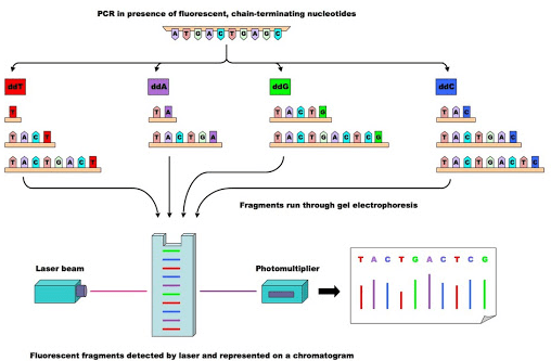
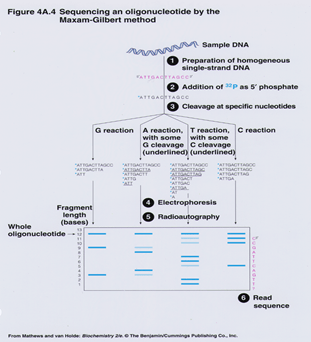
a. Study of Direct DNA Sequencing 

b. Molecular Automated Testing 

c. Drug resistance (MODS) and thin-layer agar (TLA) assays for microscopic observation 

d. Additional quick tests (e.g., BACTEC-460, luciferase reporter assays, ligase chain reaction, FASTPlaque TB-RIF) 

e. To test for potential related pulmonary findings, obtain a chest radiograph. 

Treatment : 

TB is cured with early detection and effective antibiotics. 

The correct type of antibiotic and the period of therapy will depend on: 

i. The overall health and age of the individual 

ii. If they have active or latent TB 

iii. The position of the infection 

iv. If the TB strain is immune to drugs 

v. Latent TB treatment can vary. It may mean taking an antibiotic for 12 weeks once a week or for nine months every day. 

Treatment for active TB can require 6-9 months of taking several drugs. The treatment would be more difficult if a person has a drug-resistant strain of TB. Completing the full course of treatment is significant, even if the symptoms go away. Some bacteria can survive and become immune to antibiotics if a person stops taking their medication early. The person may continue to develop drug-resistant TB in this case. 

Prevention: Ways of preventing anyone from being infected with TB include: 

i. Having an early diagnosis and treatment 

ii. Staying away from other individuals until the risk of infection is no longer present 

iii. Wearing a mask, shielding the mouth and rooms with ventilation 

iv. Vaccinating against TB 

In individual nations, as part of a routine immunization program, children receive an anti-TB injection, the bacillus Calmette-Guérin ( BCG) vaccine. 

The live strain of Mycobacterium bovis developed by Calmette and Guérin for use as an attenuated vaccine to prevent tuberculosis ( TB) and other mycobacterial infections is Bacille Calmette-Guérin (BCG). The vaccine was first given to humans in 1921 and remained the only vaccine for general use against TB. 

The BCG vaccine is the world’s most commonly administered vaccine; it has been given to over three billion people, mainly in the form of compulsory newborn immunization (as dictated by World Health Organization guidelines)[1]. Several BCG vaccines are used worldwide, manufactured by various manufacturers, and administered under different schedules. The BCG vaccine is also effective against other diseases, including leprosy and Buruli ulcer, for safety. Moreover, it is used in the treatment of superficial bladder carcinoma as an immunostimulant.

BACTERIAL DISEASE : LEPROSY

BY: SAI MANOGNA (MSIWM014)

Introduction : 

A chronic infection caused by the acid-fast, rod-shaped Mycobacterium leprae bacillus is leprosy. Leprosy is also known as Hansen’s disease. Two related diseases that mainly affect superficial tissues, especially the skin and peripheral nerves, maybe leprosy. A mycobacterial infection initially triggers a broad array of cellular immune responses. The second component of the condition, peripheral neuropathy with possible long-term effects, is then elicited by these immunologic events.

As a highly infectious and debilitating disease, leprosy was once feared, but nowadays, it does not spread quickly, and treatment is very successful. However, nerve damage can result in hands and feet being crippled, paralysis, and blindness if left untreated.

Classification of Leprosy :

The number and sort of skin sores have determined leprosy. The type of leprosy depends on specific symptoms and treatment. The forms are: 

Tuberculoid:  A moderate type of leprosy, which is less severe. People with this type only have one or a few patches (paucibacillary leprosy) of flat, pale-colored skin. Owing to nerve damage underneath, the affected region of the skin can feel numb. It is less infectious to tuberculoid leprosy than other types. 

Lepromatous:  Come on. A more extreme form of the disorder. It brings widespread skin bumps and rashes, numbness, and muscle weakness (multibacillary leprosy). It can also impact the nose, kidneys, and male reproductive organs. It is more infectious than leprosy caused by tuberculosis. 

Borderline: The tuberculoid and lepromatous symptoms are present for people with borderline leprosy. 

You can hear physicians use this simplified classification as well: 

Paucibacillary – single lesion (SLPB): One lesion 

Paucibacillary (PB): lesions from two to five 

Multibacillary (MB): Six lesions or more

Incubation period :

The incubation period is called the interval between contact with the bacteria and the appearance of symptoms. Symptoms typically take about 3 to 5 years to appear after coming into contact with the leprosy-causing bacteria. Up to 20 years later, some individuals do not show symptoms. The long incubation period of leprosy makes it very hard for doctors to determine when and where a person with leprosy has been infected.

Pathophysiology :

Depending on the host’s reaction to the organism, leprosy can manifest in various ways. 

1. The tuberculoid type of the disease that usually affects the skin and peripheral nerves are present in individuals who have a robust cellular immune response to M leprae. 

2. They tend to be dry and hypoesthetic, and the number of skin lesions is small. 

3. Typically, nerve activity is asymmetric. Owing to the low number of bacteria in the skin lesions, i.e., < 5 skin lesions, with no organisms on the smear, this type of the disease is often referred to as paucibacillary leprosy. 

4. In these people, the findings of skin tests with antigens from killed species are positive.

5. The lepromatous type of the disease, characterized by extensive skin involvement, is for individuals with limited cellular immune response. 

6. Infiltrated nodules and plaques are often identified as skin lesions, and nerve involvement appears to be symmetric in distribution. 

7. The organism grows best at 27-30 ° C; therefore, skin lesions tend to develop in the body’s colder areas, with sparing of the groin, axilla, and scalp. 

8. Owing to no small number of bacteria present in the lesions, i.e.,> 6 lesions, with potential bacilli visualization on the smear, this type of disease is often referred to as multibacillary leprosy. 

9. Skin test results for antigens from killed species are non-reactive.

Patients can also have symptoms of both types (indeterminate or borderline leprosy) but typically evolve to one or the other over time. Interestingly, most people exposed to leprosy never contract the disease, possibly because more than 95 percent of individuals are naturally immune to this disease.

Epidemiology :

In 2018, 208,619 new leprosy cases were reported globally, according to WHO estimates based on data from 159 countries. The worldwide prevalence was 184,212 cases (rate, 0.2/10,000) reported at the end of 2018. In 2018, 79.6 percent of all new leprosy cases were in Brazil, Indonesia, and India. Furthermore, in 2018, 23 priority countries accounted for 96 percent of cases globally.

a. Mortality/Morbidity :

Leprosy is never lethal. The nerve damage and crippling sequelae are the main consequences of infection. 33-56 percent of newly diagnosed patients have already demonstrated symptoms of compromised nerve function[11], according to one report. According to reports, three million individuals who have undergone multidrug treatment for leprosy have suffered impairment due to nerve damage. While the skin and peripheral nerves are involved in both lepromatous leprosy and tuberculoid leprosy, tuberculoid leprosy has more severe manifestations. Nerve involvement contributes to sensory and motor loss of control, leading to repeated trauma and amputation. Most generally, the ulnar nerve is involved. 

Damage to the following nerves is related to characteristic leprosy impairment: 

i. Ulnar and Median-Hand Clawed 

ii. Tibial posterior-Plantar insensitivity and clawed toes 

iii. Peroneal Common-Foot Drop 

iv. Radial nerves of the cutaneous, nasal, and greater auricular

b. Race : 

Leprosy was once globally endemic, although no predilection for the race is recognized. The incidence of leprosy fell significantly in northern Europe and North America in the late 1800s, and the disease is now recorded mainly in tropical areas. 

c. Gender :

In males, leprosy is typically more common than in females, with a 2:1 male-to-female ratio. The prevalence of leprosy among females is equal to or greater than that of males in some areas of Africa. 

d. Age :

Leprosy can occur at any age, but the age-specific occurrence of leprosy in developed countries peaks in children younger than ten years, accounting for 20 percent of leprosy cases. In infants, leprosy is very rare; however, they are at relatively high risk of maternal leprosy, particularly in cases of lepromatous leprosy or mid borderline leprosy.

e. HIV Coinfection :

Preliminary data suggest that, unlike tuberculosis, HIV coinfection does not appear to affect leprosy. Besides, coinfection with HIV does not seem to affect the lepromatous to tuberculoid leprosy ratio.

Symptoms of Leprosy 

Leprosy mostly affects the skin and nerves, called the peripheral nerves, beyond the brain and spinal cord. 

Disfiguring skin sores, bumps, or lumps that do not go down for several weeks or months is leprosy’s principal symptom. The sores on the skin are pale-colored. Nerve damage leads to loss of sensation in legs and arms, weakness of muscles.

Causes :

Exactly how leprosy is transmitted is not clear. When an individual coughs or sneezes with leprosy, they can spread droplets containing M. The leprosy bacteria that someone else breathes in. For leprosy to be transmitted, near physical contact with an infected individual is required. Shaking hands, kissing, or sitting beside them during a meal on a bus or at a table, do not transmit the disease.

Pregnant leprosy mothers are unable to pass it on to their newborn babies. Neither is it spread through sexual touch.

Risk Factors :

Leprosy can permanently damage skin, nerves, arms, legs, feet, and eyes without care. 

Leprosy complications may include: 

Glaucoma or blindness 

Iritis 

Loss of hair 

For infertility 

Facial disfigurement (including permanent swelling, bumps, and lumps) 

Erectile dysfunction in males and infertility 

Failure of the kidney 

Weakness in the muscles contributing to claw-like hands or not being able to stretch feet 

The inside of the nose is permanently damaged, which can lead to nosebleeds and a chronic stuffy nose 

Permanent nerve damage, including those in the arms, legs, and feet, outside the brain and spinal cord 

Damage to the nerves can lead to a dangerous loss of feeling. Do not experience pain when one gets cuts, burns, or other injuries to hands, legs, or feet if they have leprosy-related nerve damage.

Diagnosis :

It is possible to distinguish Hansen’s disease by the presence of skin patches that may look lighter or darker than normal skin. The skin areas affected can often be reddish. The lack of sensation is prominent in these skin patches. For a needle, one cannot feel a light brush or a prick. 

The physician will take a sample of skin or nerve (through skin or nerve biopsy) and check for bacteria under the microscope and confirm your diagnosis, and may even conduct tests to rule out any skin diseases.

Treatment :

It is possible to treat leprosy. 16 million people with leprosy have been healed in the last 2 decades. The World Health Organization provides free care for all people with leprosy. 

Therapy relies on the type of leprosy they have. For treatment, antibiotics are used. Doctors recommend treatment on a long-term basis, usually for 6 months to a year. You may need to take antibiotics longer if you are suffering from severe leprosy. The nerve damage that comes with leprosy cannot be treated with antibiotics. 

A standard treatment for leprosy that combines antibiotics is multidrug therapy (MDT). That means you are going to take two or more drugs, mostly antibiotics: 

Paucibacillary leprosy: Two antibiotics, such as dapsone, are used every day, and rifampicin is used once a month. 

Multibacillary leprosy: In addition to daily dapsone and monthly rifampicin, you can take a daily dose of the antibiotic clofazimine. For 1-2 years, you will undergo multidrug treatment, and then you will be healed.

Antibiotics: The bacteria causing leprosy can be destroyed by antibiotics used during treatment. However, though medication may cure the condition and keep it from getting worse, nerve damage or physical disfigurement may have existed before a diagnosis is not reversed. Thus, before any irreversible nerve damage occurs, the condition must be detected as soon as possible.

Transmission : 

The exact way Hansen’s disease spreads between people is not understood. Scientists claim that it can happen if a person with Hansen’s disease coughs or sneezes, and a healthy person breathes in the droplets that contain the bacteria. Prolonged close contact is required with someone with untreated leprosy for several months. 

You do not get leprosy from casual contact with a person who has Hansen’s disease, such as: shaking or hugging palms, sitting for a meal together, sitting on the bus next to each other. 

During pregnancy, Hansen’s disease is also not transmitted from a mother to her unborn baby, and it does not spread by sexual contact. It is often challenging to locate the infection source due to the bacteria’s slow-growing nature and the long time to cause symptoms of the disease.

For general health purposes, if possible, avoid contact with armadillos. Speak to your physician if you have come into touch with an armadillo and are anxious about having Hansen’s disease. Over time, the doctor will follow up with you and conduct annual skin tests to see if the disease progresses. 

Prevention : 

In high-risk areas, awareness campaigns on leprosy are essential to enable patients and their families, who have been traditionally shunned from their communities, to come forward and receive care. 

Early diagnosis and multidrug therapy treatment is the most effective way to avoid leprosy disabilities and avoid further disease spread. However, the Bacille Calmette-Guérin ( BCG) vaccine is partially protective against leprosy.

BACTERIAL DISEASE – TYPHOID

BY: SAI MANOGNA (MSIWM012)

Introduction :

Any disease caused by bacteria involves bacterial diseases. Bacteria, which are small types of life that can only be seen through a microscope, are microorganisms. Viruses, some fungi, and some parasites include other types of microorganisms. Millions of bacteria usually reside in the skin, intestines, and genitals. The vast majority of bacteria cause no disease, and many bacteria are beneficial and even required for good health. Often, these bacteria are referred to as good bacteria or healthy bacteria. 

Pathogenic bacteria are considered dangerous bacteria that cause bacterial infections and illnesses. When these invade the body and begin to replicate and crowd out healthy bacteria or develop in typically sterile tissues, bacterial diseases occur. Toxins that damage harmful bacteria can also release the body.

Typhoid :

An infectious, potentially life-threatening bacterial infection is typhoid fever, also called enteric fever. Typhoid fever is caused by the Salmonella enteric serotype Typhi bacterium (also known as Salmonella Typhi), carried into the blood and digestive tract by infected humans and spreads by food drinking water contaminated with infected feces to others. Typhoid fever signs include fever, rash, and pain in the abdomen. 

Fortunately, typhoid fever, particularly in its early stages, is treatable, and if one chooses to live in or fly to high-risk areas of the world, a vaccine is available to help prevent the disease.

Incubation Period :

Typhoid and paratyphoid infections have an incubation period of 6-30 days. With steadily rising exhaustion and a fever that rises daily from low-grade to as high as 102 ° F to 104 ° F ( 38 ° C to 40 ° C) by the third to the fourth day of illness, the onset of illness is insidious. In the morning, fever is usually the lowest, peaking in the late afternoon or evening. 

Pathophysiology :

1. When present in the gut, all pathogenic Salmonella species are swallowed up by phagocytic cells, moving them through the mucosa and presenting them to the lamina propria macrophages. 

2. Across the distal ileum and colon, nontyphoidal salmonellae are phagocytized. Macrophages identify pathogen-associated molecular patterns (PAMPs) such as flagella and lipopolysaccharides with the toll-like receptor (TLR)-5 and TLR-4 / MD2 / CD-14 complex. 

3. Macrophages and intestinal epithelial cells are then attracts the interleukin 8 (IL-8) T cells and neutrophils, inducing inflammation and suppressing the infection. 

4. Unlike the nontyphoidal salmonellae, S typhi and paratyphi penetrate mainly via the distal ileum into the host system. They have specialized fimbriae that bind to the epithelium over lymphoid tissue clusters in the ileum, the critical point of relay for macrophages moving into the lymphatic system from the stomach. 

5. The bacteria then attract more macrophages by activating their host macrophages.

6. Typhoidal salmonella co-opts the cellular machinery of the macrophages for their reproduction, as they are transported to the thoracic duct and lymphatics to the mesenteric lymph nodes and then to the reticuloendothelial tissues of the spleen, bone marrow, liver, and lymph nodes. 

7. Once there, until some critical density is reached, they pause and begin to multiply. Afterward, to reach the rest of the body, the bacteria cause macrophage apoptosis, breaking out into the bloodstream. 

8. By either bacteria or direct extension of infected bile, the bacteria then invade the gallbladder. The effect is that in the bile, the organism re-enters the gastrointestinal tract and reinfects patches of Peyer. 

9. Usually, bacteria that do not reinfect the host are shed in the stool and are then available for other hosts to invade.

Epidemiology :

The International 

Worldwide, typhoid fever occurs mostly in developing countries where sanitary conditions are low. In Asia, Latin America, Africa, the Caribbean, and Oceania, typhoid fever is endemic, but 80 percent of cases originate from Bangladesh, China, India, Indonesia, Laos, Nepal, Pakistan, or Vietnam. In underdeveloped countries, typhoid fever is the most common. About 21.6 million people are infected by typhoid fever (incidence of 3.6 per 1,000 population), and an estimated 200,000 people are killed every year. 

Most cases of typhoid fever occur among foreign travelers in the United States. The average annual incidence of typhoid fever by county or area of departure per million travelers from 1999-2006 was as follows:

Outside Canada / United States, Western Hemisphere-1.3 

Africa-7.6 Africa 

Asia-10.5. 

India-89 (in 2006 122) 

Complete (except for Canada / United States, for all countries)-2.2 

Morbidity / Mortality :

Typically, typhoid fever is a short-term febrile condition with timely and effective antibiotic care, requiring a median of 6 days of hospitalization. It has long-term sequelae and a 0.2 percent mortality risk when treated. Untreated typhoid fever is a life-threatening disease that lasts many weeks, frequently affecting the central nervous system, with long-term morbidity. In the pre-antibiotic age, the case fatality rate in the United States was 9 -13%.

Sex : 

Fifty-four percent of cases of typhoid fever recorded between 1999 and 2006 in the United States included males. Moreover, race has no predilection. 

Age : 

Many confirmed cases of typhoid fever include children of school age and young adults. The true incidence is, however, thought to be higher among very young children and babies. The presentations may be atypical, that ranges from a mild febrile disease to severe convulsions, and the infection of S.typhi may go unrecognized. In the literature, this could account for contradictory reports that this category has very high or very low morbidity and mortality rate.

Symptoms :

Typhoid fever symptoms typically occur five to 21 days after food or water infected with Salmonella Typhi bacteria is consumed and can last up to a month or longer. Typical Typhoid Fever signs include: 

i. Pressure in the abdomen and tenderness 

ii. Perplexity 

iii. Fatigue and Weakness 

iv. Trouble focusing 

v. Constipation or diarrhea

vi. Headaches 

vii. The Nosebleeds 

viii. A dry cough 

ix. Impoverished appetite 

x. Rash (small, flat, red rashes that are also known as rose spots on the belly and chest) 

xi. Lethargy 

xii. Swollen lymph ganglions 

xiii. Chills and Fever. With typhoid fever, persistent fever of 104 degrees Fahrenheit is not rare. 

Symptoms: life-threatening 

Typhoid fever, including intestinal bleeding, kidney failure, and peritonitis, may lead to life-threatening complications. If they are with anyone who has any of these signs, seek urgent medical attention : 

Bloody stools or severe rectal bleeding 

A shift in consciousness or alertness level 

Confusion, delirium, disorientation, or hallucinations 

Unexplained or chronic dizziness 

Dry, broken lips, tongue, or mouth 

Unresponsiveness or lethargy 

Not urinating tiny quantities of tea-colored urine or urinating it. 

Extreme pain in the abdomen 

Extreme diarrhea in patients 

Extreme signs in infants include sunken fontanel (soft spot) at the top of the head, lethargy, no weeping tears, little or no wet diapers, and diarrhea. Infants two months of age or younger, be especially concerned about fever.

Causes :

The Salmonella Enteric Serotype Typhi (Salmonella Typhi) bacterium is responsible for typhoid fever. Via ingestion of infected food and water, Salmonella Typhi can enter and infect the body. By being washed in polluted water or being touched by an infected person with unwashed hands, food can become contaminated with the bacteria. Drinking water can become infected with untreated Salmonella Typhi-containing sewage.

Risk factors :

A variety of variables improves the chances of contracting typhoid fever. In developing, non-industrialized countries, typhoid fever is a significant health threat, although rare in the United States, Canada, and other industrialized countries. Factors of vulnerability include: 

i. Near contact with individuals infected or recently infected 

ii. Travel to areas with more frequent and widespread outbreaks of typhoid fever, such as India, Southeast Asia, Africa, and South America

iii. Avoiding contact with a person who has or has signs of typhoid fever, such as abdominal pain, headache, and fever

iv. Residence in a developing world or continent with inadequate treatment facilities for water and sewage or poor hygiene practices 

v. Due to diseases such as HIV / AIDS or drugs such as corticosteroids, the compromised immune system 

vi. Do not eat fruits and vegetables that are unable to peel. Eating fully cooked, hot, and still steaming foods. Unless it is made from distilled water, drinking only bottled water and not using ice 

vii. Before visiting high-risk areas, having vaccinated against typhoid fever 

viii. During and after contact with an individual who has typhoid fever or with an individual who has signs of typhoid fever, such as abdominal pain, headache, rash, and fever, washing hands regularly with soap and water for 15 seconds 

ix. Washing hands regularly for at least 15 seconds with soap and water, particularly before handling food and after using the toilet, touching feces, and changing diapers

Diagnosis :

1. Salmonella bacteria infiltrate the small intestine following the ingestion of infected food or drink and temporarily enter the bloodstream. 

2. The bacteria are transported into the liver, spleen, and bone marrow by white blood cells, replicating and re-enter the bloodstream. 

3. At this point, people develop symptoms, including fever. Bacteria invade the biliary system, gallbladder, and the intestinal lymphatic tissue. 

4. Here, in high numbers, they multiply. In the digestive tract, the bacteria move and can be found in stool samples. 

5. Blood or urine samples will be used to diagnose if a test result is not exact.

Treatment :

Typhoid fever is a treatable condition, and a complete course of antibiotics, such as ampicillin, trimethoprim-sulfamethoxazole, or ciprofloxacin, may also be used to cure it. Treatment can include rehydration with intravenous fluids and electrolyte replacement therapy in some severe cases. Usually, with care, symptoms improve within two to four weeks. If they have not been treated completely, symptoms may return. One needs to take the antibiotics for as long as needed to treat typhoid fever and follow up with the doctor for a series of blood and stool tests to ensure that they are no longer infectious. 

Few people infected with Salmonella Typhi become carriers, which indicates that the bacteria are present in the intestines and bloodstream and are shed in the stool even after they no longer have disease symptoms. Because of the carrier effect, it is essential to understand that they might still transmit the disease by contaminating food and water even after receiving treatment for typhoid fever. Before traveling outside developed regions, such as the United States, Canada, northern Europe, Australia, New Zealand, and Japan, it is vital to avoid the disease by getting vaccinated. During epidemic outbreaks, immunizations are also recommended, although the vaccination is not successful.

Prevention :

A larger number of typhoid cases usually occur in countries with less access to clean water and washing facilities. 

Immunization :

Vaccination is advised while traveling to a region where typhoids are prevalent. 

It is recommended to get vaccinated against typhoid fever before traveling to a high-risk area. 

Oral treatment or a one-off injection can be done : 

Oral: an attenuated, live vaccine. It consists of 4 tablets, one of which is taken every other day, the last of which is taken one week before departure. 

Shoot, the inactivated vaccine, was given two weeks before the ride. 

Vaccines are not 100 percent successful, and when eating and drinking, caution should always be exercised. 

Two forms of typhoid vaccine are available, but a more potent vaccine is still required. The vaccine’s live, oral form is the strongest of the two. It also protects individuals from infection 73 percent of the time after three years. This vaccine has more side effects, however. If the person is currently ill or if he or she is under the age of 6 years, vaccination should not begin. The live oral dose should not be taken by someone who has HIV. There may be adverse effects of a vaccine. One in every 100 people is going to feel a fever. There may be stomach complications following the oral vaccine, nausea, and headache. For any vaccine, however, serious side effects are uncommon. 

Typhoid removal :

Even if typhoid symptoms have passed, it is still possible to bear the bacteria, making it impossible to stamp out the disease because when washing food or communicating with others, carriers whose symptoms have terminated may be less vigilant. 

Prevention of Infection :

Via touch and ingestion of contaminated human waste, typhoid is propagated. This can happen through a source of water that is tainted or when food is treated. 

Some general rules to obey while traveling to help reduce the risk of typhoid infection are the following: 

i. Drink water, preferably carbonated, in glasses. 

ii. Do not have ice for drinks. Stop raw fruit and vegetables, cut the fruit, and not eat the cut on your own. Eat only food that is still hot, and avoid eating at street food stands.

iii. If it is impossible to acquire bottled water, ensure the water is heated for at least one minute on a rolling boil before consumption. 

iv. Be wary of eating something that anyone else has dealt with. 

Related Disorders :

There may be similar symptoms of the following conditions to those of typhoid fever. For a differential diagnosis, similarities may be helpful: 

Salmonella Poisoning :

In foodborne diseases, this is the most common cause of disease. These bacteria can contaminate meat, dairy, and vegetable products. In warm weather and children under the age of seven, outbreaks are more prevalent. The most common initial symptoms are nausea, vomiting, and chills. These are accompanied by stomach pain, diarrhea, and fever that can last for several weeks to five days. Intoxication with salmonella is a type of gastroenteritis. The CDC reports about 2 to 4 million salmonellosis cases per year in the United States. 

Cholera :

Cholera is a bacterial infection characterized by extreme diarrhea and vomiting that affects the whole small intestine. A toxin produced by the bacteria Vibrio cholerae is the source of the symptoms. The disease is transmitted by drinking water or consuming fish, vegetables, and other foods contaminated with Cholera’s excrement.

Botulism :

Botulism is also a form of gastroenteritis caused by a bacterial toxin. A neuromuscular poison is this toxin. In three types, it occurs foodborne, wound, and infantile botulism. The foodborne type is the most popular. Besides nausea, vomiting, diarrhea, and stomach pain, the patient can feel exhaustion, fatigue, headache, and dizziness.

Ptomaine Poisoning in the United States’ fourth most prevalent cause of bacterial foodborne disease. It is caused by the enterotoxin protein released after consuming foods that are contaminated, usually meat products. Extreme stomach cramps and diarrhea are characteristics of the disease. Nausea also happens sometimes. Vomiting and fever are rare. 

HUMAN GENOME PROJECT

BY: SREE LAKSHMI (MSIWM012)

The Human Genome Project was an international research effort to determine the genetic makeup of humans and to identify the genes that it contains. The Human Genome Project was launched in 1984 but officially launched in October 1990. It is a major international co-operation program, with the ultimate goal of achieving a “nucleotide sequence across nuclear genetics”. The Global Research Group consisted of six different countries namely USA, UK, France, Germany, Japan and China as well as several laboratories, a large number of scientists and experts from various fields.

Objectives of the Human Genome Project:

1. Establish a complete genetic sequence and make it easy to access.

2. Improve sequencing technology by developing new and more efficient methods.

3. Analyze genetic variation in the human genome, such as single nucleotide polymorphisms (SNPs) and other DNA sequence variants.

4. Improving the performance of genomics technology. Includes the creation of additional cDNA sources and detailed genetic analysis technologies; a complete study of non-protein coding sequence activities; and to promote the development of global protein analysis technologies.

5. Learn comparative genomics by completing a sequence of a particular type of model (e.g. mouse etc.) that can enhance our understanding of human genes.

6. Consider the moral, legal and social consequences of a growing knowledge base. It is expected that the conflict between this new and advanced knowledge and existing philosophical ideas could have undesirable consequences, which need to be addressed.

7. Develop bioinformatics and computational biology to transfer advanced training to young scientists and to promote the development of academic fields in genetic research.

 Most important features of HGP:

• The human genome contains the details of 23 chromosomes

• Itcontains more than three billion nucleotides.

• The human genome is estimated to have over 30,000 genes. The average gene has 3000 bases. But gene sizes vary, and the gene dystrophin gene has 2.5 million bases.

• Only about 3% of the genome contains amino acid sequences of polypeptides and the rest is in the trash (DNA duplicated).

• Jobs are known to more than 50% of the genes found.

• Repeated sequences make up the largest part of the human genome. Repeated sequences do not use direct coding but illuminate chromosome formation, dynamics and evolution.

• Chromosome 1 has many genes (2968) and Y chromosome is very small (231).

• Almost all nucleotide bases are exactly the same for all humans. The genome sequence of different populations varies less than 0.2% of base pairs.

• Most of the differences come in the form of variations from one foundation to another. One primary variation which is called single nucleotide polymorphisms (SNPs) is derived from all ~ 1,000 bp of the human genome. About 85% of all differences in human DNA are due to SNPs.

Main Objectives:

• Find the complete sequence of DNA extracted from cells donated by several unknown donors, to determine the sequence of DNA in each chromosome.

• Genetic mapping to simplify genetic linking studies.

• Obtaining all human genes to allow for the continuation of human genetic studies.

• Develop simple and automated DNA sequencing technologies.

Updated objectives:

• Determining the sequence of human DNA

• Identify all the genes in a person’s DNA

• Store this information in archives

• Improving data analysis tools

• Transfer of technology related to the private sector

• Address ethical, legal and social issues (ELSI) that may arise in the project.

Project result:

• Has a base base pair of 3164.7

• Genetics have 3000 bases, but sizes vary widely. The gene known as dystrophin has 2.4 million bases.

• The total number of genes is estimated was 30,000.

• Jobs are known to more than 50% of the genes found

• Less than 2% of genome protein codes.

• Dense men with “urban middle-class genomes” are made up of DNA blocks of G and C.

• In contrast, genetically engineered “deserts” are rich in DNA A and T genes.

Chromosome 1 has many genes (2968), and Y chromosome has very few (231). Scientists have identified about 1.4 million places where single DNA (SNPs) mutations occur in humans, and these findings will help to link disease sequences to chromosomes. Cancer with the help of human maps (SNPs) produced in the Human Genome Project.

Applications:

• Identification of human genes and their functions.

• Understand polygenic disturbances e.g. cancer, high blood pressure, diabetes

• Advances in gene therapy

• Advanced diagnosis

• Development of pharmacogenomics

• Genetic basis for mental disorders

• Understanding the norms of the general public

• Advanced knowledge of genetic modification

POLYPHENOLS

BY: SREE LAKSHMI (MSIWM012)

 Polyphenols are one of the most important and certainly the most abundant among the phytochemicals present in the plant kingdom. Polyphenols are a large and complex group of chemical substances or phytochemicals or plant-derived foods, which contain more than one phenolic hydroxyl group. Natural compounds found mainly in fruits, vegetables, grains and beverages. Chemical composition of polyphenols. Polyphenols are compounds that contain a -OH group placed in a benzene ring. Naturally, they are a compound with structural phenolic properties, which can be associated with various organic acids and carbohydrates.

The main categories of polyphenols –

1. Phenolic acids and products

2. Flavonoids

3. Stilbenes

4. Lignans

1. Phenolic Acids and Derivatives

 Phenolic Acids are a non-flavonoid polyphenolic chemical. It is further divided into two main types – benzoic acid and cinnamic acid derivatives based on C1-C6 and C3-C6.It is found in a variety of plant foods; the seeds and skins of fruit and vegetable leaves contain very high concentrations.

Types and resources:

Hydroxybenzoic acid:

 Eg. Ellagic and gallic acid – with water-based tannins – are present in berries and nuts, tea

Hydroxycinnamic acid:

 Eg. Caffeic and ferulic acid.

• Caffeic acid – a precursor to lignin

• Caffeic and Quinic acid provide chlorogenic acid.

• Fruits, vegetables, coffee beans, grains and sunflower seeds appear.

• Other sources are coffee, blueberry, kiwis, plums, cherries, apples, red wine, and cereals: corn, whole grains, oats, and rice.

 Benefits of Phenolic acid

It is easily absorbed into the walls of your intestinal tract, and can be beneficial to health as they act as antioxidants that prevent cell damage due to excessive oxidation reactions.They can also promote anti-inflammatory properties in the body when used.

2. Flavonoids

They form the largest group of plant polyphenols.They are found in different colors of plants yellow, orange and red.They have a standard structure C6-C3-C6 where two C6 units are phenolic.Due to the hydroxylation pattern and variability in the chromane ring , flavonoids can continue to be subdivided into subgroups such as –

Anthocyanins

 Flavan-3-ols

 Flavanones

 Flavonols

 Flavones, Flavonols, Flavanones, Flavanonols

These small groups are very common and are found throughout plants.Flavones and their flavonols derived from 3-hydroxy, including glycosides, methoxides and other acetylled products in all three rings, make this the largest group among all polyphenols.The most common flavonol aglycones – Quercetin and Kaempferol

3. Stilbenes

 They are not as rich in flavonoids, lignans and phenolic acids.There are two stilbenes

Resveratrol

Pterostilbene

Resveratrol

 It is a non-flavonoid polyphenolic compound found in the skin of dark grapes and products made from such grapes, such as wine and grape juice.It belongs to the stilbene class of aromatic phytochemicals and occurs as free (cis or trans transations) aglycone (slightly soluble but active form) or as glycoside (called piceid, highly soluble form).It is transferred across the intestinal tract and the circulatory system such as glucuronide (glycoside) but can also be injected into aglycone form once it has reached the organs or body fluids where it is processed by β-glucuronidases.This compound is a well-known inhibitor of cyclooxygenase-1 and cyclooxygenase-2 , which are commonly shown in colon cancer.It also inhibits monoamine oxidase while enhancing Ab protease degradation and acts as an effective antioxidant.

4. Lignans

It is a small group of non-flavonoid polyphenols. It is widely distributed in plant kingdoms – standing in more than 55 plant families – acting as antioxidants and antibodies against germs and viruses. Lignans are  found in more than 60 families of vascular plants .Biological activity includes Antiviral, anticancer, cancer prevention, anti-inflammatory, antimicrobial, antioxidant, immunosuppressive, hepatoprotective, osteoporosis Prevention.

 Health benefits of polyphenols

1. It can lower blood sugar levels

Polyphenols can help lower blood sugar levels leading to a lower risk of type 2 diabetes. This is because polyphenols can prevent the decomposition of starch into light sugars, reducing the chances of high blood sugar after a meal.Polyphenols can also help to stimulate insulin secretion. A rich polyphenol diet can lower the blood sugar levels, high glucose tolerance, and increases insulin sensitivity which are important in reduction of type 2 diabetes.

 2. It can improve the functioning of heart

Polyphenols can improve heart health.This is mainly due to the antioxidant properties of polyphenols that help reduce chronic inflammation which is a risk factor for heart disease.Polyphenol supplementation lowers blood pressure and LDL (bad) cholesterol and increases HDL (good) cholesterol.

3. It can prevent blood clots

Polyphenols can reduce the risk of bleeding. Blood clots form when platelets circulate in the blood and begin to clot. This process is known as platelet aggregation and helps prevent excessive bleeding.Excessive platelet aggregation can cause blood clots, which can have serious health consequences, including deep vein thrombosis, stroke, and pulmonary embolism.

 4. It can protect against cancer

 Foods rich in polyphenols reduce the risk of cancer. Polyphenols have powerful antioxidant and anti-inflammatory effects, both of which can help prevent cancer.

5. It can promote healthy digestion

 Polyphenols can benefit digestion which increases the intestines of beneficial bacteria while protecting harmful ones. Polyphenol-rich tea extracts can promote beneficial bifidobacterial growth.Green tea polyphenol can help fight harmful germs, including C. Difficile, E.coli, Salmonella and improvement in symptoms of peptic ulcer disease (PUD) and inflammatory bowel disease (IBD).

 6. It can improve brain function

Polyphenol-rich foods can increase concentration and memory. Grape juice – rich in polyphenols – helps increase memory in older adults with mild mental retardation. Cocoa flavanols can increase blood flow to the brain and link these polyphenols to improve working memory and attention. The extraction of rich Gypsum polyphenols Biloba plants seems to increase memory, learning, and concentration. It also linked improved mental functioning and short-term memory loss.

VECTOR-BORNE DISEASES

BY: SAI MANOGNA (MSIWM013)

Vector-borne infections, diseases caused by insects- and tick-borne pathogens, have long affected human affairs. The bite of a tiny mosquito carrying malaria parasites in the marshes of what is now called Iraq defeated Alexander the Great, the conqueror of many nations. To this day, vector-borne diseases remain influential, flooding the hospitals of sub-Saharan Africa with malaria victims, suppressing nations’ economies, and destroying industrial operations where they remain endemic. Specific, less common agents cause blindness and terrible disfigurement. Together, they form an alarming set of possible health and livelihood risks to those who travel and work or live in the tropics where they have the most significant effect.

Vectors :

Arthropod (insect) vector transmitted pathogens are some of the most harmful and volatile on earth. They are perhaps the hardest to stop or manage because they are so immune to intrusion and so deeply rooted in the habitats and environments of the regions they infest. In this equation, vectors make all the difference since they increase the range and transmissibility of pathogens exponentially over those that rely on transmission through direct human touch. Vectors help pathogens cross the gap between humans and various host animals (mice, rodents, monkeys, birds, prairie dogs, pigs). Over cycles less favorable to transmission (winters, dry seasons), some harbor reservoirs of pathogens. Without approaching the vector directly, vectors are facilitators of several harmful disease-causing species, the prevention, and treatment of which can not be successful for long.

For their entire lives, vectors stay infected, which is longer than others. 

Individuals think. For example, a mosquito does not have a set lifespan. In their first week of life, many die, but some will live almost forever. They are restricted by the harm that accumulates on their wings and appendages that are not repairable and do not degrade as soon as they wear out. In water, predation, desiccation, and trapping are likely to kill more mosquitoes than any other source. West Nile vectors that emerge in August of one year on the East Coast of the United States can be active over winter and in May of the following year for a life span of at least nine months.

The bulk of transmissions are due to mosquitoes and ticks. Large vector-borne diseases are involved, including sand flies and black flies. Even within a closely related community, each of these species has specific habitat requirements and feeding habits, which can differ significantly. For instance, around the globe, dozens of species of Anopheles mosquitoes can spread malaria. Some of them bite at night, some only at dawn and at dusk, in bright sunshine, some breed. Others never come out of the forest’s deep shade. Saltwater, for others, is lethal. In water with a saline content approaching that of seawater, some excel. Generalization, generally, fails when it comes to vector biology. The unique vector behaviors provide the keys to managing them and preventing infection from spreading.

The insect-borne disease of industrial significance :

Mosquitoes: Mosquitoes, by far the most important vector of disease, number more than 3,000 species worldwide. Only the female mosquito can spread the disease since only she, and not the male, has the knife-like part of the mouth required to extract blood from her victims. She requires a blood meal to allow protein for egg creation.

Sand Flies: Closely related to mosquitoes, sandflies are blood-feeders and breed in caves, rodent burrows, manure piles, and other dark areas that hold moisture and are rich in organic matter. They are weak fliers, tending to travel from host to host in fast “hopping” flights. Their bodies are so tiny (3 mm) that they are hard to detect before they begin to bite. Their bite has been creating extreme pain for a few days.

Black Flies: Black flies are yet another relative of mosquitoes specializing in flowing water from small trickles to large rivers. Unlike mosquitoes, black flies eat through the skin and never eat indoors. They can attack such large quantities that their salivary fluids alone can cause a person to get sick, causing a disease called “black fly fever.” They can also vector a nematode that can live in the human body for up to 15 years, killing tissue in the internal organs, most notably in the skin, causing blindness in the eyes.

Ticks: Ticks usually have a much longer life span than a mosquito. Hard ticks eat just a few times during their lifetime, limiting their risk of being infected. However, the persistence and host selectivity of hard ticks allow them to be relatively useful vectors. Soft ticks are long-lived nests and burrow dwellers. Like mosquitoes, they will feed several times during their lifetime.

Important Vector-borne Diseases:

A. Malaria : 

Malaria occurs in every tropical and subtropical landscape across the globe, often allowing seasonal excursions to temperate areas. The genomes, metabolisms, and life cycles of protozoan parasites are more complex than almost any other vector-borne threat. This makes it a difficult target for medicines and vaccines because the parasite’s shape-shifting pathways make it possible to avoid chemical and immunological defenses. They are also a moving target, deliberately modifying their outer coating during each step of their life cycle and producing a diverse antigenic and metabolic wardrobe through sexual recombination, a generator of diversity that is not available to simpler microbes such as viruses and bacteria.

1. Four parasite species affect humans, but more than 95 percent of cases include two of them, Plasmodium falciparum and P. vivax. The most dangerous pair, P. falciparum, crosses Africa’s deep tropics towards South America and Asia.

2. P. vivax, which may grow in mosquitoes at colder temperatures, has a broader range, spreading beyond the fringes of P. falciparum’s distribution and coexisting with P. falciparum in many regions.

3. P. falciparum malaria poses the greatest threat to the industry of any vector-borne disease, as it can kill an unprotected person very quickly and can reinfect and repeatedly weaken even those who develop semi-protective immunity. 4. Most deaths in local communities occur in children between 6 months and 2 years of age. 

5. Their weakness stems from a lack of immunological defense. Immune evasiveness of malaria parasites prevents full immunity production, but older children and adults who have undergone multiple infections enjoy some degree of defense against the most extreme manifestations of the disease.

Prevention: Prevention of malaria needs serious care while visiting areas where it is transmitted. While no vaccine is currently available, prophylactic drugs and steps to minimize exposure to night-biting Anopheles mosquitoes, such as bed nets and repellents, could be beneficial. Unlike other diseases, malaria patients also never get a second chance.

B. Dengue Fever :

1. Dengue virus tends to be the epithet of malaria in several respects. While malaria transmission is most prevalent in rural areas, Dengue is a city disease. 

2. Although Anopheles ‘malaria vectors bite mostly at night, Aedes’ dengue vectors bite mostly during the day. While the initial malaria infection usually causes the most severe symptoms, a second dengue infection may be much more severe than the first if it includes a different virus’s different serotype. 

3. Dengue fever can be painful (hence the nickname of “breakbone fever”) and Weakening, but generally not life-threatening when first obtained. 

4. However, there are significant manifestations in places where more than one of the virus coexists’ four significant strains. 

5. Being exposed to second, different strains of the virus may cause a severe immune reaction called Dengue Hemorrhagic Fever (DHF), leading to a significant risk of death, especially in children and younger adults. 

6. Currently, about five percent of the hundreds of thousands of people who receive DHF die, while timely and successful medical attention can dramatically reduce this case’s fatality rate.

Prevention: There are no prophylactic drugs or vaccines available to prevent Dengue, but steps that restrict or prevent biting, such as repellents or elimination of water-bearing containers in which mosquito vectors can be created, are helpful.

C. Arboviruses : 

There is a wide range of often-hazardous viruses almost everywhere on the globe where mosquitoes are found. Each of these individual viruses typically has limited ranges, and many affect relatively small populations; we consider them to be a group because of their mutual importance. 

More infamous pathogens and those currently in the news due to the revival or extension of their borders. In many places, these infections may account for poorly reported: “Fever of Unknown Origin” (FUOs) typically occurring in seasons when mosquitoes are most involved.

i. Chikungunya :

Due to its recent revival, Chikungunya tops this list in places including India, Sri Lanka, Mauritius, and Europe’s countries engaged in regular tourism to these destinations. Concern has recently emerged that it will soon expand its range in Europe due to the spread of Asian tiger mosquitoes. (Aedes albopictus), which can serve as a significant vector for this infection. 

Africa and Southeast Asia are also part of the traditional range of this virus. 

Chikungunya infection may be severe and temporarily crippling but is usually not life-threatening in otherwise healthy people. There is currently no vaccine or curative drug treatment available. Prevention must be focused solely on interventions that reduce mosquito bite exposure.

ii. Yellow Fever :

Yellow Fever is a deadly illness that has a worldwide prevalence. 

Because of the availability of an efficient vaccine, this has been significantly reduced. It is primarily Aedes mosquitoes spread by day-biting and can cycle in both urban and rural areas. Monkeys act as maintenance hosts in its rural incarnation, and tree-hole and bromeliad breeding mosquitoes move it on. No one should fly to an area where, without being vaccinated, Yellow Fever remains endemic. Some nations also need evidence of vaccination for admission.

iii. West Nile Virus (WNV) 

The virus has now spread to the New World, previously confined to Africa, the Middle East, and Southern Europe. It has become an omnipresent fixture of North America’s summer landscape and continues to make incursions into South America. Since most individuals who get infected experience nothing more than flu-like symptoms. The occurrence of a potentially lethal cerebral hemorrhage and irreversible neurological damage is a small percentage. Transmission occurs primarily through urban vectors since the most responsible mosquito (Culex pipiens) for amplifying the virus prefers relatively polluted bird populations environments. There is currently no available vaccine or curative drug treatment available.

iv. Tick-Borne Encephalitis (TBE) 

TBE differs from all the other arboviruses mentioned so far in that ticks rather than mosquitoes pass it on. TBE is found from China to Europe in temperate regions. Although infections can often be mild, in around 10-20 % of patients, permanent or long-lasting neurological damage can occur. Only 1-2% of the cases are fatal. For this infection, rodents are the primary maintenance hosts, and hard ticks in the Ixodes genus act as the primary vectors. No vaccine or curative drug treatment available at present.

Life cycle of TBE Virus : For all three life phases of the Ixodes tick, i.e. the larvae, the nymph and the adult tick, the dog will serve as the host. As with humans, the nymphs and the adults that feed on dogs are rather more numerous. There is also evidence of human dietary infection through TBE virus-contaminated milk. Although this sometimes triggers human infection clusters, we are not aware of such an infection path for dogs.

v. Rift Valley Fever (RVF) :

In North Africa and sub-Saharan, RVF is transmitted to animals and humans by 

Aedes mosquitoes day-biting. Humans also acquire infections through direct contact with contaminated animals’ blood during slaughter. Most RFV cases are relatively mild, but there is a case fatality rate of more than 50 percent in the hemorrhagic type of this disease. The overall case fatality rate due to human infection is probably less than 1 percent. RFV is one of the few viral pathogens transmitted to infected mosquito larvae, causing individual mosquitoes to be infected before they bite a host. Currently, no vaccine or curative drug treatment is available.

D. Lyme Disease :

Lyme Disease is also spread by the same forms of hard ticks that transmit TBE, but Lyme Disease has a broader range in North America. Lyme disease has never killed someone, but if left untreated, it can still be crippling. Upon reaching the synovial (joint) fluid or entering the central nervous system by the spirochete bacteria (Borrelia) that cause it, routine antibiotics can no longer enter it. The pathogen can cause symptoms such as arthritis, memory loss, and other neurological problems. 

Prevention: Prevention includes limiting tick bite contact with repellents, clothing treated with insecticides, and basic knowledge of tick habitats and their presence in the body. It takes at least two days for LD spirochetes to become triggered, and the risk of infection would be negligible if an attached tick can be removed before that time. A vaccine was previously available but, due to incomplete effectiveness, is no longer on the market.

E. Leishmaniasis :

Leishmaniasis involves many protozoan infections that can cause severe organ damage to anything from skin sores (in its mildest form). In almost every part of the tropics, certain types of leishmaniasis can be found. 

Nevertheless, the main areas of concern include North Africa, the Middle East (Iraq is a significant problem), and Southwest Asia. It is difficult to treat infections, and the treatments commonly used can be hazardous to humans and cause many side effects. Currently, no vaccine or curative drug treatment is available. 

The vectors act as Sand Flies, a relative of mosquitoes, which breed in caves, animal burrows, and manure piles. Weak, nocturnal fliers. When the wind is high, they will not be involved. Bed nets and repellents include preventive measures. Not as effective in preventing sand fly bites as they are in defending against mosquitoes, several commercial insecticide-treated bed nets have been identified.

F. African Trypanosomiasis (Sleeping Sickness) :

Like those present in Latin America, African trypanosomes are transmitted by Tsetse flies, which are present only in Africa. African Sleeping Sickness “is induced by this pathogen, which can induce coma by invading the central nervous system.” Especially common in mixed savannah/woodland environments are Tsetse flies. Ranges of Trypanosoma brucei gambiense 

Although Trypanosoma brucei rhodesiense is found in East and Southern Africa, it is mainly found in West and Central Africa. A more rapidly advancing and the Rhodesian type creates acute infection, but both will kill people if left untreated. Currently, no vaccine or curative drug treatment is available.

G. Lymphatic Filariasis (Elephantiasis) :

Generally, filariasis does not kill but can cause severe impairment. Multiple nematode worms cause this mosquito-borne infection that invades the lymphatic system, causing swelling and tissue accumulation in different parts of the body but affecting the legs in particular. This condition causes gross distortion of appendages known as “elephantiasis” in its most extreme manifestation. Major surgery and thorough tissue removal provide the only treatment for infections that reach this degree of severity. It requires several years of prolonged infection to develop more severe symptoms, so it does not present a significant concern to staff in the short term. On-the-job exposure, however, can cause several years of deteriorating health and pain. 

Throughout India, Africa, and parts of Southeast Asia and Oceania, filariasis is found. Wuchereria bancrofti or Brugia malayi are responsible for most cases. In the genus Culex, night-biting mosquitoes act as the primary vectors, so bed nets are an efficient way to reduce exposure to this parasite. Currently, no vaccine or curative drug treatment is available.

H. Onchocerciasis (River Blindness): 

Nematodes also cause onchocerciasis, but it’s vectors are black flies that breed in clean, flowing water and are linked to mosquitoes. The adult worms that cause onchocerciasis cause the body to develop fibrotic tissue under the skin to develop hard lumps or nodules. Clinically, the most significant concern occurs from the millions of microfilariae pre-larval worms shed from these nodules which migrate into the skin, causing pruritus and blindness when they enter the eyes in some instances (hence the word ‘river blindness’). Ivermectin, a well-tolerated medication, can destroy microfilaria and temporarily inhibit adult female worms’ ability to reproduce. Black flies only strike outdoors and during daylight hours, so bed nets are not useful for exposure prevention. However, the repellents that function on mosquitoes are typically successful. They are against black flies.

Onchocerciasis is restricted to Africa and Latin America. River-inhabited black flies. The primary vectors in Africa serve as (Simulium damnosum). The significant vectors in Latin America are black flies called Simulium ochraceum and metallicum. At present, no vaccine is available.

MONOCLONAL ANTIBODIES

BY: SAI MANOGNA (MSIWM014)

Most antigens include multiple epitopes, thereby inducing the proliferation and differentiation of several B cells’ clones, each derived from a B-cell that recognizes a specific epitope. The serum antibodies resulting from this are heterogeneous, consisting of a mixture of antibodies, each unique to one epitope. This polyclonal antibody response facilitates the localization, phagocytosis, and complement-mediated lysis of the antigen; it thus has strong in vivo benefits for the organism. Sadly, the antibody heterogeneity that improves in vivo immune defense also decreases an antiserum’s effectiveness for different in vitro uses. These monoclonal antibodies derived from a single clone and, therefore, unique to a single epitope are preferable for most research, diagnostic, and therapeutic purposes.

1. It is not possible to specifically biochemically purify a monoclonal antibody from a polyclonal antibody preparation.

2. Georges Kohler and Cesar Milstein created a system for the preparation of monoclonal antibodies in 1975, which soon became a key technology in immunology.

3. They were able to create a hybrid cell called a hybridoma by fusing a regular activated, antibody-producing B-cell with a myeloma cell that possessed the myeloma cell’s immortal growth properties secreted the antibody formed by the B cell.

4. As a result, hybridoma cell clones that secrete massive amounts of monoclonal antibodies can be cultured indefinitely.

5. The invention of monoclonal antibody production techniques has provided immunologists with a robust and flexible research method. When each was awarded a Nobel prize, Köhler and Milstein’s work was remembered.

Catalyze enzymes of Monoclonal Antibodies :

In certain aspects, an antibody’s binding to its antigen is identical to the binding of an enzyme to its substrate. The binding includes weak, noncovalent interactions in both cases and shows high specificity and also high affinity. What separates an antibody-antigen interaction from an enzyme-substrate interaction is that the antibody does not alter the antigen. At the same time, a chemical alteration in its substrate is catalyzed by the enzyme. However, like enzymes, the transition state of a bound substrate can be stabilized by antibodies of approximate specificity, thus reducing the activation energy for substrate chemical modification.

Production of monoclonal antibodies :

The issue of whether some antibodies could act like enzymes and catalyze chemical reactions was raised by the similarities between antigen-antibody interactions and enzyme-substrate interactions. A hapten-carrier complex was synthesized to investigate this possibility, in which the hapten structurally resembled the transition state of an under-going hydrolysis ester.

1. To produce monoclonal anti-hapten monoclonal antibodies, spleen cells from mice immunized with this transition state equivalent were fused with myeloma cells.

2. Some of them accelerated hydrolysis by about 1,000-fold when these monoclonal antibodies were incubated with an ester substrate; that is, they behaved like the enzyme that usually catalyzes the hydrolysis of the substrate.

3. These antibodies catalytic activity was highly specific; i.e., only esters whose transition-state structure closely resembled the transition-state equivalent used in the immunizing conjugate as hapten was hydrolyzed.

4. About their dual function as antibody and enzyme, these catalytic antibodies have been called abzymes.

5. The central goal of catalytic antibody research is the derivation of a battery of abzymes that break peptide bonds at unique amino acid residues, as well as restriction enzymes that cut DNA at particular sites.

6. In both structural and functional analysis of proteins, such abzymes will be invaluable instruments.

7. Besides, it may be possible to manufacture abzymes with the potential to dissolve blood clots or break viral glycoproteins at specific locations, thus blocking viral infectivity.

8. Unfortunately, it has been challenging to derive catalytic antibodies that cleave peptide bonds of proteins.

Most of the latest research in this area is dedicated to solving this critical yet challenging problem.

Clinical Uses :

a. Clinical Medicine: In clinical medicine, monoclonal antibodies prove to be very useful as diagnostic, imaging, and therapeutic reagents. Initially, they were used primarily as in vitro diagnostic reagents. Products for detecting pregnancy, diagnosing various pathogenic microorganisms, measuring the blood levels of different medications, matching histocompatibility antigens, and detecting antigens shed by certain tumors are among the several monoclonal antibody diagnostic reagents now accessible.

b. Radiology: Radiolabeled monoclonal antibodies may also be used in vivo to identify or locate tumor antigens, allowing certain primary or metastatic tumors in patients to be detected early. For example, to detect a tumor’s spread to regional lymph nodes, monoclonal antibodies to breast cancer cells are labeled with iodine-131 and introduced into the blood. This monoclonal imaging technique will expose breast cancer metastases that would be undetected by other, less sensitive scanning techniques.

c. Immunology: Potentially useful therapeutic reagents are immunotoxins consisting of tumor-specific monoclonal antibodies coupled to lethal toxins.

Shigella toxin, diphtheria toxin, and ricin, all of which inhibit protein synthesis, are used in preparing immuno-toxins. These toxins are potent that a single molecule has been shown to kill a cell. Each of these toxins consists of two types of functionally different polypeptide components, an inhibitory chain and one or more binding chains that interact on cell surfaces with receptors; without the binding polypeptides, the toxin does not reach cells and is therefore harmless. By replacing the binding polypeptides with a monoclonal antibody unique to a specific tumor cell, an immuno-toxin is prepared.

The attached monoclonal antibody will, in principle, deliver the toxin chain directly to tumor cells, where it will cause death by protein synthesis inhibition. Initial clinical responses to such immunotoxins have shown promise in patients with leukemia, lymphoma, and other cancer forms. Further research is ongoing to improve and demonstrate their safety and efficacy.

DNA SEQUENCING

BY: SREE LAKSHMI (MSIWM012)

Maxam-Gilbert sequence:

In 1976-1977, Allan Maxam and Walter Gilbert developed DNA sequences based on chemical reactions and subsequent purification. It depends on the associated chemical bond of different nucleotide bonds. Chemical degradation is also known. Chain jumping method is the most widely used method due to its speed and ease. This process requires the installation of radiation labels on one side and the cleaning of the DNA fragment to be followed. The following chemical treatments are:

• G reaction: Dimethyl Sulfate + Piperidine treatment

• A + G Response: Dimethyl Sulfate + Piperidine + Formic Acid Treatment

• T + C reaction: Hydrazine + Piperidine

• C reaction: Hydrazine + Piperidine + 1.5M Sodium Chloride

• Dimethyl sulfate attacks the purine ring (A & G)

• Attack of Hydrazine ring pyrimidine (C&T)

• Piperidine restores the binding capacity of the phosphodiester where the base is removed

 Chemical therapy creates breaks with a small amount of one or two four nucleotides based on each of the four reactions (G, A + G, C, C + T). So a series of labelled pieces were made, from the end of the radio beams to the first ‘cut’ site in each molecule. The four reaction fragments were arranged separately on the gel electrophoresis for size separation. These fragments are shown using a gel shown on an X-ray film of autoradiography showing a series of black bands attached to each radiolabelled DNA fragment, respectively.

Benefits

  • Pure DNA can be read directly
  • Homopolymeric DNA run is best followed as a unique DNA sequence
  • It can be used to analyze the interaction of DNA proteins
  • Can be used to analyze the formation of nucleic acids and epigenetic changes in DNA

Disadvantages:

  • Requires extensive use of hazardous chemicals.
  • He has advanced technology.
  • It is difficult to “grow” and cannot be used to analyze more than 500 basic pairs.
  • Learn reading length decreases from incomplete response to completion.
  • It is difficult to make Maxam-Gilbert’s DNA kits based on sequence.

Sanger sequence

Also known as chain termination method or video sequencing method. The terminator process or video sequencing process of DNA sequences in two layers of DNA polymerases:

  • Their ability to faithfully integrate a complimentary copy of a single-tailed DNA template.
  • Their ability to use 2 ‘, 3’-dideoxynucleotide as substrates

 When the analog is embedded in a growing DNA sequence, the 3 ‘end has no hydroxyl group and is no longer a substrate for chain expansion. Thus, the growing DNA sequence is broken, meaning that dideoxynucleotide acts as terminators. In practice, the Klenow piece of DNA polymerase is used because this does not have 5 ‘→ 3’ remission functions associated with an incomplete enzyme. DNA synthesis regeneration needs to be done first and this is usually the chemical oligonucleotide bound to the sequence of analyzes. dideoxynucleoside triphosphate. Thus, in each reaction there are fragments of active DNA fragments that are slightly divided, each with the same end of 5, but each varies in length to the end of a particular 3. After a good incubation period, the DNA in each structure was shown to be electronically converted into a successive gel. The terminator chain method works very well and uses fewer toxic chemicals and a lower amount of radioactivity than the Maxam and Gilbert method. The main purpose of the Sanger method was to use dideoxynucleotide triphosphates (ddNTPs) as breakers in DNA chains.

WORKING

This chain-breaking process requires a single-stranded DNA template, DNA primer, DNA polymerase, radiotide or shiny nucleotides, and modified nucleotides that complete the DNA strand expansion. The DNA samples was divided into four sequences, which included all four deoxynucleotides dATP, dGTP, dCTP, dTTP and also DNA polymerase. In each reaction only dideoxynucleotide (ddATP, ddGTP, ddCTP, and ddTTP) is added to the nucleotide terminals, lacking the 3′-OH group needed to form a phosphodiester bond between the two nucleotides, thus ending DNA expansion strand and lead to DNA fragments of various lengths. Freshly formed and transcribed DNA fragments are burned, and measured with gel electrophoresis in a gel that explains the polyacrylamide-urea gel for each of these four processes is performed in one of four ways (lines A, T, G, C). DNA strands are then detected by autoradiography or UV light, and DNA sequences can be read directly from an X-ray film or gel image. The black band on the track shows a DNA strip that leads to the removal of chains after the introduction of dideoxynucleotide (ddATP, ddGP, ddCTP, or ddTTP). chain fragmentation involves the marking of nucleotides containing radios with a phosphorus label, or the use of a labeled primer at the end of 5 in a fluorescent color.Dye-primer sequences help optical system study to speed up analysis and cost.

BIOCHIPS

BY: SREE LAKSHMI (MSIWM012)

Biochip development began with the first work of sensory technology. An American company called Affymetrix developed the first biochip, called Gene chip, which contains a large number of DNA sensors used to detect errors. Biochip is a small laboratory version, which uses more than hundreds of simultaneous chemical reactions. They are specially designed to work in the natural environment, especially within living organisms. It is not an electronic device. Biochips contain millions of biosensors, which act as a micro reactor used to detect certain analytics such as enzymes, proteins, biological molecules, and antibodies.

Working of the Biochip

Biochip has different processes such as DNA, RNA, protein fragments, etc., represented by a point on a chip. These probes bind the existing targets in the sample to be tested. Due to hybridization a link between the investigations and their purpose is made. Biochip scanners and microarray image analysis software and used for target identification and signal testing. The results are calculated at the mathematical level and interpreted into the biological context.

 Components in a Biochip:

Biochip has two components which includes a transponder and a student.

Transponder:

Biochips contain an inactive transponder which means that these transponders require a small amount of electricity to operate. The transponder contains the following four components.

  • Antenna Coil – It is the smallest base used to send and receive signals from a scanner.
  • Computer microchip –It maintains a unique identification number ranging from 10-15 digits.
  • Tuning capacitor –It is charged with a very small signal sent by the operator.
  • Glass capsule – It is made from materials that are incompatible with soda-lime glass. It is used to hold an antenna coil, capacitor and microchip.

 Student:

It contains a coil called exciter used to create an electromagnetic field (emf) with the help of radio signals. It provides the power needed to start the chip. The receiving coil is there to receive the shipping code backed from the excited chip inserted.

 Three types of Biochips are available: –

DNA Microarray

It contains a large number of tiny DNA dots that are fixed on a solid surface. It is used to calculate speech levels with a large number of genes. Each DNA tag contains probes which is Pico moles of some kind. Typically, a probe-target hybridization is detected and calculated by the detection of a fluorophore which refers to the fluorescent chemical compound that can emit light over a luminous spectrum. It is set to determine the equal amount of nucleic acid series in a target. The new macromolecule arrangement was a macro range about nine X-12 inches and the first machine which is a based animation analysis was unveiled in 1981.

Microfluidic Chip

They are the site of a chemical laboratory. They are used for a wide range of responses such as DNA analysis, molecular biological processes and many other chemical reactions. These chips are quite complex because they contain thousands of substances. These parts are physically designed as a bottom-up full-custom Set, which can be very large staff.

Microarray Protein

These chips are used to track activity and protein synthesis, and to find their performance on a large scale. Its main advantage is that it can be used to track large amounts of protein in the same way. This protein chip has a support surface such as a microtiter plate or beads, nitrocellulose membrane, glass slide. This is automatic, fast, economical, highly sensitive, consumes a small number of samples. The first method of protein chips was introduced in antibody microarrays in scientific literature in 1983. The technology that supports this chip was very easy to develop DNA microarrays, which have become the most widely used microarrays.

Benefits:

Biochips have the following benefits –

  • They are very small in size and strong and fast.
  • It can make thousands of organisms in just seconds.
  • Biochip and help with various diseases.

Disadvantages:

Biochips have the following disorders –

  • They are expensive.
  • They can be repaired inside the human body or without their permission.
  • They can raise major issues of personal privacy.

Applications:

  • The biochip can be used to track any person or animal anywhere in the world.
  • It can be used in various fields such as the BP sensor, the oxygen sensor in the medical field.
  • Biochips can be used to store his medical and financial information.

NUCLEIC ACIDS

BY: SREELAKSHMI S (MSIWM011)

Nucleic acid molecules maintain details of cell growth and reproduction. These are polymers that contain long chains of monomers called nucleotides. Nucleotide has a base of nitrogenous, pentose sugar and phosphate group .There are two types of nucleic acids: deoxyribonucleic acid (DNA) and ribonucleic acid (RNA).

  • Purines: Adenine (A) and Guanine (G)
  • Pyrimidine: Cytosine (C), Thymine (T) and Uracil (U)
  • Pentose Sugars: There are two related pentose sugars:
  • RNA contains ribose
  • DNA contains deoxyribose

Sugar contains its carbon atoms composed of primes to separate them from nitrogen bases

Nucleosides

The nucleoside contains a nitrogen base attached to the glycosides bond to C1 ’of ribose or deoxyribose.Nucleosides are named for the substitution of the nitrogen base ending in -osine purines and -idsine pyrimidine

Nucleotides

Nucleotide is a nucleoside that forms a phosphate ester with the C5 ‘OH group of ribose or deoxyribose.Nucleotide is named after the nucleoside term followed by 5’-monophosphate. Additional phosphate groups can be added to nucleoside 5’-monophosphates to form triphosphates and triphosphates. ATP is a major source of energy for cellular activity

The main structure of Nucleic Acids:

The main structure of nucleic acid is the nucleotide sequence. Nucleotides in nucleic acids are associated with phosphodiester bonds. Group 3’-OH sugar in one nucleotide forms an ester bond in the phosphate group to 5’-carbon sugar for the next nucleotide. The nucleic acid polymer has a free 5′-phosphate group at one end and a free 3′-OH group on the other. Sequences are learned from 5′-end free using base characters.

 In RNA, A, C, G, and U a 3′-5 ‘ester bond is connected between ribose and phosphate

In DNA, A, C, G, and T are linked by 3’-5’ ester bonds between deoxyribose and phosphate


DNA Double Helix

In 1953 Watson and Crick wrote a three-dimensional model of the DNA structure (The Double Helix) In DNA there are two strands of polynucleotides that combine around the same axis to form a double right helix. The hydrophilic nuclei of deoxyribose groups interacting with phosphates are outside the double helix, facing the surrounding water. The strands work on opposite sides of the bases arranged in pairs such as steps .two bases are held together by binding hydrogen. The pairing of the bases from both ropes is very clear

Two basic compliments are A-T and G-C

The two forms of hydrogen bond are between A and T

The three forms of hydrogen bond are between G and C

Each pair contains purine and pyrimidine, so they have the same width, keeping the two strands the same distance from each other. In the first type proposed by Watson and Crick, the adjacent bases are separated by 3.4Å.

In eukaryotic cells (animals, plants, fungi) DNA is stored in the nucleus, which is separated from the cell by an entire insignificant membrane. DNA is organized only by chromosomes during cell duplication. During reproduction, DNA is stored in a compound ball called chromatin, and it is wrapped in a protein called histones to form nucleosomes

DNA types:

The Watson-Crick structure is also called B form DNA, or B-DNA. Form B is the most stable structure. The two structural variations that are best represented in crystal structures are types A and Z. Form A is popular in many waterless solutions. DNA is still arranged with a double helix of the right hand, but the helix is ​​wider and the number of base pairs per helical turn is 11, rather than 10.5 as in B-DNA. The base pair of A-DNA pairs is approximately 200 in relation to the helix axis. These structural changes deepen the large groove while making the small groove less shallow. The Z-form DNA difference is within the left helical exchange. There are 12 basic pairs per helical curve, and the structure appears very small and compact. The DNA sequence assumes a zigzag pattern. The large groove is hardly visible in Z-DNA, while the small groove is small and deep. Whether A-DNA is derived from cells is uncertain, but there is evidence of some simple Z-DNA strands in both prokaryotes and eukaryotes.

Differences between RNA and DNA:

o pentose sugar in RNA is ribose, in DNA is deoxyribose

o In RNA, uracil replaces basic thymine (U pairs in A)

o RNA is left alone while DNA is doubled

RNA molecules are much smaller than DNA molecules

Three main types of RNA:

Ribosomal (rRNA), messenger (mRNA) and transmission (tRNA)

Ribosomal RNA

It is part of the RNA of the ribosome

It is a large part of the ribosomes. Protein synthesis is important. Ribosomes are areas of protein synthesis. They contain ribosomal rRNA (65%) and protein (35%).Ribosomal RNAs make up two subunits, a large subunit (LSU) and a small subunit (SSU).

Messenger RNA

      They are RNA strands that attach to the DNA of a component so that the protein is synthesized .They carry details (genetic code) of protein synthesis from DNA in a fraction of the nucleus to ribosomes.

Transfer RNA

RNA transfer translates genetic code from the RNA messenger and delivers certain amino acids to the ribosome for protein synthesis. Each amino acid known as one or more tRNA.tRNA has an L-shaped higher education structure. The structure is compacted and reinforced with foundation bonding and foundation installation. One end binds to amino acids and the other binds to mRNA in a favorable 3-base sequence.