BACTERIA-REPRODUCTION & GENE TRANSFER

bacteria: reproduction and gene transfer

Content:

  • Binary fission
  • Transformation
  • Transduction
  • Conjugation

Reproduction and growth:

  • In unicellular organism cell growth and reproduction are two tightly linked processes unlike multicellular organism.
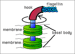
  • After gaining a fixed size bacteria reproduce through binary fission, budding and fragmentation.
  • Bacteria in optimum condition grow and divide rapidly and double its population in every 9.8 minutes.

Binary fission:

  • It is the most common mode of cell division and growth cycle of bacterial population.
  • In binary fission single cell divides into two identical cells with development of transverse septum (cross wall).
  • Two daughter cells contains nucleus of its own which is identical to the parent cell.
  • Cytoplasm divides leads to production of two equal sized cells.

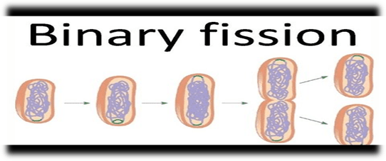
Process of binary fission:

  • Before the division DNA in the bacterial cell is tightly coiled
  • DNA is then uncoiled and duplicated.
  • Each copy of the DNA is pulled to the separate poles.
  • Synthesis of new cell wall begins
  • Once the new cell wall is synthesised fully it results in complete split of bacterium.
  • New daughter cells now have tightly coiled DNA, plasmids and ribosomes.
Types of binary fissionExample
TransverseParamecium
ObliqueCeratium
LongitudinalEuglena
Irregularamoeba

Gene transfer:

  • Gene transfer means movement of genetic information in organisms.
  • There are two types of gene transfer method one is vertical in which gene is transferred from parents to offspring and another one is horizontal in which gene is transferred in between two organisms.
  • In prokaryotes vertical gene transfer is by the means of binary fission and horizontal gene transfer method consist of three process i.e. transformation, transduction and conjugation.

Transformation:

  • In 1928 Fred Griffith discovered this method of horizontal gene transfer.
  • In this process naked DNA molecule or fragment from surrounding environment is uptake by the recipient and incorporated in its chromosome.
  • It is of two types natural and artificial, natural transformation is very rare event and observed in both gram negative and gram positive bacteria.
  • Ability of bacteria to uptake DNA fragment and get transformed is known as competence.

Process of transformation:

  • Competent bacteria naturally pull DNA fragment into their cell from the environment.
  • These DNA fragment naturally released in the environment after a bacterial cell die.
  • Ds DNA once crosses the membrane in cytoplasm the 3’ end is leading.
  • The translocated strand interested in the chromosome of recipient bacteria by homologous recombination.
  • Now the recipient bacteria undergoes replication and the cells acquired new phenotype are said to be transformed.
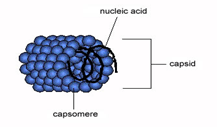
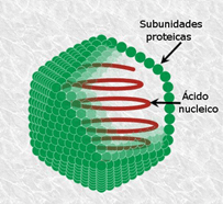
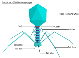
Transduction:

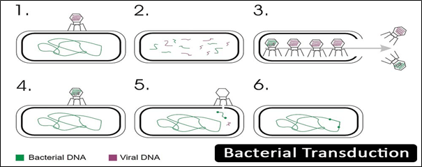
  • In transduction, DNA is transfer from donor bacteria to recipient bacteria by bacteriophage (functions as vector).
  • It was discovered by Lederberg and Zinder in 1951.
  • Bacteriophage due to high specificity of surface receptors has narrowest host range.
  • Transduction has one advantage over conjugation is that it doesn’t require physical contact of donor to recipient cell.
  • Transduction process is resistant to the DNase enzyme.

Steps:

  • The phage infects the host and inserts its phage DNA into the cytoplasm of the host.
  • During lytic cycle the phage DNA along with the bacterial chromosome is broken down into pieces
  • Bacterial chromosome packed into the viral capsid is released by the lysis of the bacterium.
  • Now the transducing phage with bacterial chromosome is ready to infect another bacterium in this way donor’s DNA enters into the cytoplasm of second bacterium.
  • Host recombinase recA is present in the cell due to which donor DNA recombines with homologous bacterial DNA and produces transductants.

Conjugation:

  • The process of transfer of plasmid or other transmissible DNA element from donor to recipient via sex pilus or conjugation tube.
  • Recipient of conjugation is known as transconjugants.
  • Is can transfer DNA regions of hundreds to thousands of kilobases and has board host range fro DNA transfer.
  • Occur in between many species of gram negative and gram positive bacteria even occurs between plants and bacteria.
  • Conjugation involves F plasmid is most common.

Steps:

  • F+ structure contains tra locus which has pilin gene with some regulatory proteins responsible for the formation of pili on surface.
  • Proteins present on pili attach to the F- cell surface and responsible for making contact between them but doesn’t transfer plasmid.
  • The traD enzyme on the base of the pili makes the membrane to fuse.
  • After the conjugation initiated the enzyme relaxes attached to the conjugative plasmid and make  a nick at oriT.
  • The nicked strand is now transferred to the recipient cell
  • F+ cell carry such integrated F element is known as Hfr cell.
  • The F element of Hfr cell is replicated along with the bacterial chromosome and in this way transmitted from one to next generation.

CORONA VIRUS

Content

  • Introduction
  • History
  • Structure
  • COVID-19
  • Transmission
  • Symptoms
  • Prevention and control

Introduction

  • Coronaviruses are RNA viruses that cause diseases in mammals and birds derived from Latin word corona which means ‘crown’, named by June Alimedia and David Tyrrell who first observed human coronaviruses.
  • Coronavirus cause respiratory tract infection in human that range from mild to lethal, mild illness include common cold while lethal verities cause MERS(middle east respiratory syndrome), SARS(severe acute respiratory syndrome) and COVID-19.
  • Constitute subfamily orthocoronavirinae. The virus is enveloped and has a positive sense single-stranded RNA genome and helical symmetry nucleocapsid.
  • Coronaviruses are among the largest virus, size ranges between 26-32 kilobases.

History

  • Arthur Schalk and M. C. Hawn in 1931 described a new respiratory infection of chicken in North Dakota. The mortality rate was 40-90% in new born chicks.
  • Six years later Fred Beaudette and Charles Hudson successfully isolate and cultivated IBV (infectious bronchitis virus) which cause disease.
  • In 1960s human coronaviruses were discovered. E. C. Kendall, Malcom Byone, and David Tyrrell working at British Medical Research Council isolated a novel common cold virus B814 from a boy. In n1965 Tyrell and Byone cultivated the novel virus in organ culture of human embryonic trachea by serially passing through it, the virus cannot be cultivated by standard techniques used for rhinoviruses and other common cold viruses.
  • At the same time Dorothy Hamre and John Procknow at the University of Chicago from a medical student isolated a novel cold virus 229E grew in kidney tissue culture.
  • In 1967 June Almeida imaged this two novel strain by electron microscopy and said that the two strains were morphologically related by their club-like spikes.
  • Many human coronaviruese have been identified SARS (2003), MERS (2012) and SARS cov-2 (2019).

Structure:

  • Large, spherical particles with bulbous surface projection
  • The virus diameter is around 125nm, envelope diameter is 85nm and the spikes are 20nm long.
  • The envelope is made up of lipid bilayer in which the membrane, envelope and spike structural protein are anchored in the ratio of 1:20:300(E:S:M).
  • A single particle on an average has 74 surface spikes.
  • The spikes are homotrimers of S protein composed S1 and S2 subunit. S protein is the class 1 fusion protein responsible for receptor binding and membrane fusion with the host cell.
  • S1 subunit forms head and has RBD (receptor binding domain), S2 forms the stem which anchor the spikes in the viral envelope and enable fusion on protease activation
  • E and M contribute in envelope formation and maintaining the shape.

COVID-19

  • Novel coronavirus is a new strain which has not been identified in humans yet.
  • Coronaviruses are zoonotic which means they transfer from animal to human. Example SARS cov was transmitted from civet cats to human and MERS cov from dromedary camels to humans. Many coronaviruses strains are circulating in animals have not infected humans.


Transmission

  • Infection carriers shed the viruses into environment while sneezing, coughing or sometimes speaking.
  • When these droplets come in contact with other person it gets inside the body through mouth, nose and eyes.

 Symptoms

Most common symptoms:

  • Fever
  • Tiredness
  • Dry cough

Less common symptoms:

  • Diarrhoea
  • Aches and pains
  • Headache
  • Sore throat
  • Conjunctivitis
  • Loss of taste or smell

Serious symptoms:

  • Chest pain or pressure
  • Difficulty breathing or shortness of breath
  • Loss of speech or movement

Prevention and Control

It is better to prevent from any disease than cure

  • Prevention of coronaviruses is to avoid being exposed to it
  • The major source of transmission is transfer of droplets, this can be controlled by respiratory hygiene

Following measures to be adopted to avoid transmission of these droplets

  • Always wash hand with soap and water for at least 20 second or if no dirty notice alcohol based rub can be used, especially after traveling or after sneezing, coughing and blowing nose
  • Avoid close contact with people who are sick or in contact with sick person because it may take few days to show symptoms and may be asymptomatic.
  • Wearing mask or cover moth with cloth when come contact with other people
  • Keeping a distance of at least 1 meter from other person
  • In any person doesn’t have cloth or mask covering their moth they should use tissue or under elbow while sneezing and frequently dispose tissue in the dustbin.
  • Regular cleaning and disinfection of the frequently touched surfaces i.e. Doorknobs, light switches, table, tapes

Precaution taken in hospitals for the patient and healthcare worker

  • Patient should be placed in a single room with proper ventilation if single rooms are not available the suspected patients should grouped together.
  • Health care staff should were proper medical mask, facial protection or eye protection to avoid contamination
  • In hospitals beds of the patient should be placed at least 1 meter apart
  • The equipment which are being shared among the patient should be disinfect after each use
  • Awareness should be generated among the patient, the general public families about the symptoms prevention and precautions

VIRUS

Virus-I

Structure, Characteristics and classification

Introduction:

  • Virus is a parasite of sub-microscopic level on all the organisms. They infect all type of life forms like plants, animals, bacteria.
  • They are ambiguous in nature i.e. weather living or non-living, they are non-living in free-state but behave like living organism in a host.
  • In 1892  Dmitri Ivanovsky’s described about virus when he was working on bacterial pathogen infecting tobacco plants.
  • When virus invades a host organism it forced to replicate rapidly to produce thousands of copies of the virus. In contrast when virus is not inside a host or in the process of infecting it, virus exists in independent form known as ‘virions’ consists of a genetic material (RNA or DNA), a protein coat – capsid and the outer envelope of lipids.
  • Virus can be of various shapes ranging from helical to the icosahedral form.
  • Virus are much smaller than bacteria, size of the most virus that have been studied is between 20-300 nm.
  • They contain only one type of nucleic acid at a time either DNA or RNA but never both.
  • Virus are responsible for various infectious diseases in plant and animals rabies, AIDS (HIV), avian influenza, Ebola virus disease. Transmission of viral disease is through ‘vector’ (diseases bearing organism).

Structure:

Virus shows a wide variety of shape and size, a complete virus particle know as virions contain nucleic acids surrounded by the protein coat capsid, capsid is made up of small subunits of protein knows as capsomers and an outer layer called envelope made up of lipid which is derived from host cell membrane.

The main morphological structures are:

Helical

  • Composed of single type of capsomeres stacked around the central axis of helical structure having central cavity results in the formation of rod shape structure which may be short and rigid or long and flexible
  • Genetic material (typically ssRNA or ssDNA) bound to the protein helix by the interaction between the negatively charged nucleic acid and the positively charged capsomer protein.

Example: tobacco mosaic virus

Icosahedral

  • Most animal viruses exhibit icosahedral symmetry. The minimum no of triangular faces is three which gives rise to the 60 more capsomers. Rotavirus has more than 60 capsomers. Regular icosahedron is the optimum way to form closed shell symmetry with identical subunits.
  • Capsomers attaches with 5 other capsomers at the apices called pentons and on the triangular faces attaches with 6 capsomers called hexon.
  • Pentons and hexon may be of same protein or may be of other proteins.

Prolate

  • Elongation of icosahedron along the fivefold axis, it is common in bacteriophage and composed of a cap with cylinder structure.

Envelope

  • In some species, the virus modify their cell membranes to envelope them self it may be either the outer membrane of the host or the internal nuclear membrane or endoplasmic reticulum making a outer lipid bilayer known as viral envelope.
  • The infectivity are depend on the envelope of the virus, the membrane i
  • Has protein coded by the viral and host genome and lipid membrane with any carbohydrates originate by the host.

Characteristics:

  • Non- cellular organism enclosed by a protective envelope
  • Virus attaches to their host by the help of spikes
  • Having nucleic acid (DNA and RNA) in the core which is surrounded by the protein coat
  • Considered as both living and non-living i.e. inactive when present outside the host and became active within the host cells
  • Virus uses host mechanism and enzymes to reproduce itself.

Classification :

On the basis of genetic material:

DNA virus:

  • DNA as genetic material
  • They attack on both humans and animals
  • Example: papillomavirus, parovirus and herpesvirus

RNA virus:

  • RNA as genetic material
  • Example: polio virus, ebola virus, hepatitis C virus

DNA-RNA viruses:

  • Having both DNA and RNA as genetic material
  • Example: leukoviruses, rous’s viruses

On the presence of number of strands:

TypeExample
Double-stranded DNAherpes viruses, adenoviruses
Single-stranded DNAbacteriophagesφ, X, 74 bacteriophages
Single-stranded RNAinfluenza virus, poliomyelitis, bacteriophage MS-2
Double-stranded RNArice dwarf viruses, wound tumour virus

On the basis of envelope:

Enveloped:

DNA viruses:  poxviruses, herpesviruses

RNA viruses: toga virus, coronavirus

Non-enveloped:

DNA viruses: adenovirus, papovirus

RNA viruses: hepatitis A and E virus

On the basis of capsid structure:

TypeExample
Naked icosahedralPoliovirus, hepatitis A virus
Enveloped icosahedralRubella virus, HIV-1
Naked helicalTMV
Enveloped helicalMumps virus, measles virus
ComplexSmallpox virus, hepatitis B virus

On the basis of shape:

TypeExample
Space vehicle shapedAdenovirus
Filamentous shapedEbola virus
Brick shapedPoxvirus
Bullet shapedRabies virus

On the basis of type of host:

Animal viruses:

  • Viruses infect and live inside the animal cell
  • Example: rabies virus, mumps virus

Plant viruses:

  • Their genetic material is RNA remains enclosed in the protein coat and infect plants
  • Example: potato virus, TMV, turnip yellow viruses

Bacteriophages:

  • Virus infect bacteria are known as bacteriophage, contain DNA as genetic material

On the basis of mode of transmission:

Transmitted throughExample
Respiratory routeRhino virus, swine flu
Faeco-oral routePolio virus, rota virus
Blood transfusionHIV, hepatitis B virus
Sexual contactRetro virus
Zoonotic virusAlpha virus, flavi virus

Baltimore Classification:

  • In early 1970s Nobel prize winner David Baltimore developed the most commonly used virus classification system
  • Baltimore focus on how mRNA is produced during replication and classify virus into various groups

Group I: mRNA is produced as the same way as their cellular DNA by transcription, contain ssDNA as their genetic material

Group II: convert their ss genome dsDNA before transcription of mRNA occur, have ssDNA as genetic material

Group III: uses RNA dependent RNA polymerase to generate mRNA from the one of the strand used as template, genome is dsRNA.

Group IV: genetic material is ssRNA . Genomic RNA is with positive polarity means that is directly serve as mRNA

Multiple full length RNA strand with negative polarity are formed from the intermediate of dsRNA (replicative intermediates) made in the genomic copying process.

These serve as template form the production of positive polarity RNA

Group V: contain ssRNA with negative polarity; means that sequence is complementary to mRNA. Negative-strand is converted into mRna

Group VI: virus have two copies of genome i.e. ssRNA  conveted using reverse transcriptase enzyme to dsDNA

dsDNA transported to the host’s nucleus in inserted in its genome viral DNA is produced by transcription with host DNA

group VII: genetic material is dsDNA which make ssRNA as intermediate acts as mRNA and also converted back to dsDNA  by reverse transcriptase enzyme.

BACTERIA

Introduction

  • Bacteria (singular bacterium) are unicellular microorganism which are of microscopic size and cannot be seen with unaided eyes.
  • Constitute large domain-prokaryotes.
  • They are among the first life form evolve on the earth and present in most of the habitat.
  • Bacteria inhabit normal to the extreme habitat like air, water, soil, radioactive waste, hot springs, deep seas, even in human gut, etc.
  • Also live in symbiotic and parasitic relationship with plants and animals. Example rhizobium associate with leguminous plants.
  • Length of bacteria ranges in few micrometres. E.coli (1.0-2.0 micrometre long and 0.5 micrometre in radius), Mycobacterium tuberculosis (2-4 micrometre long and 0.2-0.5 micrometre width), V cholerae (1-3 micrometre long and 0.5-0.8 micrometre radius).
  • The study of this discipline of microbiology is called bacteriology.
  • Bacteria are beneficial for human and other animals in a way that they produce various kind of vitamins, enzyme and food products. Example- vitamin B12, lactic acid, alcohol.
  • They are key components of our biosphere playing important role in biogeochemical cycles, removal of toxic substance and decomposition of waste materials.
  • Involve in nitrogen fixation hence improve soil fertility.
  • With beneficial characteristics several bacteria are pathogenic and cause various kind of disease in human, plants and animals. Example- cholera, tuberculosis, syphilis, anthrax, and more.
  • In industries bacteria are useful in waste water treatment and industrial fermentation for cheese and yogurt production.

Structure

  • Bacteria is a prokaryotic organism their body lacks nucleus and cellular components.
  • Bacteria are covered by a membrane called cell wall chiefly made up peptidoglycan (murein layer).
  • Peptidoglycan layer mainly constitute of polysaccharide which are cross linked by peptide bonds.
  • Peptidoglycan layer is made up of two glucose derivative N-acetylglucosamine (NAG) and N-acetylmuramic acid (NAM) chain. The chain is linked with tertapeptide bonds
  • Four protein in tetrapeptide bond are L-alanine, D-alanine, L-lysine or meso-diaminopimelic acid (DPA) and D-glutamine.
  • Cell wall contributes to the survival of the bacteria, protection from harsh environment and antibiotics.
  • Peptidoglycan layer in gram positive bacteria is 20-80 nm thick in contrast gran negative bacteria contain 2-7 nm thick layer.
  • Gram negative bacteria contain acidic substance known as teichoic acids which provide rigidity to the cell wall.

Outer Membrane

  • Over the cell wall gram negative bacterium contain an external layer called outer membrane it contain lipopolysaccharide, phospholipids, lipoprotein and proteins.

Glycocalyx

  • It’s the carbohydrate enriched layer that covers the outside of the bacteria.
  • It provides protection against host
  • This glycocalyx layer associate with the pathogenic property to the bacteria.
  • Glycoclayx in a tightly packed form is called as capsule, in contrast in loose packing it is called as slime layer.

Surface Auxiliary

FLAGELLA

FLAGELLA
  • Hairlike structure, attach on the surface of the cell, main function is to provide mobility to the bacteria
  • Both gram positive and gram negative bacteria contain flagella. Consist of three parts filament, hook and basal body.

PILLI

PILLI
  • Thin hairlike structure on the surface of gram negative bacteria
  • Play an Important role in conjugation process

Fimbrae

  • Present on both kind of cells gram negative an gran positive
  • Helps in attachment to the surface

Classification

  • Shape
TypeExample
Bacillus (Rod-Shaped)Escherichia coli (E. coli)
Coccus (Sphere)Streptococcus pneumoniae
Vibrio (Comma Shaped)Vibrio cholerae
Spirilla or spirochete (Spiral)Spirillum volutans
  • Mode of Nutrition
TypeExample
Autotrophic BacteriaPurple bacteria
Heterotrophic BacteriaAll disease-causing bacteria
  • Cell Wall
TypeExample
Gram positiveStaphylococcus aureus
Gram negativeEnterobacteriaceae
  • Mode of Respiration
TypesExample
AerobicPsuedomonas aeruginosa
AnaerobicClostridium

Reproduction

  • The mode of replication in bacterium is binary fission.
  • In this process the parent bacterial cell divided into two identical daughter cells.
  • Replication of DNA starts in the parent cell and each copy is transfer into the daughter cell.
  • Rate of reproduction is depend on the conditions like temperature, nutrient availability, moisture this is called favourable condition. E.coli generation rate is 2 million bacteria in 7 hrs.
  • In some rare cases they undergo sexual reproduction by conjugation, transduction and transformation. Helps in genetic modification in bacteria which results in the antibiotic resistant property.

SCOPE

Scope and appliance of microbiology

About

  • Microbiology is a discipline of biology which deals with the study of microscopic organism, their interaction with other organisms and with environment.
  • It includes microscopic level organisms like bacteria, algae, fungi, protozoa and the infectious agent viruses too.
  • Microorganisms present all over the globe form high altitude to the deep seas include hot springs, extremely cold climates, and high pressure even in the salty lakes.
  • Microorganisms are both beneficial and harmful to human. I.e. required in the industrial production of food stuff (bread, yogurt, beer, wine, etc), antibiotics(penicillin, chloromycetin, streptomycin) vaccine, enzymes, vitamins and many more products along with it is harmful in the way by causing fatal disease like small pox, plague, malaria, cholera, HIV, influenza and more.
  • They plays important role in maintaining the stability of ecosystem by recycling the organic and inorganic substance in carbon, nitrogen, sulphur and phosphorus cycle.
  • There were many events in history that tells us how infectious microbes put the human population in danger. Like black death(1346),  yellow fever(1793), Spanish flu(1918), SARS(2002), H1N1 flu(2009), MERS(2014), ebola(2014), etc.
  • In addition to the disease outbreaks microorganism plays major role in food spoilage, detoriation on materials like paper, wood, metal and plastics.
  • In agriculture nowadays genetically improved crops are used to get more yields and disease resistant crop which can be obtained by the involvement of microorganism.
  • As microbes are present everywhere it enhances the scope and contribute in many fields like pharma, agriculture, dairy, food industries, research, nanotechnology, water industry, chemical industry.
  • Microbiologists are the person who studies these microorganisms and their morphology, behaviour, metabolic activity, habitat, reproduction, nutritional requirement, their application and pathogenicity, improvemnet and modifications which leads to the high demand of microbiologist globally.

Fields:

Dairy and food industry:

  • Deals with the microbial production for the food stuff, prevention of spoilage of food and transmission of food borne disease.  

Agricultural microbiology:

  • It include the study of microbial strains which are used to obtain genetically modified crops which are resistant to many diseases and higher yields. Production of bio-fertilizers and maintenance of the rhizo-flora.

Medical microbiology:

  • Study of disease their causative agents, prevention, diagnosis and treatment. In addition in includes various clinical appliance of microbes oh human health.  

Environmental microbiology:

  • The study of microbes and their interaction with environment, role in geochemical cycles, microbial diversity, bio-remediation

Genetic engineering:

  • It deals with the study of modifying microorganism at gene level and engineered microbes are used to produce hormones, enzymes, vaccines, vitamins, antibiotic and other products.

Microbial physiology:

  • Includes the study of microbial morphological structure, metabolism and growth.

Industrial microbiology:

  • Deals with the production of antibiotic, fermented food, aminoacids, vitamins, steroids, enzymes, alcohol. In addition with the strain improvement and process of enhancing product quantity.

Soil microbiology:

  • The study of soil flora and role of microorganism in soil fertility

Water microbiology:

  • Major part of this field is the waste water management as is it a challenging condition for the world with industrial waste discharged in the water bodies and leading to the pollution causing threat for aquatic life.

Applications:

The diversity of microbes on the globe makes microbiology the most complex and largest discipline.

Food:

  • There are a majority of microbes used in the food and dairy industries for the production of food from wine, beer through the cheese, yogurt to manufacturing of bread.
  • Include Process of fermentation, pasteurization, industrial production, processing of food its packaging, food preservation and storage.
  • Microbial spoilage of food production and their prevention.

Environmental microbiology:

  • working of biogeocycle(carbon, nitrogen, sulphur and phosphorus) done by microorganism
  • microorganism are present in free living state and in association with plants in symbiotic relationship.
  • Maintaining the soil fertility without exhausting soil nutrients.
  • Responsible for cleaning toxic substance from the environment. 
  • Some are pathogenic to the plant but there are few strain which act as biological control agents and protect plant against this diseases.

Medical microbiology:

  • Disease causing microbes i.e. bacteria, algae, fungi, protozoa and virus responsible for causing numerous types of diseases ranging from  acute to severe life threatening.
  • Examples are cholera, influenza, malaria, HIV, tuberculosis, plague, etc
  • Their diagnosis, transmission, prevention and cure are the major part of the medical microbiology
  • In contrast to the pathogenicity there are some strains inhibit the growth of other diseases causing microbes by producing antibiotic, hence, used for the production of antibiotics.

Biotechnology:

  • Genetically engineered strain used for the production of therapeutic substance like human growth hormone, insulin, etc
  • Also contribute in the commercial production of  acetone, alcohol, drugs etc

Research:

  • Diversity and unicellular structure of microbes make them easy to study and research over multicellular structure.
  • In addition they can produce millions of copies from a single cell rapidly with very low cost which is good for experiments performed.
  • Short generation time leads to quick result analysis.

Future aspects of microbiology:

  • Due to population explosion in the world there could be scarcity of food in near future in that condition single cell protein can be an alternative.
  • Newly and highly resistant species of diseases causing microbes is a challenge for the present and in the future so r DNA technology is useful to overcome this problem.
  • Treatment of cancer and HIV like diseases
  • Food preservation methods for highly perishable food items

History of Microbiology


Louis Pasteur

  • He was a French microbiologist.
  • He Known for his work in the field of Microbial fermentation, vaccination, and pasteurization

 Born– 27 December 1822

Died– 28 December 1895

Award’s

  • Legion of Honor Grand Cross (1881)
  • Rumford Medal (1856)
  • Foreign members of the Royal society (1869)
  • Copley Medal (1874)
  • Albert Medal (1882)
  • Leeuwenhoek Medal (1895)

Contribution:

  • He was one of the leading microbiologist during the golden age of microbiology(1860-1910) .
  •  He is considered as the father of modern microbiology.
  • Pasteur was a French microbiologist gave the theory of Bio-genesis is most powerful of spontaneous generation, using swan neck flask experiment his work on the subject was published in 1861 as  memory of the organised bodies which exist in the atmosphere.
  • He gave  Microbial theory of fermentation in 1857.
  • He observed the fermentation of lactic acid from sugar by several different kind of yeast, bacteria and noticed microscopic globules in the very deposit of fermentation vessels, when these globules was transferred to a fresh nutrient consisting sugar yeast extract, the globule greatly lactic acid was formed.
  •  In 1867 Pasteur, was suggested that mild heating at 62.8°C  for 30 minutes rather that boiling was enough to destroy or kills the undesirable microorganisms without ruining the  taste of product, this process was referred as Pasteurization.
  • Pasteur developed Anthrax vaccine in 1881 5 years later, he was successful in preparing vaccine against Rabies.
  • Pasteur work seems to be demonstrate that microbes may  be cause of diseases or if they  may spoil the wine, perhaps they may also makes the body sick. This developed the germ theory of disease.

ROBERT KOCH   

Full name: Robert Heinrich Hermann Koch.

Born: 11 December 1843.

Died: 27 May 1910.

Discoveries:

  • Koch’s postulates.
  • Mycobacterium tuberculosis.
  • Asiatic cholera.
  • Anthrax bacterium.

Awards:

  • For MemRS [1897].
  • Nobel Prize in medicine [1905].
  • The first direct demonstration of the role of bacteria is causing disease was provided by Robert Koch.
  • A German physician who first of all isolated Anthrax bacillus [i.e. Bacillus anthracic] the cause of anthrax in 1876.
  • In 1882 he discovered Mycobacterium tuberculosis.
  • The most notable contribution of was the establishment of the casual relationship between the microorganism and a specific disease by applying a set of criteria referred to as Koch’s postulates.
  • Koch’s postulates published in 1884 and are the cornerstone of germ theory of disease and are still in use today to prove the Etiology [specific cause] of an infection disease.5
  • The postulates are : –
  • The suspected microorganisms must always be found in diseased but never in healthy individuals.
  •  The microorganism must be isolated in a pure or nutrient medium.
  •  The same disease must result when the isolated microorganisms is inoculated into a healthy host.
  •  The same organism must be re-inoculated from the experimentally infected host.
  •  Also demonstrated Vibrio Cholera that is the causing agent of the disease Cholera in 1883.

ANTONIE VAN LEEUWENHOEK   

Full name: Antoine Philips Van Leeuwenhoek.

Born: 24 October 1632.

Died: 26 August 1723.

Subject of study:

  • Bacteria
  •  Protozoa,
  • Microscope,
  •  red blood cell,
  • weevil.
  • He  was the first to observe bacteria and protozoa.
  • Van Leeuwenhoek was the first to experiment by using single lens microscope of his own design with microbes, which he originally referred to as ANIMALCULES.
  • He is commonly known as the father of Microbiology [Ancient].
  • He assembled simple microscope in 1674 and made more than 500 optical lenses. He also made at least 25- single lens microscope of different types out of which only nine survived.
  • He was also examined the blood and other tissues of human including his own tooth scrapping, minerals and plant materials.
  • Leeuwenhoek was the first person to give precise and accurate description of bacteria and protozoa using microscope ,he made himself because of this extraordinary contribution to microbiology. He is referred as the Father of Bacteriology and Protozoology.

Main Discoveries:

  • Infusoria in 1674
  • Bacteria [e.g. large Selenomonas from the human mouth] in 1683.
  • The vacuole of the cell.
  • Spermatozoa in 1677.
  • The banded of Muscle of fibers in 1682.