BACTERIAL DISEASE – TYPHOID

BY: SAI MANOGNA (MSIWM012)

Introduction :

Any disease caused by bacteria involves bacterial diseases. Bacteria, which are small types of life that can only be seen through a microscope, are microorganisms. Viruses, some fungi, and some parasites include other types of microorganisms. Millions of bacteria usually reside in the skin, intestines, and genitals. The vast majority of bacteria cause no disease, and many bacteria are beneficial and even required for good health. Often, these bacteria are referred to as good bacteria or healthy bacteria. 

Pathogenic bacteria are considered dangerous bacteria that cause bacterial infections and illnesses. When these invade the body and begin to replicate and crowd out healthy bacteria or develop in typically sterile tissues, bacterial diseases occur. Toxins that damage harmful bacteria can also release the body.

Typhoid :

An infectious, potentially life-threatening bacterial infection is typhoid fever, also called enteric fever. Typhoid fever is caused by the Salmonella enteric serotype Typhi bacterium (also known as Salmonella Typhi), carried into the blood and digestive tract by infected humans and spreads by food drinking water contaminated with infected feces to others. Typhoid fever signs include fever, rash, and pain in the abdomen. 

Fortunately, typhoid fever, particularly in its early stages, is treatable, and if one chooses to live in or fly to high-risk areas of the world, a vaccine is available to help prevent the disease.

Incubation Period :

Typhoid and paratyphoid infections have an incubation period of 6-30 days. With steadily rising exhaustion and a fever that rises daily from low-grade to as high as 102 ° F to 104 ° F ( 38 ° C to 40 ° C) by the third to the fourth day of illness, the onset of illness is insidious. In the morning, fever is usually the lowest, peaking in the late afternoon or evening. 

Pathophysiology :

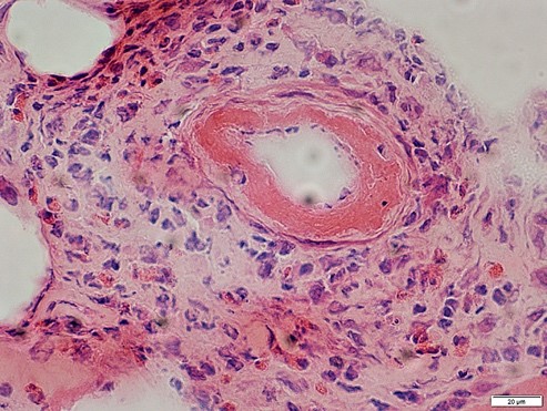
1. When present in the gut, all pathogenic Salmonella species are swallowed up by phagocytic cells, moving them through the mucosa and presenting them to the lamina propria macrophages. 

2. Across the distal ileum and colon, nontyphoidal salmonellae are phagocytized. Macrophages identify pathogen-associated molecular patterns (PAMPs) such as flagella and lipopolysaccharides with the toll-like receptor (TLR)-5 and TLR-4 / MD2 / CD-14 complex. 

3. Macrophages and intestinal epithelial cells are then attracts the interleukin 8 (IL-8) T cells and neutrophils, inducing inflammation and suppressing the infection. 

4. Unlike the nontyphoidal salmonellae, S typhi and paratyphi penetrate mainly via the distal ileum into the host system. They have specialized fimbriae that bind to the epithelium over lymphoid tissue clusters in the ileum, the critical point of relay for macrophages moving into the lymphatic system from the stomach. 

5. The bacteria then attract more macrophages by activating their host macrophages.

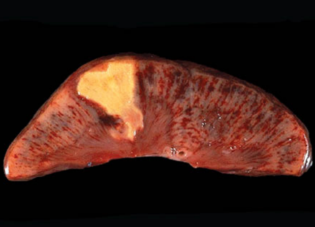
6. Typhoidal salmonella co-opts the cellular machinery of the macrophages for their reproduction, as they are transported to the thoracic duct and lymphatics to the mesenteric lymph nodes and then to the reticuloendothelial tissues of the spleen, bone marrow, liver, and lymph nodes. 

7. Once there, until some critical density is reached, they pause and begin to multiply. Afterward, to reach the rest of the body, the bacteria cause macrophage apoptosis, breaking out into the bloodstream. 

8. By either bacteria or direct extension of infected bile, the bacteria then invade the gallbladder. The effect is that in the bile, the organism re-enters the gastrointestinal tract and reinfects patches of Peyer. 

9. Usually, bacteria that do not reinfect the host are shed in the stool and are then available for other hosts to invade.

Epidemiology :

The International 

Worldwide, typhoid fever occurs mostly in developing countries where sanitary conditions are low. In Asia, Latin America, Africa, the Caribbean, and Oceania, typhoid fever is endemic, but 80 percent of cases originate from Bangladesh, China, India, Indonesia, Laos, Nepal, Pakistan, or Vietnam. In underdeveloped countries, typhoid fever is the most common. About 21.6 million people are infected by typhoid fever (incidence of 3.6 per 1,000 population), and an estimated 200,000 people are killed every year. 

Most cases of typhoid fever occur among foreign travelers in the United States. The average annual incidence of typhoid fever by county or area of departure per million travelers from 1999-2006 was as follows:

Outside Canada / United States, Western Hemisphere-1.3 

Africa-7.6 Africa 

Asia-10.5. 

India-89 (in 2006 122) 

Complete (except for Canada / United States, for all countries)-2.2 

Morbidity / Mortality :

Typically, typhoid fever is a short-term febrile condition with timely and effective antibiotic care, requiring a median of 6 days of hospitalization. It has long-term sequelae and a 0.2 percent mortality risk when treated. Untreated typhoid fever is a life-threatening disease that lasts many weeks, frequently affecting the central nervous system, with long-term morbidity. In the pre-antibiotic age, the case fatality rate in the United States was 9 -13%.

Sex : 

Fifty-four percent of cases of typhoid fever recorded between 1999 and 2006 in the United States included males. Moreover, race has no predilection. 

Age : 

Many confirmed cases of typhoid fever include children of school age and young adults. The true incidence is, however, thought to be higher among very young children and babies. The presentations may be atypical, that ranges from a mild febrile disease to severe convulsions, and the infection of S.typhi may go unrecognized. In the literature, this could account for contradictory reports that this category has very high or very low morbidity and mortality rate.

Symptoms :

Typhoid fever symptoms typically occur five to 21 days after food or water infected with Salmonella Typhi bacteria is consumed and can last up to a month or longer. Typical Typhoid Fever signs include: 

i. Pressure in the abdomen and tenderness 

ii. Perplexity 

iii. Fatigue and Weakness 

iv. Trouble focusing 

v. Constipation or diarrhea

vi. Headaches 

vii. The Nosebleeds 

viii. A dry cough 

ix. Impoverished appetite 

x. Rash (small, flat, red rashes that are also known as rose spots on the belly and chest) 

xi. Lethargy 

xii. Swollen lymph ganglions 

xiii. Chills and Fever. With typhoid fever, persistent fever of 104 degrees Fahrenheit is not rare. 

Symptoms: life-threatening 

Typhoid fever, including intestinal bleeding, kidney failure, and peritonitis, may lead to life-threatening complications. If they are with anyone who has any of these signs, seek urgent medical attention : 

Bloody stools or severe rectal bleeding 

A shift in consciousness or alertness level 

Confusion, delirium, disorientation, or hallucinations 

Unexplained or chronic dizziness 

Dry, broken lips, tongue, or mouth 

Unresponsiveness or lethargy 

Not urinating tiny quantities of tea-colored urine or urinating it. 

Extreme pain in the abdomen 

Extreme diarrhea in patients 

Extreme signs in infants include sunken fontanel (soft spot) at the top of the head, lethargy, no weeping tears, little or no wet diapers, and diarrhea. Infants two months of age or younger, be especially concerned about fever.

Causes :

The Salmonella Enteric Serotype Typhi (Salmonella Typhi) bacterium is responsible for typhoid fever. Via ingestion of infected food and water, Salmonella Typhi can enter and infect the body. By being washed in polluted water or being touched by an infected person with unwashed hands, food can become contaminated with the bacteria. Drinking water can become infected with untreated Salmonella Typhi-containing sewage.

Risk factors :

A variety of variables improves the chances of contracting typhoid fever. In developing, non-industrialized countries, typhoid fever is a significant health threat, although rare in the United States, Canada, and other industrialized countries. Factors of vulnerability include: 

i. Near contact with individuals infected or recently infected 

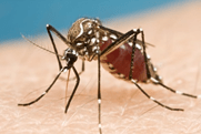
ii. Travel to areas with more frequent and widespread outbreaks of typhoid fever, such as India, Southeast Asia, Africa, and South America

iii. Avoiding contact with a person who has or has signs of typhoid fever, such as abdominal pain, headache, and fever

iv. Residence in a developing world or continent with inadequate treatment facilities for water and sewage or poor hygiene practices 

v. Due to diseases such as HIV / AIDS or drugs such as corticosteroids, the compromised immune system 

vi. Do not eat fruits and vegetables that are unable to peel. Eating fully cooked, hot, and still steaming foods. Unless it is made from distilled water, drinking only bottled water and not using ice 

vii. Before visiting high-risk areas, having vaccinated against typhoid fever 

viii. During and after contact with an individual who has typhoid fever or with an individual who has signs of typhoid fever, such as abdominal pain, headache, rash, and fever, washing hands regularly with soap and water for 15 seconds 

ix. Washing hands regularly for at least 15 seconds with soap and water, particularly before handling food and after using the toilet, touching feces, and changing diapers

Diagnosis :

1. Salmonella bacteria infiltrate the small intestine following the ingestion of infected food or drink and temporarily enter the bloodstream. 

2. The bacteria are transported into the liver, spleen, and bone marrow by white blood cells, replicating and re-enter the bloodstream. 

3. At this point, people develop symptoms, including fever. Bacteria invade the biliary system, gallbladder, and the intestinal lymphatic tissue. 

4. Here, in high numbers, they multiply. In the digestive tract, the bacteria move and can be found in stool samples. 

5. Blood or urine samples will be used to diagnose if a test result is not exact.

Treatment :

Typhoid fever is a treatable condition, and a complete course of antibiotics, such as ampicillin, trimethoprim-sulfamethoxazole, or ciprofloxacin, may also be used to cure it. Treatment can include rehydration with intravenous fluids and electrolyte replacement therapy in some severe cases. Usually, with care, symptoms improve within two to four weeks. If they have not been treated completely, symptoms may return. One needs to take the antibiotics for as long as needed to treat typhoid fever and follow up with the doctor for a series of blood and stool tests to ensure that they are no longer infectious. 

Few people infected with Salmonella Typhi become carriers, which indicates that the bacteria are present in the intestines and bloodstream and are shed in the stool even after they no longer have disease symptoms. Because of the carrier effect, it is essential to understand that they might still transmit the disease by contaminating food and water even after receiving treatment for typhoid fever. Before traveling outside developed regions, such as the United States, Canada, northern Europe, Australia, New Zealand, and Japan, it is vital to avoid the disease by getting vaccinated. During epidemic outbreaks, immunizations are also recommended, although the vaccination is not successful.

Prevention :

A larger number of typhoid cases usually occur in countries with less access to clean water and washing facilities. 

Immunization :

Vaccination is advised while traveling to a region where typhoids are prevalent. 

It is recommended to get vaccinated against typhoid fever before traveling to a high-risk area. 

Oral treatment or a one-off injection can be done : 

Oral: an attenuated, live vaccine. It consists of 4 tablets, one of which is taken every other day, the last of which is taken one week before departure. 

Shoot, the inactivated vaccine, was given two weeks before the ride. 

Vaccines are not 100 percent successful, and when eating and drinking, caution should always be exercised. 

Two forms of typhoid vaccine are available, but a more potent vaccine is still required. The vaccine’s live, oral form is the strongest of the two. It also protects individuals from infection 73 percent of the time after three years. This vaccine has more side effects, however. If the person is currently ill or if he or she is under the age of 6 years, vaccination should not begin. The live oral dose should not be taken by someone who has HIV. There may be adverse effects of a vaccine. One in every 100 people is going to feel a fever. There may be stomach complications following the oral vaccine, nausea, and headache. For any vaccine, however, serious side effects are uncommon. 

Typhoid removal :

Even if typhoid symptoms have passed, it is still possible to bear the bacteria, making it impossible to stamp out the disease because when washing food or communicating with others, carriers whose symptoms have terminated may be less vigilant. 

Prevention of Infection :

Via touch and ingestion of contaminated human waste, typhoid is propagated. This can happen through a source of water that is tainted or when food is treated. 

Some general rules to obey while traveling to help reduce the risk of typhoid infection are the following: 

i. Drink water, preferably carbonated, in glasses. 

ii. Do not have ice for drinks. Stop raw fruit and vegetables, cut the fruit, and not eat the cut on your own. Eat only food that is still hot, and avoid eating at street food stands.

iii. If it is impossible to acquire bottled water, ensure the water is heated for at least one minute on a rolling boil before consumption. 

iv. Be wary of eating something that anyone else has dealt with. 

Related Disorders :

There may be similar symptoms of the following conditions to those of typhoid fever. For a differential diagnosis, similarities may be helpful: 

Salmonella Poisoning :

In foodborne diseases, this is the most common cause of disease. These bacteria can contaminate meat, dairy, and vegetable products. In warm weather and children under the age of seven, outbreaks are more prevalent. The most common initial symptoms are nausea, vomiting, and chills. These are accompanied by stomach pain, diarrhea, and fever that can last for several weeks to five days. Intoxication with salmonella is a type of gastroenteritis. The CDC reports about 2 to 4 million salmonellosis cases per year in the United States. 

Cholera :

Cholera is a bacterial infection characterized by extreme diarrhea and vomiting that affects the whole small intestine. A toxin produced by the bacteria Vibrio cholerae is the source of the symptoms. The disease is transmitted by drinking water or consuming fish, vegetables, and other foods contaminated with Cholera’s excrement.

Botulism :

Botulism is also a form of gastroenteritis caused by a bacterial toxin. A neuromuscular poison is this toxin. In three types, it occurs foodborne, wound, and infantile botulism. The foodborne type is the most popular. Besides nausea, vomiting, diarrhea, and stomach pain, the patient can feel exhaustion, fatigue, headache, and dizziness.

Ptomaine Poisoning in the United States’ fourth most prevalent cause of bacterial foodborne disease. It is caused by the enterotoxin protein released after consuming foods that are contaminated, usually meat products. Extreme stomach cramps and diarrhea are characteristics of the disease. Nausea also happens sometimes. Vomiting and fever are rare. 

VECTOR-BORNE DISEASES

BY: SAI MANOGNA (MSIWM013)

Vector-borne infections, diseases caused by insects- and tick-borne pathogens, have long affected human affairs. The bite of a tiny mosquito carrying malaria parasites in the marshes of what is now called Iraq defeated Alexander the Great, the conqueror of many nations. To this day, vector-borne diseases remain influential, flooding the hospitals of sub-Saharan Africa with malaria victims, suppressing nations’ economies, and destroying industrial operations where they remain endemic. Specific, less common agents cause blindness and terrible disfigurement. Together, they form an alarming set of possible health and livelihood risks to those who travel and work or live in the tropics where they have the most significant effect.

Vectors :

Arthropod (insect) vector transmitted pathogens are some of the most harmful and volatile on earth. They are perhaps the hardest to stop or manage because they are so immune to intrusion and so deeply rooted in the habitats and environments of the regions they infest. In this equation, vectors make all the difference since they increase the range and transmissibility of pathogens exponentially over those that rely on transmission through direct human touch. Vectors help pathogens cross the gap between humans and various host animals (mice, rodents, monkeys, birds, prairie dogs, pigs). Over cycles less favorable to transmission (winters, dry seasons), some harbor reservoirs of pathogens. Without approaching the vector directly, vectors are facilitators of several harmful disease-causing species, the prevention, and treatment of which can not be successful for long.

For their entire lives, vectors stay infected, which is longer than others. 

Individuals think. For example, a mosquito does not have a set lifespan. In their first week of life, many die, but some will live almost forever. They are restricted by the harm that accumulates on their wings and appendages that are not repairable and do not degrade as soon as they wear out. In water, predation, desiccation, and trapping are likely to kill more mosquitoes than any other source. West Nile vectors that emerge in August of one year on the East Coast of the United States can be active over winter and in May of the following year for a life span of at least nine months.

The bulk of transmissions are due to mosquitoes and ticks. Large vector-borne diseases are involved, including sand flies and black flies. Even within a closely related community, each of these species has specific habitat requirements and feeding habits, which can differ significantly. For instance, around the globe, dozens of species of Anopheles mosquitoes can spread malaria. Some of them bite at night, some only at dawn and at dusk, in bright sunshine, some breed. Others never come out of the forest’s deep shade. Saltwater, for others, is lethal. In water with a saline content approaching that of seawater, some excel. Generalization, generally, fails when it comes to vector biology. The unique vector behaviors provide the keys to managing them and preventing infection from spreading.

The insect-borne disease of industrial significance :

Mosquitoes: Mosquitoes, by far the most important vector of disease, number more than 3,000 species worldwide. Only the female mosquito can spread the disease since only she, and not the male, has the knife-like part of the mouth required to extract blood from her victims. She requires a blood meal to allow protein for egg creation.

Sand Flies: Closely related to mosquitoes, sandflies are blood-feeders and breed in caves, rodent burrows, manure piles, and other dark areas that hold moisture and are rich in organic matter. They are weak fliers, tending to travel from host to host in fast “hopping” flights. Their bodies are so tiny (3 mm) that they are hard to detect before they begin to bite. Their bite has been creating extreme pain for a few days.

Black Flies: Black flies are yet another relative of mosquitoes specializing in flowing water from small trickles to large rivers. Unlike mosquitoes, black flies eat through the skin and never eat indoors. They can attack such large quantities that their salivary fluids alone can cause a person to get sick, causing a disease called “black fly fever.” They can also vector a nematode that can live in the human body for up to 15 years, killing tissue in the internal organs, most notably in the skin, causing blindness in the eyes.

Ticks: Ticks usually have a much longer life span than a mosquito. Hard ticks eat just a few times during their lifetime, limiting their risk of being infected. However, the persistence and host selectivity of hard ticks allow them to be relatively useful vectors. Soft ticks are long-lived nests and burrow dwellers. Like mosquitoes, they will feed several times during their lifetime.

Important Vector-borne Diseases:

A. Malaria : 

Malaria occurs in every tropical and subtropical landscape across the globe, often allowing seasonal excursions to temperate areas. The genomes, metabolisms, and life cycles of protozoan parasites are more complex than almost any other vector-borne threat. This makes it a difficult target for medicines and vaccines because the parasite’s shape-shifting pathways make it possible to avoid chemical and immunological defenses. They are also a moving target, deliberately modifying their outer coating during each step of their life cycle and producing a diverse antigenic and metabolic wardrobe through sexual recombination, a generator of diversity that is not available to simpler microbes such as viruses and bacteria.

1. Four parasite species affect humans, but more than 95 percent of cases include two of them, Plasmodium falciparum and P. vivax. The most dangerous pair, P. falciparum, crosses Africa’s deep tropics towards South America and Asia.

2. P. vivax, which may grow in mosquitoes at colder temperatures, has a broader range, spreading beyond the fringes of P. falciparum’s distribution and coexisting with P. falciparum in many regions.

3. P. falciparum malaria poses the greatest threat to the industry of any vector-borne disease, as it can kill an unprotected person very quickly and can reinfect and repeatedly weaken even those who develop semi-protective immunity. 4. Most deaths in local communities occur in children between 6 months and 2 years of age. 

5. Their weakness stems from a lack of immunological defense. Immune evasiveness of malaria parasites prevents full immunity production, but older children and adults who have undergone multiple infections enjoy some degree of defense against the most extreme manifestations of the disease.

Prevention: Prevention of malaria needs serious care while visiting areas where it is transmitted. While no vaccine is currently available, prophylactic drugs and steps to minimize exposure to night-biting Anopheles mosquitoes, such as bed nets and repellents, could be beneficial. Unlike other diseases, malaria patients also never get a second chance.

B. Dengue Fever :

1. Dengue virus tends to be the epithet of malaria in several respects. While malaria transmission is most prevalent in rural areas, Dengue is a city disease. 

2. Although Anopheles ‘malaria vectors bite mostly at night, Aedes’ dengue vectors bite mostly during the day. While the initial malaria infection usually causes the most severe symptoms, a second dengue infection may be much more severe than the first if it includes a different virus’s different serotype. 

3. Dengue fever can be painful (hence the nickname of “breakbone fever”) and Weakening, but generally not life-threatening when first obtained. 

4. However, there are significant manifestations in places where more than one of the virus coexists’ four significant strains. 

5. Being exposed to second, different strains of the virus may cause a severe immune reaction called Dengue Hemorrhagic Fever (DHF), leading to a significant risk of death, especially in children and younger adults. 

6. Currently, about five percent of the hundreds of thousands of people who receive DHF die, while timely and successful medical attention can dramatically reduce this case’s fatality rate.

Prevention: There are no prophylactic drugs or vaccines available to prevent Dengue, but steps that restrict or prevent biting, such as repellents or elimination of water-bearing containers in which mosquito vectors can be created, are helpful.

C. Arboviruses : 

There is a wide range of often-hazardous viruses almost everywhere on the globe where mosquitoes are found. Each of these individual viruses typically has limited ranges, and many affect relatively small populations; we consider them to be a group because of their mutual importance. 

More infamous pathogens and those currently in the news due to the revival or extension of their borders. In many places, these infections may account for poorly reported: “Fever of Unknown Origin” (FUOs) typically occurring in seasons when mosquitoes are most involved.

i. Chikungunya :

Due to its recent revival, Chikungunya tops this list in places including India, Sri Lanka, Mauritius, and Europe’s countries engaged in regular tourism to these destinations. Concern has recently emerged that it will soon expand its range in Europe due to the spread of Asian tiger mosquitoes. (Aedes albopictus), which can serve as a significant vector for this infection. 

Africa and Southeast Asia are also part of the traditional range of this virus. 

Chikungunya infection may be severe and temporarily crippling but is usually not life-threatening in otherwise healthy people. There is currently no vaccine or curative drug treatment available. Prevention must be focused solely on interventions that reduce mosquito bite exposure.

ii. Yellow Fever :

Yellow Fever is a deadly illness that has a worldwide prevalence. 

Because of the availability of an efficient vaccine, this has been significantly reduced. It is primarily Aedes mosquitoes spread by day-biting and can cycle in both urban and rural areas. Monkeys act as maintenance hosts in its rural incarnation, and tree-hole and bromeliad breeding mosquitoes move it on. No one should fly to an area where, without being vaccinated, Yellow Fever remains endemic. Some nations also need evidence of vaccination for admission.

iii. West Nile Virus (WNV) 

The virus has now spread to the New World, previously confined to Africa, the Middle East, and Southern Europe. It has become an omnipresent fixture of North America’s summer landscape and continues to make incursions into South America. Since most individuals who get infected experience nothing more than flu-like symptoms. The occurrence of a potentially lethal cerebral hemorrhage and irreversible neurological damage is a small percentage. Transmission occurs primarily through urban vectors since the most responsible mosquito (Culex pipiens) for amplifying the virus prefers relatively polluted bird populations environments. There is currently no available vaccine or curative drug treatment available.

iv. Tick-Borne Encephalitis (TBE) 

TBE differs from all the other arboviruses mentioned so far in that ticks rather than mosquitoes pass it on. TBE is found from China to Europe in temperate regions. Although infections can often be mild, in around 10-20 % of patients, permanent or long-lasting neurological damage can occur. Only 1-2% of the cases are fatal. For this infection, rodents are the primary maintenance hosts, and hard ticks in the Ixodes genus act as the primary vectors. No vaccine or curative drug treatment available at present.

Life cycle of TBE Virus : For all three life phases of the Ixodes tick, i.e. the larvae, the nymph and the adult tick, the dog will serve as the host. As with humans, the nymphs and the adults that feed on dogs are rather more numerous. There is also evidence of human dietary infection through TBE virus-contaminated milk. Although this sometimes triggers human infection clusters, we are not aware of such an infection path for dogs.

v. Rift Valley Fever (RVF) :

In North Africa and sub-Saharan, RVF is transmitted to animals and humans by 

Aedes mosquitoes day-biting. Humans also acquire infections through direct contact with contaminated animals’ blood during slaughter. Most RFV cases are relatively mild, but there is a case fatality rate of more than 50 percent in the hemorrhagic type of this disease. The overall case fatality rate due to human infection is probably less than 1 percent. RFV is one of the few viral pathogens transmitted to infected mosquito larvae, causing individual mosquitoes to be infected before they bite a host. Currently, no vaccine or curative drug treatment is available.

D. Lyme Disease :

Lyme Disease is also spread by the same forms of hard ticks that transmit TBE, but Lyme Disease has a broader range in North America. Lyme disease has never killed someone, but if left untreated, it can still be crippling. Upon reaching the synovial (joint) fluid or entering the central nervous system by the spirochete bacteria (Borrelia) that cause it, routine antibiotics can no longer enter it. The pathogen can cause symptoms such as arthritis, memory loss, and other neurological problems. 

Prevention: Prevention includes limiting tick bite contact with repellents, clothing treated with insecticides, and basic knowledge of tick habitats and their presence in the body. It takes at least two days for LD spirochetes to become triggered, and the risk of infection would be negligible if an attached tick can be removed before that time. A vaccine was previously available but, due to incomplete effectiveness, is no longer on the market.

E. Leishmaniasis :

Leishmaniasis involves many protozoan infections that can cause severe organ damage to anything from skin sores (in its mildest form). In almost every part of the tropics, certain types of leishmaniasis can be found. 

Nevertheless, the main areas of concern include North Africa, the Middle East (Iraq is a significant problem), and Southwest Asia. It is difficult to treat infections, and the treatments commonly used can be hazardous to humans and cause many side effects. Currently, no vaccine or curative drug treatment is available. 

The vectors act as Sand Flies, a relative of mosquitoes, which breed in caves, animal burrows, and manure piles. Weak, nocturnal fliers. When the wind is high, they will not be involved. Bed nets and repellents include preventive measures. Not as effective in preventing sand fly bites as they are in defending against mosquitoes, several commercial insecticide-treated bed nets have been identified.

F. African Trypanosomiasis (Sleeping Sickness) :

Like those present in Latin America, African trypanosomes are transmitted by Tsetse flies, which are present only in Africa. African Sleeping Sickness “is induced by this pathogen, which can induce coma by invading the central nervous system.” Especially common in mixed savannah/woodland environments are Tsetse flies. Ranges of Trypanosoma brucei gambiense 

Although Trypanosoma brucei rhodesiense is found in East and Southern Africa, it is mainly found in West and Central Africa. A more rapidly advancing and the Rhodesian type creates acute infection, but both will kill people if left untreated. Currently, no vaccine or curative drug treatment is available.

G. Lymphatic Filariasis (Elephantiasis) :

Generally, filariasis does not kill but can cause severe impairment. Multiple nematode worms cause this mosquito-borne infection that invades the lymphatic system, causing swelling and tissue accumulation in different parts of the body but affecting the legs in particular. This condition causes gross distortion of appendages known as “elephantiasis” in its most extreme manifestation. Major surgery and thorough tissue removal provide the only treatment for infections that reach this degree of severity. It requires several years of prolonged infection to develop more severe symptoms, so it does not present a significant concern to staff in the short term. On-the-job exposure, however, can cause several years of deteriorating health and pain. 

Throughout India, Africa, and parts of Southeast Asia and Oceania, filariasis is found. Wuchereria bancrofti or Brugia malayi are responsible for most cases. In the genus Culex, night-biting mosquitoes act as the primary vectors, so bed nets are an efficient way to reduce exposure to this parasite. Currently, no vaccine or curative drug treatment is available.

H. Onchocerciasis (River Blindness): 

Nematodes also cause onchocerciasis, but it’s vectors are black flies that breed in clean, flowing water and are linked to mosquitoes. The adult worms that cause onchocerciasis cause the body to develop fibrotic tissue under the skin to develop hard lumps or nodules. Clinically, the most significant concern occurs from the millions of microfilariae pre-larval worms shed from these nodules which migrate into the skin, causing pruritus and blindness when they enter the eyes in some instances (hence the word ‘river blindness’). Ivermectin, a well-tolerated medication, can destroy microfilaria and temporarily inhibit adult female worms’ ability to reproduce. Black flies only strike outdoors and during daylight hours, so bed nets are not useful for exposure prevention. However, the repellents that function on mosquitoes are typically successful. They are against black flies.

Onchocerciasis is restricted to Africa and Latin America. River-inhabited black flies. The primary vectors in Africa serve as (Simulium damnosum). The significant vectors in Latin America are black flies called Simulium ochraceum and metallicum. At present, no vaccine is available.

ANTIBIOTICS

BY: SAI MANOGNA (MSIWM013)

Introduction :

Antibiotics are used in the treatment of preventing bacterial infection. They work by destroying or stopping bacteria from reproducing and spreading. Antibiotics do not work against respiratory infections like the common cold, flu, most coughs, and sore throats. The immune system can also cleanse several mild bacterial infections without using antibiotics, but they are not regularly prescribed. Antibiotics must be administered and appropriately taken to help prevent the development of antibiotic resistance.

Antibiotics, also known as antibacterials, are medicines that destroy or slow bacterial growth. In 1929, Alexander Fleming identified penicillin, the first antibiotic compound. Penicillins, Aminoglycosides, Quinolones,  Macrolides, Sulfonamides, Cephalosporins, Carbapenems, and Tetracyclines are common antibiotics. General antibiotic prescription principles are used: first-line antibiotics, reserve wide-spectrum antibiotics only for indicated conditions, prescribe antibiotics for bacterial infections if symptoms are severe or extreme.

Antibiotics can be given in several ways:

Oral antibiotics: pills, capsules, or a liquid drink to treat most cases of mild to severe body infections

Topical antibiotics: creams, lotions, sprays, or drops sometimes used to treat skin infections

Antibiotic injections: these may be administered or infused directly into the blood or muscle and are typically reserved for more extreme infections

Types Of Antibiotics :

There are hundreds of different forms of antibiotics: but most can be grouped into six classes. They are listed below.

Penicillins (such as penicillin and amoxicillin)

Cephalosporins (such as cephalexin)

Aminoglycosides (such as gentamicin and tobramycin)

Tetracyclines (such as tetracycline and doxycycline)

Macrolides (such as erythromycin and clarithromycin)

Fluoroquinolones (such as ciprofloxacin and levofloxacin)

1. Penicillins :

a. Penicillins are an antibiotic of penicillium fungi. An antibiotic is a drug that prevents bacteria ‘s growth or destroys.

b. The 1928 accident discovered penicillin G (also called benzylpenicillin). Alexander Fleming, a Scottish physicist, developed a form of bacteria called Staphylococcus Aureus on an uncovered petri dish when infected with mold spores. He noticed the bacteria near the mold dying. He isolated the mold material that destroyed the bacteria and named it penicillin.

c. Natural penicillins (penicillin G, penicillin V) are active only against gram-positive bacteria. Penicillin V is acid-resistant than penicillin G

d. Penicillin V was isolated from the same mold. All other penicillins are semi-synthetic, made by changing the structure of the original naturally occurring penicillins.

e. Classic semi-synthetic penicillins include ampicillin and oxacillin. These have some degree of beta-lactamase resistance and are effective against some gram-negative bacteria.

f. Most bacteria can be categorized as gram-positive or gram-negative based on variations in their cell wall structure, which can be microscopically differentiated using the dye form.

g. One of the main distinctions is that gram-positive bacteria are more susceptible to antibiotics, whereas gram-negative bacteria are more antibiotic-resistant.

Penicillins such as piperacillin and ticarcillin are penicillins with additional activity against some hard-to-kill types of gram-negative bacteria (Pseudomonas, Klebsiella, and Enterococcus).

A beta-lactamase inhibitor incorporates certain penicillins. A beta-lactamase inhibitor blocks beta-lactamase enzyme activity but tends to have little antibiotic activity. Some penicillins (like oxacillin, dicloxacillin, and nafcillin) are naturally resistant to certain beta-lactamases and are called penicillin-resistant. Others, such as amoxicillin, ampicillin, and piperacillin, may be extended by combining them with a beta-lactamase inhibitor. Clavulanate, sulbactam, and tazobactam all inhibit beta-lactamase.

Penicillins function by preventing bacterial cell wall cross-linking of amino acid chains. This does not affect pre-existing bacteria, but new bacterial cells have fragile cell walls that rupture easily.

Uses: Penicillins are used to treat

 i. Dental abscess

ii. Ear infections

iii. Gonorrhea

iv. Pneumonia

v. Rheumatic fever

vi. Skin infections

vii. Urinary tract infections

2. Tetracyclines:

a. The first tetracyclines were derived from Streptomyces bacteria in the 1940s.

b. These can be used to treat infections caused by susceptible microorganisms such as gram-positive and gram-negative bacteria, chlamydiae, mycoplasma, protozoans and rickettsiae.

c. Tetracyclines inhibit protein synthesis in microbial RNA (an essential molecule for DNA messenger). They are mainly bacteriostatic, thereby stopping bacteria from spreading but not necessarily killing them.

d. Although tetracyclines all function the same way, there are variations between the four tetracyclines (demeclocycline, doxycycline, minocycline, and tetracycline).

Doxycycline is the most commonly used tetracycline. It causes photosensitivity or binds to calcium, causing dental discoloration or bone growth retardation.

Uses: Tetracyclines are used to treat

i. Malaria

ii. Treatment of moderate to severe acne

iii. Anthrax

iv. Infections of an eye, gastrointestinal tract, respiratory tract, and skin

v. infections caused by Campylobacter, Yersinia pestis, Vibrio cholerae, Chlamydiae, and other atypical organisms

3. Cephalosporins :

a. Cephalosporins are a broad group of Acremonium-derived antibiotics (formerly Cephalosporium).

b. Cephalosporins are bactericidal (kill bacteria), similar to penicillins. They bind and block enzyme activity responsible for producing peptidoglycan, an essential bacterial cell wall component. They are called broad-spectrum antibiotics because they affect a wide range of bacteria.

c. Since the first cephalosporin was discovered in 1945; scientists have refined cephalosporin structure to make it more effective against broader bacteria.

d. Whenever structure changes, a new “generation” of cephalosporins is made. There are five generations of cephalosporins.

e. Both cephalosporins begin with cef, ceph, or kef. Notice that this classification scheme is not used consistently across countries.

Currently, there are five “generations” of cephalosporins, each generation varying slightly in their antibacterial range ( i.e., how successful they are in destroying certain types of bacteria). There are variations in administration (such as oral or intravenous administration), absorption, excretion, and how long the body’s cephalosporin activity lasts.

First-generation :

First-generation cephalosporins are the first group of cephalosporins found. Their optimal activity against gram-positive bacteria like staphylococci and streptococci. They have little anti-gram-negative activity.

Second-generation :

Second-generation cephalosporins are more active against gram-negative bacteria, with less gram-positive bacteria.

Third-generation :

The second-generation cephalosporins preceded third-generation cephalosporins. No third-generation cephalosporin treats all scenarios of infectious disease.

Cefotaxime and ceftizoxime provide the best gram-positive coverage of all third-generation agents; ceftazidime and cefoperazone are unique in providing antipseudomonal coverage.

Fourth generation :

Fourth-generation cephalosporins are structurally similar to third-generation cephalosporins. However, they have a different ammonium group that enables them to penetrate the outer membrane of gram-negative bacteria, enhancing their activity. They are also active against β-lactamase generating Enterobacteriaceae that may inactivate cephalosporins of third-generation.

Some fourth-generation cephalosporins have excellent activity against gram-positive bacteria such as staphylococci, penicillin-resistant pneumococci, and streptococci group viridans.

Fifth-generation :

Ceftaroline is currently the only cephalosporin of next-generation available in the U.S. It acts against methicillin-resistant Staphylococcus aureus ( MRSA) and gram-positive bacteria. It also retains later-generation cephalosporin activity and works against susceptible gram-negative bacteria.

Uses: Cephalosporins are used to treat

i. Bone, skin, and ear infections

ii. Urinary tract infections

4. Quinolines:

a. Quinolones are antibiotics that kill or inhibit bacteria.

b. Five separate quinolone groups exist. Another antibiotic class, called fluoroquinolones, was derived from quinolones by modifying their fluorine structure.

c. Quinolones and fluoroquinolones have some common and some differences, such as against which organisms they work.

d. Quinolones and fluoroquinolones affect the role of topoisomerase IV and DNA gyrase so that they can no longer fix DNA or assist in its development.

e. Quinolones and fluoroquinolones vary in their action against the two enzymes produced by bacteria, topoisomerase IV and DNA gyrase.

f. Those more active against topoisomerase IV have more effect on gram-positive bacteria; those active against DNA gyrase are more active against gram-negative bacteria.

g. Quinolones and fluoroquinolones also vary in body absorption, metabolization, and excretion.

Uses: Quinolines are used to treat

i. Unusual infections such as plague and anthrax

5. Macrolides :

a. Macrolide derivatives are either a macrolide or macrolide-related antibiotics.

b. Macrolides are antibiotics found in streptomycetes.

c. They are natural lactones with a complete ring of 14-20 atoms.

d. Macrolides bind to the bacterial ribosome 50S subunit and inhibit ribosomal translocation, leading to bacterial protein synthesis inhibition.

e. Their action is primarily bacteriostatic, but at high concentrations, depending on the type of microorganism.

Uses: Macrolides are used to treat uncomplicated skin infections, pneumonia, pertussis, and other susceptible infections.

6. Aminoglycosides :

a. Aminoglycosides are a class of antibiotics used primarily to treat aerobic gram-negative bacilli infections

b. These are effective against other bacteria such as Mycobacterium tuberculosis Staphylococci. They are often used with other antibiotics.

c. Aminoglycosides can function by inhibiting protein synthesis within bacteria.

d. Bacteria kill rates are increased when higher aminoglycoside concentrations are present.

e. Kidney impairment and hearing loss are the most common side effects of aminoglycosides.

f. Aminoglycosides are usually used when other antibiotics are contraindicated or ineffective.

g. Aminoglycosides are not well absorbed by mouth, so healthcare workers need to be injected.

Others :

a. Carbapenems

These injectable beta-lactam antibiotics have a wide range of bacteria-killing ability. They can be used for mild to life-threatening bacterial infections such as stomach infections, pneumonia, kidney infections, hospital-acquired multidrug-resistant infections, and many other severe bacterial diseases. They are often saved or used as “last-line” agents to help prevent resistance.

b. Antibiotic – Glycopeptide

Members can be used to treat methicillin-resistant staphylococcus aureus (MRSA) infections, complicated skin infections, C. Difficult-associated diarrhea, enterococcal infections such as beta-lactam-resistant endocarditis, and other antibiotics.

c. Sulphonamides

Sulfonamides function against some gram-positive and many gram-negative bacteria, but resistance is widespread. Sulfonamide uses include urinary tract infections ( UTIs), pneumonia treatment or prevention, or ear infections (otitis media).

d. Lincomycins :

Lincomycins have activity against gram-positive aerobes and anaerobes, as well as some gram-negative anaerobes. Lincomycin derivatives may be used to treat severe infections such as pelvic inflammatory disease, lower respiratory tract infections, intra-abdominal infections, and bone and joint infections. Some forms are also used topically to treat acne.

Antimicrobial resistance:

Overuse of antibiotics in recent years means they become less successful, and “superbugs” have arisen. There are bacterial strains that withstand several different forms of antibiotics, including:

Methicillin Staphylococcus aureus (MRSA)

Clostridium difficile (CD)

Multidrug – resistant tuberculosis (MDR-TB) bacteria

Carbapenemase Enterobacteriaceae (CPE)

These infections can be extreme and challenging to manage and are rapidly causing disability and death worldwide.

Antibiotic side-effects

Like any drug, antibiotics can cause side-effects. Most antibiotics do not cause problems if adequately used, and severe side effects are rare.

The most popular side-effects are:

Bloating, indigestion, diarrhea

Some people may be allergic to antibiotics, particularly penicillin, and a form called cephalosporins. This may lead to an extreme allergic reaction (anaphylaxis), a medical emergency in scarce circumstances.

MHC Molecules

BY- SAI MANOGNA (MSIWM014)

Each mammalian species studied to date has a closely linked gene cluster.  The major histocompatibility complex (MHC) plays a role in intercellular recognition and self-nonself-discrimination. The MHC is involved in the production of immune responses that are both humoral and cell-mediated. While antibodies may respond on their own with antigens, most T cells only recognize antigens when combined with an MHC molecule. Also, since MHC molecules act as antigen-presenting structures, the unique collection of MHC molecules expressed by an individual influences the antigen repertoire to which the TH and TC cells of that individual may respond. For this reason, an individual’s response to antigens from infectious organisms is partly determined by the MHC and has therefore been involved in disease susceptibility and autoimmunity production.

General Organization and Inheritance of the MHC:

1. The theory that foreign tissue rejection is the result of an immune reaction to cell-surface molecules, now called antigens of histocompatibility, arose from Peter Gorer ‘s work in the mid-1930s.

2. To recognize blood-group antigens, Gorer used inbred strains of mice. He identified four groups of genes, designated I through IV, that encoded blood-cell antigens in the course of these studies.

3. Work carried out by Gorer and George Snell in the 1940s and 1950s established that gene-encoded antigens in the group designated II were involved in the rejection of transplanted tumors and other tissue. Snell called these genes “histocompatibility genes”; their current designation was about Gorer ‘s group II blood-group antigens as histocompatibility-2 (H-2) genes.

4. Although Gorer died before his contributions were fully recognized, Snell was awarded the 1980 Nobel Prize for this work.

A series of genes arranged inside a long, continuous stretch of DNA on chromosome 6 in humans and chromosome 17 in mice is the main histocompatibility complex. The MHC is referred to in humans as the HLA complex and in mice as the H-2 complex. Although the arrangement of genes is quite different, the MHC genes are grouped into regions encoding three groups of molecules in both cases.

Class I MHC Genes: Glycoproteins expressed on the surface of almost all nucleated cells are encoded by class I MHC genes; the critical feature of class I gene products is the presentation to TC cells of peptide antigens.

Class II MHC Genes: Glycoproteins expressed predominantly in antigen-presenting cells (macrophages, dendritic cells, and B cells) are encoded by class II MHC genes, where they present processed antigenic peptides to TH cells.

Class III MHC Genes: In addition to other products, Class III MHC genes encode different secreted proteins which have immune functions, including components of the complement system and molecules that are involved in inflammation.

Both MHC molecules of class I and class II are membrane-bound glycoproteins closely related in both structure and function. These molecules have been isolated and purified, and x-ray crystallography has determined their extracellular domains’ three-dimensional structures. Both membrane glycoprotein types act as highly specialized antigen-presenting molecules that form extremely stable antigenic peptide complexes, displaying them on the cell surface for T cell recognition. MHC molecules of class III, on the other hand, are a group of unrelated proteins which do not share structural similarities and common function with molecules of class I and II.

Class I MHC Molecules:

1. A 45-kilodalton (kDa) alpha chain-linked noncovalently with a 12-kDa beta2-microglobulin molecule is found in Class I MHC molecules.

2. The alpha chain is a transmembrane glycoprotein encoded within the A, B, and C regions of the human HLA complex by polymorphic genes and within the K and D/L regions of H-2 complex of the mouse.

3. Beta2-Microglobulin is a protein encoded on a distinct chromosome by a strongly conserved gene.

4. For the expression of class-I molecules on cell membranes, the alpha chain association with beta2-microglobulin is required.

5. With its hydrophobic transmembrane part and hydrophilic cytoplasmic tail, the alpha chain is anchored in the plasma membrane.

Structure of Class I MHC:

1. Structural analyses have shown that the class I MHC alpha chain molecules are divided into three external domains (alpha1, alpha2, and alpha3. Each contains approximately 90 amino acids; a transmembrane domain-containing approximately 25 hydrophobic amino acids, followed by a short stretch of charged amino acids; and a 30 amino acid cytoplasmic anchor segment.

2. The beta2-microglobulin is similar in size and organization to the alpha3 domain; it does not contain a transmembrane region and is noncovalently bound to the class I glycoprotein.

3. Sequence data shows homology in immunoglobulins between the alpha3, beta2-microglobulin, and constant-region domains.

4. The enzyme papain cleaves the alpha chain just 13 residues proximal to its transmembrane domain, releasing the extracellular portion of the molecule, consisting of a1, a2, a3 beta2-microglobulin.

5. Two pairs of interacting domains were revealed by purification and crystallization of the extracellular portion: a membrane-distal pair composed of the alpha-1 and alpha-2 domains and a membrane-proximal pair composed of the alpha-3 domain and beta2-microglobulin.

6. The alpha-1 and alpha-2 domains combine to form a platform of eight antiparallel beta-strands spanned by two long alpha-helical regions.

7. With long alpha-helices as sides, and beta-strands of the beta-sheet as the bottom, the structure forms a deep groove, or cleft, about 25Å ×10Å ×11Å.

8. On the top surface of class I MHC molecule, this peptide-binding cleft is located, and it is wide enough to bind a peptide of 8-10 amino acids.

9. The discovery of tiny peptides in the cleft that had co-crystallized with the protein was the big surprise in the x-ray crystallographic study of class I molecules. These peptides are processed antigen and self-peptides bound to the alpha-1 and alpha-2 domains in this deep groove.

10. The a3 domain and beta2-microglobulin are organized into two beta-pleated sheets, each formed by antiparallel beta-strands of amino acids. As defined, this structure, known as the immunoglobulin fold, is characteristic of immunoglobulin domains.

11. Class I MHC molecules and beta2-microglobulin are known as immunoglobulin members due to this structural similarity.

12. Among class I MHC molecules, the alpha-3 domain appears highly conserved and contains a sequence interacting with the CD8 membrane molecule present on TC cells.

13. Beta2-Microglobulin deeply interacts with the alpha-3 domain and also interacts with the a1 and a2 domain amino acids.

For the Class I molecule to achieve its fully folded conformation, the interaction of beta2-microglobulin and a peptide with a class I alpha chain is necessary. The assembly of class I molecules is assumed to occur by the initial interaction of beta2-microglobulin with the folding class I alpha chain. The binding of a suitable peptide to form the native trimeric class I structure consisting of the class I alpha chain, beta2-microglobulin, and a peptide stabilizes this metastable “empty” dimer. Ultimately, this full molecular complex is transferred to the cell surface.

Class II MHC Molecules:

1. There are two distinct polypeptide chains in Class II MHC molecules, a 33-kDa alpha chain and a 28-kDa beta chain connected by noncovalent interactions.

2. Class II MHC molecules, including class I alpha chains, are membrane-bound glycoproteins containing external domains, a segment of the transmembrane, and a segment of the cytoplasmic anchor.

3. In a class II molecule, each chain comprises two external domains: the domains alpha-1 and alpha-2 in one chain and the domains beta-1 and beta-2 in the other.

4. Like the membrane-proximal alpha-3 / beta-2-microglobulin domains of class I MHC molecules, the membrane-proximal alpha-2, and beta-2 domains bear sequence resemblance immunoglobulin-fold structure; thus, class II MHC molecules are also known as super-family immunoglobulins.

5. The membrane-distal part of the class II molecule consists of the domain’s alpha-1 and beta-1 and forms the antigen-binding cleft for the antigen being processed.

6. The X-ray crystallographic analysis indicates that class II and class I molecules are identical, which is remarkably obvious when the molecules are superimposed.

7. Like that in class I molecules, the peptide-binding cleft of HLA-DR1 is composed of a floor of eight antiparallel beta-strands and sides of antiparallel alpha-helices.

8. The class II molecule lacks conserved residues that bind short peptides to the terminal residues and form an open pocket instead; class I offers more of a socket, class II, an open-ended groove.

Adaptive Immunity

BY: SAI MANOGNA (MSIWM013)

Adaptive immunity is capable of identifying unique foreign microorganisms and molecules (i.e., foreign antigens) and selectively eliminating them. In comparison to innate immune responses, in all members of a species, adaptive immune responses are not the same but are reactions to particular antigenic challenges.

Four characteristic attributes represent adaptive immunity:

a. Antigen specificity

b. Diversity

c. Immunologic memory

d. Recognition of Self/non-self

The immune system’s antigenic specificity enables it to discern subtle distinctions between antigens. Antibodies can differentiate between two molecules of protein that differ by only one amino acid. In its recognition molecules, the immune system can produce immense complexity, enabling it to identify billions of unique structures for foreign antigens. If an antigen has been recognized and reacted to by the immune system, it exhibits immunological memory; that is, a second encounter with the same antigen causes an increased immune reactivity state. Because of this feature, after an initial experience, the immune system will confer life-long immunity to several infectious agents. Finally, the immune system typically only responds to foreign antigens, suggesting that it can recognize itself. The immune system’s ability to differentiate oneself from non-self and react only to non-self molecules is necessary. The result of an inappropriate reaction to self-molecules may be fatal.

Adaptive immunity is not autonomous from innate immunity. In initiating the particular immune response, the phagocytic cells critical to non-specific immune responses are closely involved. Conversely, it has been shown that various soluble factors released by a specific immune reaction increase these phagocytic cells’ activity. For instance, as an inflammatory response grows, soluble mediators are created that attract immune system cells. In exchange, the immune reaction may help to monitor the strength of the inflammatory response. The two mechanisms function together to remove a foreign invader through the carefully controlled inter-play of adaptive and innate immunity.

Lymphocytes and Antigen-presenting cells Co-operate in Adaptive immunity :

Two main classes of cells are involved in a successful immune response: T lymphocytes and antigen-presenting cells. Lymphocytes are formed by the hematopoiesis process in the bone marrow. Lymphocytes leave the bone marrow, circulate and remain in various lymphoid organs in the blood and lymphatic systems. Lymphocytes mediate the distinguishing immunological characteristics of specificity, diversity, memory, and self/non-self recognition since they generate and exhibit antigen-binding cell-surface receptors.

B LYMPHOCYTES

1. B lymphocytes mature within the bone marrow. Each expresses a specific antigen-binding receptor on its membrane when they leave it.

2. A membrane-bound antibody molecule is this antigen-binding or B-cell receptor. 3. Glycoproteins consisting of two identical heavy polypeptide chains and two similar light polypeptide chains are antibodies.

4. Disulfide bonds join each heavy chain with a light chain, and additional disulfide bonds hold the two pairs together.

5. A cleft within which antigen binds forms the amino-terminal ends of the pairs of heavy and light chains.

6. The antigen-binding to the antibody causes the cell to divide rapidly when a naive B cell (one that has not previously experienced antigen) first encounters the antigen that matches its membrane-bound antibody. Its progeny differentiates into memory B cells, and plasma cells called effector B cells.

7. Compared to naive cells, memory B cells have a longer life span, and parent B cells release the same membrane-bound antibody.

8. The antibody is formed by plasma cells in a form that can be secreted and has little to no membrane-bound antibodies. While plasma cells live for only a few days, they secrete enormous amounts of antibodies during this period.

9. It has been calculated that more than 2000 antibody molecules per second can be secreted by a single plasma cell. The main effector molecules of humoral immunity are secreted antibodies.

T LYMPHOCYTES

1. T cells move to the thymus gland to mature, unlike B cells, which mature within the bone marrow.

2. The T cell expresses a unique antigen-binding molecule, called the T-cell receptor, on its membrane during its maturation within the thymus.

3. T-cell receptors can recognize only antigens connected to cell-membrane proteins called major histocompatibility complex (MHC) molecules, unlike membrane-bound antibodies on B cells that can recognize antigen alone.

4. The polymorphic (genetically diverse) glycoproteins found on cell membranes are MHC molecules that act in this recognition case called “antigen presentation.”

MHC molecules are of two types:

  1. Class I MHC Molecules
  2. Class II MHC Molecules

Class I MHC molecules consist of a heavy chain linked to a small invariant protein called 2-microglobulin, expressed by almost all vertebrate organisms’ nucleated cells. Only antigen-presenting cells release Class II MHC molecules, consisting of an alpha and a beta glycoprotein chain. T cells proliferate and differentiate into memory T cells and multiple effector T cells when a naive T cell encounters an antigen mixed with an MHC molecule on a cell.

5. Two well-defined subpopulations of T cells exist T helper cells (Th) and T cytotoxic cells (Tc). While the third type of T cell has been postulated, called a T suppressor (Ts) cell

6. The presence of CD4 or CD8 membrane glycoproteins on their surfaces will differentiate T helper and T cytotoxic cells from one another.

7. T cells that display CD4 typically act as Th cells, whereas those that express CD8 usually operate as Tc cells.

8. The cell is activated after a Th cell recognizes and interacts with an antigen-MHC class II molecule complex, which becomes an effector cell that collectively secretes different growth factors known as cytokines.

9. In activating Tc cells, B cells, macrophages, and numerous other cells involved in the immune response, the secreted cytokines play an essential role.

10. Various types of immune reactions arise from variations in the pattern of cytokines generated by activated Th cells.

11. Tc cell recognizes an antigen-MHC class I molecule complex proliferates and differentiates into an effector cell called a cytotoxic T lymphocyte (CTL), under the influence of Th-derived cytokines.

12. The CTL does not typically secrete many cytokines, unlike the Tc cell, and exhibits cell-killing or cytotoxic activity instead.

13. The CTL plays a crucial role in tracking the body’s cells and removing any antigen-showing cells, such as virus-infected cells, tumor cells, and foreign tissue graft cells.

14. Cells exhibiting foreign antigen complexed with an MHC molecule of class I are referred to as altered self-cells; these are CTL targets.

Necrosis

BY – SAI MANOGNA (MSIWM014)

Irreversible cell damage leads invariably to cell death as a result of interactions with noxious stimuli. Infectious agents, oxygen deprivation or hypoxia, and extreme environmental factors such as heat, radiation, or exposure to ultraviolet irradiation are all included in these noxious stimuli. The subsequent death is referred to as necrosis, commonly distinct from the other significant consequence of permanent damage, referred to as apoptosis cell death. Apoptosis is a cell death that is programmed or structured and may be physiological or pathological. Additional knowledge is beyond the scope of this chapter about this type of cell death. A pathological process is almost always associated with necrosis as a form of cell death. 

They show two main types of microscopes or macroscopic appearance as cells die from necrosis. The first is liquefactive necrosis, also referred to as colliquative necrosis, characterized by a partial or total breakdown of dead tissue and liquid, viscous mass transformation. The tissue and cellular profile degradation occur in liquefactive necrosis within hours. Coagulative necrosis, the other significant pattern, is distinguished, in contrast to liquefactive necrosis, by the preservation of typical necrotic tissue architecture for several days after cell death. 

The slimy, liquid-like essence of tissues undergoing liquefactive necrosis results in liquefaction. In part, this morphological appearance is due to the activities of hydrolytic enzymes that cause cellular organelles to dissolve in a cell undergoing necrosis. Liquefaction enzymes are derived either from bacterial hydrolytic enzymes or from lysosomal hydrolytic enzymes.

Types of necrosis :

Six types of necrosis are identified based on the morphological patterns associated with cell death.

a. Liquefactive necrosis

b. coagulative necrosis

c. Caseous Necrosis

d. Fat Necrosis

e. Gangrenous Necrosis

f. Fibrinoid necrosis

  1. Liquefactive necrosis :

The necrosis pattern that is seen with infections. The pattern is also seen in the brain following ischemic injury. The release of digestive enzymes and neutrophil constituents causes liquefaction in infections, although there is a poor understanding of the cause of liquefactive necrosis following ischemic injury in the brain.

Gross Appearance: Due to pus formation, the tissue is in a liquid state and sometimes creamy yellow. 

  • Coagulative necrosis :

Occurs in any organ in the body but the brain; this is the default necrosis trend associated with ischemia or hypoxia. 

Gross Appearance: tissue is solid, and for days after cell death, architecture is preserved. 

  • Caseous necrosis :

A rare form of tuberculosis-related cell death. 

Gross appearance: white, fluffy, cheesy-looking material (case-looking).

A granuloma is known as the entire structure formed in response to tuberculosis. 

  • Fat necrosis :

Acute inflammation affecting tissues with multiple adipocytes, such as the pancreas and breast tissue, causes fat necrosis. Digestive enzymes that break down lipids to produce free fatty acids are released by damaged cells. 

Gross Appearance: Whitish deposits as a consequence of calcium soap formation. 

  • Gangrenous Necrosis : 

Medical usage in the description of lower limb ischemic necrosis (sometimes upper limbs or digits). 

Gross appearance: varying degrees of putrefaction on black skin. 

  • Fibrinoid necrosis :

Vascular damage (autoimmunity, immune complex deposition, infections (viruses, spirochetes, rickettsiae)) is a pattern associated with this. 

Gross Appearance: Not necessarily grossly discernible. 


All of these reflect morphological patterns, grossly, and microscopically evident. Typically, fibrinoid necrosis is evident only microscopically. In the following paragraphs, we examine the characteristic gross and microscopic effects of liquefactive necrosis.

Difference between Apoptosis and Necrosis:

CharacteristicsApoptosisNecrosis
DefinitionProgrammed cell deathPremature cell death
Process Occurs through shrinkage of cytoplasm, followed by chromatin condensationOccurs through swelling of cytoplasm along with mitochondria followed by cell lysis
Cause Naturally occurring physiological processThe pathological process, caused by external agents such as toxins, trauma, and infections.
Membrane integrityPlasma membrane blebbing is observed without losing integrityThe membrane integrity is loosened.
ChromatinAggregation of chromatinNo structural changes in chromatin
OrganellesMitochondria become leaky often by forming membrane pores, while lysosomes kept their integrity. Organelles still function even after cell death.Lysosomes become leaky, while mitochondria kept their integrity. Organelles are disintegrated by swelling and do not function after cell death.
Vesicle formationMembrane-bound vesicles called apoptotic bodies fragments the cell into small bodiesNo vesicle formation. Complete cell lysis occurs and releases contents into the extracellular fluid.
RegulationTightly regulated by its activation pathway of enzymesUnregulated process
CaspaseCaspase dependent pathwayCaspase independent pathway
Energy requirementThe active process occurs at 40cThe inactive process does not occur at 40c
Digestion of DNANon-random mono and oligonucleosomal length fragmentation of DNA and show band pattern in Agarose gel electrophoresis.DNA in the cell is randomly digested and shows a smear in Agarose gel electrophoresis.
Timing of DNA digestionPrelytic DNA fragmentation.Postlytic DNA digestion.
OccurrenceThe localized process involves destroying individual cellsAffects contiguous cell groups
PhagocytosisEither by phagocytes or adjacent cellsBy phagocytes
SymptomsNeither inflammation nor tissue damageA significant inflammatory response is generated. May cause tissue damage
InfluenceOften beneficial, but abnormal activity may lead to diseases.Always harmful. If necrosis is untreated- it may be fatal.
FunctionInvolved in regulation of the number of cells in multicellular organisms. Involved in tissue damage and induction of the immune system, defending the body from pathogens as well.

IMMUNOLOGY

BY- SREELAKSHMI (MSIWM012)

Immunology is the branch of science dealing with the study of immunity. Louis Pasteur is considered as the Father of Immunology.

HISTORY OF IMMUNOLGY

 Immunology started from the observation of people who recovered from certain infectious diseases and who never got infected with the same.

  • The earliest written evidence on immunology is by Thucydides during 430 BC .He was describing about a plague in Athens where he mentioned that people who recovered from plague could only nurse the sick because they won’t get the disease again.
  • The first recorded attempt was by Chinese and turks in the 15 century. Dried crusts of from smallpox pustules was inhaled through nostrils or inserted into cuts in the skin. They used in this technique called variolation  to prevent the deadly and fatal smallpox.
  • Variolation technique was later improved by Edward Jenner in 1718.
  • Next major advancement was that success of Louis Pasteur in growing bacterium responsible for fowl cholera in chicken. After completing, he concluded that ageing weekend the virulence of pathogen. He called the attenuated strain as vaccine .he named it so in honor of Jenner’s technique of cowpox inoculation.
  • Next decade various researchers demonstrated that an active component from the serum of immune animals are capable neutralizing toxins, precipitating toxins and occlude rich bacteria. They were termed as angio toxin precipitating and agglutinating respectively. Gamma –globulin present in serum is responsible for this activities. This active molecule is called as antibody.

DIFFERENT TYPES OF IMMUNE RESPONSE

  1. Inherent Immunity

It’s a first line of defense mechanism and non-specific. Inherent immunity include physical barriers (e.g., skin, saliva etc.)and cells (e.g. Macrophages, neutrophils, basophils, mast cells etc.).It is active for first few days during infection period.

  • Adaptive Immunity

It is the second line of defense. It responds to anything that is foreign and also remembers it.It involves antibodies and lymphocytes. Active and passive immunity comes under Acquired immunity

ANTIGEN

It can be any substance that can be recognized by immunoglobulin receptor of B-cells or by the T-cell receptor when complexes with MHC. Antigens include toxins, bacteria, foreign blood and the cells of transplanted organs.

   TYPES OF ANTIGENS

  • Exogenous Antigen: Antigens that have entered the body from outside either by inhalation, ingestion or injection .Immune response to these antigens is often sub-clinical. Some Exogenous Antigen later become endogenous Antigens.
  • Endogenous Antigens: They are generated within an individual normal cells as a result of cell metabolism. Endogenous antigens include xenogenic, autologous and idiotypic antigens.
  • Tumour Antigens: They are present on the surface of tumor cells. They can sometimes be presented only by tumour cells and never by the normal ones due to some tumour specific mutations, such antigens are called Tumour specific Antigens (TSAs).Commonly these antigens are presented by both tumour cells and normal cells, and they are called Tumour Associated Antigens.

       ANTIBODIES (IMMUNOGLOBULINS)


They are group of glycoproteins which are present in the serum and tissue fluids of all mammals. They are produced by the immunocompetent B-cells called as plasma cells .Some of these antigen-binding proteins are carried on the surface of B-cells, where they act as receptors for specific antigens and thus, confers antigenic specificity on B-cells.

Structure of Antibodies: It is Y-shaped in appearance whose arms can swing at an angle of 180 degree. It consist of two identical light chains and heavy chains which are linked by disulphide bonds and non-covalent interactions such as hydrogen bonds, salt bridges and hydrophobic bonds in the form of heterodimer.

TYPES OF ANTIBODIES

  • Immunoglobulin G (IgG): It is a major immunoglobulin present in serum. It is the major Ig produced during the secondary response. It is the only Ig which can cross placenta. It also helps in the activation of classical compliment pathway.
  • Immunoglobulin A (IgM): It accounts for approximately 5%-10% of the total serum immunoglobulin with an average serum concentration of 1.5 mg/mL is the first immunoglobulin to be synthesized by the newborn. It is confined to the intravascular pool only. IgM are capable of agglutinating the antigen as well as it can neutralize the viral particles.IgM is also more efficient activator of the classical complement pathway.
  • Immunoglobulin a (IgA): It constitutes only 10%-15% of the total immunoglobulin in serum. It served as a first line of defense against the microbial invasion at the mucosal surfaces. Secretory IgA present in breast milk protect the newborn against infection during the first month of life.
  • Immunoglobulin E (IgE): It’s present in extremely low in serum. It mediate the immediate hypersensitivity reactions or allergic reactions. On the exposure of allergen, IgE will be produced which binds to Fc receptors present on the membranes of blood basophils and tissue mast cells. It also plays a major role in parasitic infections
  • Immunoglobulin D (IgD): It constitutes only 0.2% of the total immunoglobulin in serum. It is expressed by mature B-cells on its surface together with IgM.

SCOPE OF IMMUNOLOGY

Immunology is a diverse and growing discipline. It plays an important role in the development vaccines. Immunology is associated with the treatment of allergy and asthma. It plays a major role in the disciplines of medicine especially for organ transplantation, oncology, virology, bacteriology. Immunoinformatics is special stream which link immunology and bioinformatics.Majorily for vaccine design.

Radioimmunoassay (RIA)

BY- SAI MANOGNA (MSIWM014)

Radioimmunoassay (RIA):

Radioimmunoassay (RIA) is one of the most responsive antigen or antibody detection techniques. In 1960, this procedure was first developed by two endocrinologists A. Berson and Rosalyn Yalow, to evaluate levels in diabetics of insulin-anti-insulin complexes. While their technique addressed some skepticism, at concentrations of 0.001 micrograms per millilitre or less, it soon proved its usefulness for testing hormones, serum proteins, medicines, and vitamins. The importance of the technique was recognised in 1977, some years after Berson ‘s death, by the granting of the Nobel Prize to Yalow.

Principle : RIA’s technique involves the competitive binding to a high-affinity antibody of radiolabeled antigen and unlabeled antigen. The labeled antigen is mixed with an antibody at a concentration that saturates the antigen binding sites of the antibody.. Then, in increasingly greater quantities, test samples of unlabeled antigen of unknown concentration are added. The antibody does not differentiate between labelled and unlabeled antigen, so the two kinds of antigen compete against the antibody for available binding sites. If the concentration of unlabeled antigen increases, it will displace more labelled antigen from the binding sites. In order to assess the amount of antigen present in the test sample, the decrease in the quantity of radiolabeled antigen bound to a particular antibody in the presence of the test sample is measured.

Procedure :

A gamma-emitting isotope such as 125I is commonly labelled with the antigen, but beta-emitting isotopes such as tritium (3H) are often used as labels. The radiolabeled antigen is part of the assay mixture; a complex mixture, such as serum or other body fluids, containing the unlabeled antigen (test sample).

1. The first step in setting up an RIA is to decide the amount of antibody required in the assay mixture to bind 50 percent to 70 percent of a fixed amount of radioactive antigen.

2. Unlabeled antigen applied to the sample mixture would also compete for the limited supply of antibodies with radiolabeled antigen. (Even a small amount of unlabeled antigen added to the labelled antigen and antibody assay mixture will cause the amount of radioactive antigen bound to decrease, and this decrease will be proportional to the amount of unlabeled antigen added).

3. The Ag-Ab complex is precipitated to distinguish it from free antigen (antigen not bound to Ab) to assess the quantity of labelled antigen bound, and the radioactivity in the precipitate is calculated.

4. Using unlabeled antigen samples of a known concentration (in place of the test sample), a standard curve can be produced and the amount of antigen in the test mixture can be accurately calculated from this map.

For the separation of the bound antigen from the free antigen in RIA, several methods have been established. One strategy involves precipitating the complex of Ag-Ab with a secondary antiserum anti-isotype.

For example, if rabbit IgG antibodies are included in the Ag-Ab complex, then goat anti-rabbit IgG will bind to rabbit IgG and precipitate the complex.

Another technique makes use of the fact that Staphylococcus aureus protein A has a high IgG affinity. If the Ag-Ab complex produces an IgG antibody, it can be combined with formalin-killed S to precipitate the complex. The amount of free labeled antigen remaining in the supernatant can be determined in a radiation counter after removal of the complex by one of these methods; subtracting this value from the total amount of labelled antigen added yields the amount of labeled antigen bound.

Advantages:

  1. Used to detect very small amounts of serum antigens and antibodies.
  2. Used to quantify hormones, pharmaceutical products, HBsAg and other viral antigens.
  3. Analysis of nanomolar and picomolar concentrations in biological fluids.

Disadvantages:

  1. Cost effective
  2. Radio-labeled compounds have short shelf life
  3. The concerns surrounding the handling of radioactive (nuclear) wastes.

FLOW CYTOMETRY

BY- SAI MANOGNA (MSIWM014)

Flow Cytometry : Flow cytometry is an efficient technique, because it enables the physical and chemical properties of cells up to thousands of particles per second in a heterogenous population. Multiple parameters of single cells can be analyzed simultaneously using this approach. This makes it a quick and quantitative technique for the analysis and purification of suspended cells. We can decide the phenotype and work using flow cytometry and even sort live cells.

It is primarily used to measure the fluorescence intensity provided by the protein- detecting fluorescent labelled antibodies, or ligands that bind to specific cell associated molecules such as DNA-binding propidium iodide. The staining process involves creating a single cell suspension from samples of cell culture or tissue. Cells are then incubated and tested on the flow cytometer in tubes or microtiter plates with unlabelled or fluorochrome- labelled antibodies.

Principle : Flow cytometry is used when a large number of different cell types need to be profiled in a population. On the basis of variations in size and morphology, the cells are separated. In addition to recognizing and segregating different subpopulations, fluorescently-tagged antibodies targeting the particular antigens on the cell surface may be used.

Components in flow cytometer :

  1. A flow cell
  2. A measuring system
  3. A detector
  4. An amplification system
  5. Computer analysis of the signals.
  1. A flow cell : this has a liquid stream containing the cells so they pass only single cells through the beam for sensing.
  2. Measuring system : This uses the measurement of conductivity and optical system lamps such as mercury and xenon, high power water cooled lasers such as argon and krypton, diode lasers such as blue, green. Red, violet resulting in light signals.
  3. Detector : The detector converts the measurement of forward scattered light and side scattered light as well as dye specific fluorescence signals into digital signals which further can be processed by computer.

Flow Cytometric Analysis :

 A.  Functional analysis : This method can determine the biological activity of cells including reactive oxygen species production, changes to the mitochondrial membranes during apoptosis, rates of phagocytosis in labelled bacteria, indigenous calcium content and changes in metal content during drug reaction, etc.

B.  Cell cycle analysis : The amount of DNA present in each phase of cell cycle varies. The fluorescent dyes which bind to DNA or monoclonal antibodies which can detect antigen expression, can evaluate this variation in the content of DNA. This approach can also be used to quantify other factors including cell pigment content such as chlorophyll, DNA copy count variance, intracellular antigen, enzyme activity, oxidative bursts, glutathione and cell adherence.

C.  Apoptosis and Necrosis Assessment : Apoptosis or programmed cell death is followed by typical changes in morphology of cells, structure loss, cell detachment, cytoplasm condensation, cell shrinkage, cell residue phagocytosis and nuclear envelope changes. Oncosis is a necrotic occurence in which a cell tends to swell instead of to decrease in its size. The plasma membrane breaks down and proteolytic enzymes are released, which can also damage the surrounding tissue. These changes can be analysed by flow cytometry in plasma membrane and cell type.

D. Determination of cell viability : This approach can also be used to test cell viability following addition of pathogens or drugs. Any defects in the integrity of a cell membrane can be assessed with the usage of dyes that can penetrate the cell membrane. Fluorescent samples like bis-oxonol will bind to proteins on the cell membrane, so that different stages of necrosis are established.

Working of Flow cytometry :

During the flow cytometry, a sheath fluid concentrates the cell suspension hydrodynamically through a small nozzle, so that only one cell can pass the laser at a time. A detector is positioned in front of the laser beam to capture the forward scattered light from the cells, while multiple detectors are also mounted on to the sides to determine the amount and intensity of scattered light in each direction.

As of now we understood that there are two ways in which the light signals can be analysed;

They are:

  1. Scattering
  2. Fluorescence emission
  1. Scattering :

    i. Forward scattering : forward scattering refers to the light refracted by a cell which is moving in the same direction as it was initially moving. The proportion of light scattered i.e, forward scattered determines the cell size, where larger particles emit more forward scattered light than the smaller particles, and also larger cells will have a stronger forward scatter signal.

    ii. Side scattering : Side scattering refers to the refracted light that is orthogonal to the light path direction. This provides information about granularity in which highly granular cells emit more light than cells with low granularity.

    For instance, cells with large granules, emit high forward scattered and high side scattered light. Monocytes that show low granularity emits high forward scattered light but low side scattered light. Therefore, based on the forward and side scattered light proportions, different types of populations can be differentiated.


Forward scattered light is directly proportional to cell size and refractive index.
Side scattered light depends on the shape and granularity of the cells.


  1. Fluorescence emission :  apart from the forward and side scattered light, various cell types may also be segregated by fluorescent molecules. Fluorescent light may be emitted by fluorescent molecules after excitation by a compatible wavelength laser. Fluorescent light may originate from naturally occurring fluorescence materials in the cell such as NADPH and FAD; (this mechanism is termed as autofluorescence), or may originate from the fluorescent dyes or fluorescence-tagged antibodies that have been used to label a specific structure of the cell.

Data Analysis : Each cell passing through the ;laser light is detected as a separate event. A distinct channel is often assigned to various forms of scattered light i.e, forward-scattered, side-scattered and fluorescence emission wavelengths. The data is separately plotted for each of these occurrences and can be interpreted by two methods: Histograms and Dot-plots.

Histograms :

  • Fast to read and easy to understand
  • Most useful when only one parameter is important
  • Representation includes intensity of single channel on x-axis and number of detected events on y-axis.
  • Multiple overlaid histograms : used to compare single parameters from two different sample populations.

Dot-plots :

  • Most useful for multi-parametric data
  • Can be 2-D or 3-D
  • Each distinct event is represented as a single dot and intensity of each channel is represented on its own axis.
  • More complex.

Uses of Flow cytometry:

  1. Cell counting
  2. Cell sorting
  3. Determining cell characteristics and function
  4. Detecting microorganisms
  5. Protein engineering detection
  6. Biomarker detection
  7. Diagnosis of health disorders such as blood cancers

PRIMARY AND SECONDARY LYMPHOID ORGANS

BY: SREELAKSHMI (MSIWM012)

PRIMARY LYMPHOID ORGAN

Organs which provide the environment for the development, maturation and differentiation of the immature lymphocytes generated in hematopoiesis so that they become committed to a particular antigenic specificity are called as Primary Lymphoid Organ. It includes

  • Thymus
  • Bone Marrow

THYMUS

It is a flat, triangular, bilobed organ situated above the heart and is enclosed within the fibrous capsule. Its each lobe is divided into lobules, which are separated from each other by strands of connective tissue called trabeculate.The function of the thymus is to generate and select a repertoire of T-cells that will protect the body form infection. As thymocites develop T-cell receptors are produced randomly. T-cells with receptors which are capable of recognizing antigen MHC complexes and some which are incapable of recognizing antigen MHC complexes are produced. Stem cells from bone marrow is send to thymus.95% of all thymocytes die by apoptosis in the thymus without even reaching maturity.

BONE MARROW

It is the site of B-cell origin in mammals. Immature B-cells proliferate and differentiate within bone marrow. The function of bone marrow is to generate and select a repertoire of B-cells that protects the body from infection. During B-cell maturation, a selection process take place within the bone marrow just like thymic selection that ensures the extensive maturation of those B-cells which carry on non self-reactive receptors and eliminates those which possess self –reactive antibody receptors. Bone marrow is not a site for B-cell development in all species. It also produces RBC and Platelets.

SECONDARY LYMPHOID ORGAN

It includes organs where the mature lymphocytes settle. It traps the antigen either from lymph or the blood .They include lymph nodes and spleen which is organized into structures called lymphoid follicles.

LYMPH NODES

They are bean shaped structures which contain a reticular network packed with lymphocytes, macrophages and dendritic cells. They are clustered at junctions of the lymphatic vessels it is divided into three regions – cortex, paracortex and the medulla. The space just below the capsule is called as sub capsular sinus. There is a define area of organization for the B and T lymphocytes. Their function is to trap antigens or microorganisms that enter the lymph which results in the activation of lymphocytes and cause the immune response.

SPLEEN

Spleen is a large and ovoid lymphoid organ situated in the left abdominal cavity below the pancreas. It traps blood –borne antigens and thus responds to the systematic infections. It consist of a network of sinusoids enriched with macrophages and red blood cells and lymphocytes. Blood-borne antigens as well as lymphocytes enter spleen through the splenic artery. It reaches the marginal zone where antigen is trapped by dendritic cells and carried to peri arteriolar lymphoid sheath (PALS).Dendritic cells along with antigen along with MHC molecules to TH cells and thus activate them which further activate the B-cell. These activated B-cells migrate to primary follicles in the marginal zone .Thus, upon antigenic stimulus, these primary follicles develop into characteristic secondary follicles containing germinal centers with rapidly dividing B-cells and plasma cells.