MUTATIONS: TYPES, MUTAGENS AND APPLICATIONS

BY: SHREELAKSHMI

The sudden inheritable changes in the genetic materials are called mutations.

Mutations may be harmful, beneficial or neutral in their effect. Majority of mutations are harmful, because most of the organisms are already adapted and any changes would be disadvantageous. But some mutations are beneficial.

CLASSIFICATION OF MUTATION

1. GENE MUTATION OR POINT MUTATION

     These are changes that occur in the fine structure of the genetic material. These changes are also heritable. Usually it involves a single nucleotide or nucleotide pair.

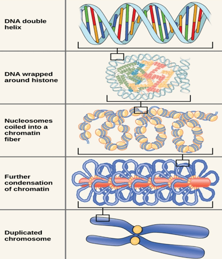
2. CHROMOSOMAL MUTATIONS

     These are changes that affect larger regions of a chromosome and the number of chromosomes. Such changes in the structure and number of chromosomes can be observed under microscope. It is associated with the appearance of new traits in organisms. Five types of chromosomal mutation:

  • Deletion-due to breakage apiece of chromosome is lost
  • Insertion-chromosomes segment breaks off, flips and reattaches.
  • Duplication-Gene sequence is repeated.
  • Translocation-Part of one chromosome is relocated with another chromosome.
  • Non Disjunction-Gametes will contain too many chromosomes due to the failure of separation during meiosis.

TYPES OF MUTATIONS

  • Somatic and Germinal Mutations: If mutation occur in the somatic cells (non-reproductive cells) it is called somatic mutation. If a mutation occurs in the reproductive cells it is called germinal or genetic mutation.
  • Dominant and recessive mutation: when a mutation produces a dominant phenotypic expression it is called dominant mutation. If a mutation produces a recessive expression, it is called recessive mutation.
  • Micro and macro mutation: When mutation produces small visible changes, e.g., white ye in Drosophila. Such mutations are called micro mutation. Mutations which produce prominent visible changes, e.g., albino maize, Ancon Sheep etc. are called macro mutations.
  • Spontaneous and Induced Mutations: Naturally occurring mutations are called spontaneous mutations. Mutations produced artificially by action of certain agents are called mutagens. Such mutations are called induced mutations.
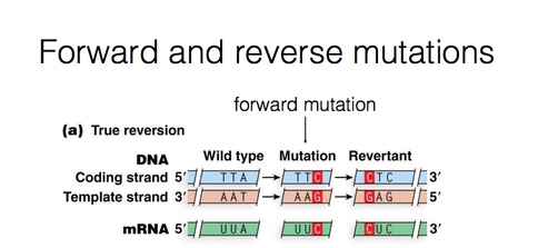
  • Forward and reverse mutations: During mutation, a change take place from the normal type (wild type) to a new form (mutant form).This is called Forward mutation. The mutants may revert to the wild forms. Such changes are called reverse or back mutations.
  • Lethal mutations: Some mutations affect the vital functions of an organisms. This leads to the death of an organism .Such mutations are called lethal mutations.

MUTAGENS

Mutagens are substances which induce mutation. Such mutations are called Induced mutation. The frequency of such mutations is higher than spontaneous mutations. Mutagens are classified into two major groups:

1. High Energy Radiations

2. Chemicals

Applications of Gene Mutation

G.J .Mendel could not have put froth the basic ideas on inheritance, had all the pea plants were only tall etc.

T.H Morgan was able to evolve his ideas about sex-linked inheritance only when he could find out the mutant white-eyed Drosophila flies.

T.Benzer could analyses the fine structure of genes with the help of mutant forms of T4 phages.

Plants with new and beneficial traits have been evolved through induced mutations. Plant breeders have produced mutants in barley, wheat, oats, soybean, tomatoes etc., which have desired characters like increased yield and resistance to disease.

The yield of Penicillium has been increased through mutation.

NUCLEAR ORGANISATION IN PROKARYOTES AND EUKARYOTES

BY: RAHUL ANDHARIA (MSIWM001)

Introduction:

Genes of an organism is encompassed by Genome. Gene is a hereditary unit, which consists of all the nucleotides and forms a part of chromosome. Spatial distribution of chromatin(DNA and protein) within a cell nucleus is called as Nuclear Organisation. There are different levels in nuclear Organisation.

History:

Chromosome organisation into distinct regions within a cell nucleus was first observed and discovered in the year 1885 by Carl Rabl. With advancement in microscopy technologies, in the year 1909, chromosome territories were discovered by Theodor Boveri when he observed that chromosomes take up separate nuclear regions.

Prokaryotic Organisation:

  • Not well defined nucleus is not present in Prokaryotes. Prokaryotes means, ‘before nucleus’. Chromosomal DNAin prokaryotes is present in structures called Nucleoid.
  •  Circular DNA is present in loops or domains in the nucleoid binded to scaffold proteins which are attached to cell membrane.
  • Bacterial chromosomes are huge, but still it gets fitted inside the cell. This occurs because of Supercoiling of DNA.

DNA Supercoiling:

  • Prokaryotes, compress their DNA into small pieces by supercoiling.
  • In bacterial genome the circular DNA is supercoiled because of addition of turns in the double helix structure of DNA.
  • Two enzymes are necessary for supercoiling in prokaryotes, Topoisomerases(type 1) and DNA gyrase.
  • Chromosomal DNA is  compacted to 1000 folds in order to fit DNA within bacterial cells.
  • DNA is organised into loops by proteins and further this loops compact the DNA by 10 folds.
  • Supercoiling folds DNA by additional 100 folds.
  • Supercoiling of DNA can be negative or it can be positive. Negative supercoiling takes place in the opposite direction to the double helix.
  • Positive supercoiling is when twisting occurs in the same direction as that of double helix DNA.

During normal growth, most of the bacterial genomes are negatively supercoiled.

Proteins involved in Supercoiling:

  • Multiple proteins are involved in DNA folding and condensation, which was reported in the year 1980s and 1990s.
  • One of the most abundant protein in nucleoid, HU, binds DNA with the help of enzyme Topoisomerase I, introducing sharp bends in the chromosome, generating tension which  is necessary for negative supercoiling.
  • One more protein called Integration Host factor(IHF) binds to specific genome sequences and produces additional bends. This was showed by Rice et.al in the year 1996.
  • Topoisomerases and DNA gyrase maintains the supercoiling, once the genome gets condensed.
  • Genes involved for modulating response to environmental stimuli can be altered in terms of their expression patterns, and this is done by H-NS which is a maintenance protein during transcription.

How these tightly packed genes gets accessed by proteins?

  • Nucleoid in Prokaryotes appears as irregular mass, but when cell is treated with chemicals for inhibiting transcription or translation, Nucleoid becomes Spherical.
  • Small regions of chromosomes project out during transcription process from nucleus to cytoplasm. In this region they unwind and gets associated with Ribosomes and thus allows easy access for various different types of transcriptional proteins.

Eukaryotic Organisation:

  • Large amount of eukaryotic DNA is packed in chromosomes present within nucleus. DNA and proteins together constitutes a chromosome.
  • Mostly histone proteins are present in abundance, but certain other proteins are also present in less amount termed as Non- histone proteins.
  • DNA-protein nuclear complex is called as chromatin.
  • Length of packaging varies, for example in humans 1.6cm shortest DNA molecule can be wrapped and as large as 8.6cm can also be wrapped.
  • Packaging of DNA into chromosomes involves folding and has different stages involved in it.

Stage 1(Nucleosome):

  • Chromosomal DNA binds to histone proteins in first stage of organisation.
  • DNA wraps around histone octamer(2copies of dimers and tetramers each) at regular intervals. DNA-histone complex is called the chromatin.
  • DNA wrapping around histone proteins forms bead like structure which is called Nucleosome.
  • Nucleosome core particle wrap DNA for about 1.67 left handed turns and contains 146base pairs of wrapped DNA.
  • DNA that connects to Nucleosome is called as Linker DNA.
  • DNA in this form is seven times shorter when compared to double helix structure without histone proteins. The size of beads is 10nm in diameter compared to double helix which is 2nm.
  • Histones are: H1, H2a H2b H3 and H4. H1 is not included in core histone and it connects to linker DNA.
  • Histone proteins are basic proteins mostly made up of lysine and arginine amino acids which are positively charged.

Stage 2(30nm fibre):

  • In this level of compaction, linker DNA and Nucleosome are coiled into a 30nm fibre.
  • Because of this coiling, the chromosome further gets shortened and becomes 50 times more shorter than the extended form.
  • Fibre is formed when H1 histone binds to linker DNA at each Nucleosome. When H1 histone interacts with each other, nucleosomes gets pulled together.

Stage 3 (Radial Loops):

  • When chromosomes are deprived of histones, they possess a central fibrous protein scaffold, to which DNA gets attached in loops.
  • The main role of these fibrous proteins is to make sure that each chromosome in non-dividing cell occupies a particular area of nucleus that doesn’t overlap with other chromosomes.

PROTIENS STRUCTURE, CLASSIFICATION AND FUNCTION

                   BY: Sreelakshmi S Nair

Proteins:

Proteins are macromolecules obtained from the one or more amino acids chain linked by peptide bonds. They are the natural polymers of amino acids. It contains nitrogen, carbon, hydrogen and oxygen. They act as a biological catalyst form structural parts of different organisms, participate in different cell reactions.

CLASSIFICATIONS OF PROTIENS

Proteins are classified on the basis of

  • Structure of Protein
  • Composition of Protein
  • Functions of Protein

Based on the structure of Protein

  • Fibrous Protein

They are linear in shape. Usually they do not have tertiary structure. They are physically strong and are insoluble in water. They perform the structural functions in the cell. Examples are: Keratin, Collagen, and Myosin.

  • Globular Protein

They are spherical or globular in shape.Teritary structure is the most functional structure. Physically they are soft while comparing to fibrous proteins. They are readily soluble in water. Most of the proteins present in the cell belongs to globular protein. It forms enzymes, antibodies and some hormones. Examples are Insulin, haemoglobin, DNA polymerase and RNA polymerase.

Based on the composition of Protein

  • Simple proteins

They are composed of only amino acids. They may be fibrous or globular. They are generally simple in structure. Examples are Collagen, Myosin, Insulin

  • Conjugated Proteins

They are complex proteins which contains one or more amino acid components. The non-protein parts are called prosthetic group. Prosthetic group may contain metals, ions, carbohydrates, lipids, nucleic acid. Conjugated proteins are generally water soluble and globular in structure. Most of the enzymes are conjugated proteins.

Based on the Function

  • Structural Proteins

Most of them are fibrous proteins. Components in connective tissue, bone, tendons, cartilage, skin, feathers, nail, hairs and horn are made of structural proteins.

  • Enzymes

They are the biological catalyst. Mostly globular conjugated proteins. Examples are DNA polymerase, Nitrogenase, and Lipase.

  • Hormones

They include proteinaceous hormone in the cells. Examples are Insulin, ACH

  • Respiratory Pigments

They are coloured proteins. All of them are conjugated proteins. Examples are haemoglobin, Mycoglobin.

  • Transport Proteins

They transport the materials in the cell. They form channels in the plasma membrane. They also form a component in blood and lymph in animals.

STRUCTURE OF PROTEIN

There are four levels

  1. Primary Structure
  2. Secondary structure
  3. Tertiary Structure
  4. Quaternary Structure
  1. Primary Structure

They give details about the amino acid sequence of a protein. It tells about the number of amino acid residues in the protein and also about the sequence of amino acids. It is stabilized by peptide bonds. Each component of an amino acid is called residue or moiety. It starts from the amino terminal (N) end and ends in the carboxyl terminal(C) end.

  • Secondary structure

It is formed by hydrogen bond between backbones atoms. Three most important secondary structure in protein are:

  • α-helix
  • β- plates
  • β-Turns

α -Helix

It is the most common secondary structure which repeat every 5.4It is the simplest arrangement of a polypeptide chain which was proposed by Puling and Corey in 1954.It is so common because it makes optimal use of internal hydrogen bond. The interactions between the amino acid chains can stabilize or destabilize the α-helix.

                    β- Plates

It is an extended form of a polypeptide chain.Polypetide backbone forms a zigzag structure. Similar to α-Helix structure is stabilized by hydrogen bond. The R-groups of adjacent amino acids protrude from the zigzag structure to the opposite direction forming an alternative pattern. It can be arranged in either parallel or anti-parallel direction.

                    β-Turns

         It’s a very common in proteins where peptide make a reverse direction. It forms a 180 degree turn involving four amino acids. The carbonyl oxygen of the first residue forms a hydrogen bond with the amino group hydrogen in the fourth amino acid in the turn. Glycine and proline allows the β-Turns frequently.

3. Tertiary Structure

                   The tertiary structure will have a single polypeptide backbone consisting of one or more    secondary structures. It can be defined by atomic coordinates. It is stabilized with the help of both covalent and non-covalent bond.

4. Quaternary Structure

Proteins which have more than one polypeptide subunit and which do not have a permanent (covalent) interaction between the subunits (like disulphide bond) are classified under quaternary structure. Bonds stabilizing quaternary structure includes hydrogen bonds, hydrophilic interactions, hydrophobic interactions, van der Waals interactions. A protein with a single subunit cannot have a quaternary structure.

Functions of Protein

  • Boosts Immune System
  •  Provides Structure
  • Maintains pH
  • Transports and stores nutrients

MEDIA FORMULATION and SCALE-UP

BY: RAHUL ANDHARIA (MSIWM001)

Growth medium is used for support of micro-organisms, cells and small plants. Microbial growth medium can be solid or liquid depending on the type of Micro-organisms to be grown and cultured. It is necessary to select an appropriate culture medium for in-vitro cultivation.

Components and media Formulation:

The requirement of media components depends on the type of cell lines used. Every component used has a specific role to perform. Basic components of media are glucose, amino acids, salts, vitamins, other nutrients, energy source, nitrogen source, water and carbon source.

Carbon and Energy Sources:

  • Carbon metabolism has a role to play in product synthesis. Formation of products directly depends on the rate at which carbon is metabolized.
  • Carbohydrates, oils and fats and hydrocarbons are some of the common carbon sources.
  1. Carbohydrates:
  2. Most commonly used source of carbon in fermentation processes.
  3. Maize, cereals and potatoes contains starch, which is an essential carbohydrate. Primarily used in fermentation of alcohol.
  4. Barely grains contains are rich in  amount of carbohydrates like sucrose, cellulose, starch and other sugars.
  5. One major source of sucrose is sugarcane and Molasses. Molasses(obtained after refining of sugarcane or sugar beets) are one of the major source of carbohydrates.

B.    Oils and Fats:

  • Oils provide more energy compared to sugars. Vegetable oil is a common source of carbon.
  • Oils and fats are generally used more as additives rather than as sole carbon source. It also has anti-foaming properties.
  • Examples include: vegetable oil, olive oil, linseed oil, soya been oil.

C.    Hydrocarbons:

  • Hydrocarbons used as carbon sources are C12- C18 alkanes.
  • They have more carbon and energy content per weight when compared to sugars. They are relatively cheap.
  • Can be used in antibiotics, organic acids, amino acids and protein fermentation.

Nitrogen Sources:

  • Most commonly used nitrogen sources includes, Ammonia, ammonium salts and urea.
  • For pH control, ammonia is used.
  • Soya meal, corn-steep liquor, peanut meal, cotton seed meal, amino acids and proteins.

Essential Metals and Minerals:

  • Na, K, Ca, P, S, Cl, Mg and Fe are known as macronutrients and are required in larger quantities.
  • Zn, Mn, Br, B, Cu, Co, No, V, and Sr are the micronutrients required in relatively less amounts.
  • Concentration of these elements used depends on the type of Micro-organisms to be cultured.

Inorganic Salts:

  • helps in maintaining osmo-regulatory balance.
  • Helps to Increase membrane potential by providing calcium, phosphate and sodium ions.

Growth Factors: common growth factors includes Vitamins, Minerals and fatty acids.

  1. Amino acids:
  2. Amino acids being building blocks of proteins, becomes an essential component of any type of culture media.
  3. Microbial cells cannot synthesise Essential amino acids, and hence it must be supplied for cell proliferation.
  4. Nitrogen for NAD and NADPH, and nucleotides is provided by L- Glutamine.
  5. As L-glutamine is an unstable amino acid, it gets converted to other form with time and hence it must be added to medium just before use.
  6. Non-essential amino acids can also be provided for those that are deprived of growth.

B.   Vitamins:

  • Cells cannot synthesise vitamins in sufficient quantities and must be supplied. Vitamins serves as important growth factor for cell proliferation.
  • B group vitamins like Thiamine, niacin Pantothenic acids are commonly added.
  • Serum is the major source of vitamin in media.

C.    Fatty acids: mostly important in serum free media, as fatty acids are commonly present in serum. Example- Linoleic acid.

Chelating Agents:

  • Insoluble metal precipitation can be prevented by using chelating Agents.
  • Chelating Agents forms complexes with metal ions present in media, and than this can be utilised by micro-organisms.
  • Example- EDTA( calcium and magnesium complex), citric acid, pyrophosphates.

Buffers: role of buffer is to regulate the pH of medium. Micro-organisms growth is affected by changes in pH and hence role of buffers is vital in any type of media Formulation.

  1. Natural Buffer systemBalance of CO2 along with co3/hco3 content of culture medium is termed as natural Buffer system. Air atmosphere has to be maintained in natural buffering system.(5-10% CO2).
  2. HEPES: can be used as buffering system. It increases the sensitivity of the media towards phototoxic effects.
  3. Phenol Red: it is used as an indicator in most of the commercially available media. pH of medium changes due to release of metabolites, as pH changes colour of the solution also changes. Phenol Red turns medium yellow at lower pH, while at higher pH it turns the medium purple.

Anti-foaming Agents:

  • Large amounts of foam is produced during microbial processes. Because of excess foaming cells gets removed from the media and leads to Autolysis.
  • Hence Anti-foaming agents are required to stop excess foaming and prevent cells from Autolysis.
  • Examples- Stearyl alcohol, cotton seed oil, linseed oil, silicones and sulphonates.

Selective agents:

  • Mostly  antimicrobials are used. This agents makes them selective for certain Micro-organisms.
  • They are added in a fixed concentration which is specific and prevents growth of unwanted Micro-organisms.
  • Examples- Selenite, bile salts, dye stuffs.

Gelling Agents:

  • Most commonly used gelling agent is Agar. It is mostly obtained from sea weeds like Gracilaria and gellidium.
  • Gelatine, polyacrylamide, carrageenan scan also be used as gelling Agents.

Serum:

  • Most important component of cell culture media. It is a complex mixture of Albumins, growth factors and growth inhibitors.
  • For cell cloning and fastidious growth of cell, fetal serum is used.
  • Due to its lower growth promoting ability, calf serum, is used for contact inhibition studies.
  • 2-10% of serum is present in normal media.
  • Serum provides multiple components like proteins(fibronectin), Albumins, amino acids, provides protease inhibitors(protects cells from proteolysis) and it can also act as buffer.

Scale Up of industrial Microbial process:

  • According to its name, Scale up simply means increasing something (process) in terms of size, production and it’s amount.
  • Scale up of industrial Microbial processes is essential to meet the customer needs and to mass produce a particular product or process for larger profit gains and for greater supply needs.
  • For scale up, any process developed in laboratory has to be converted into full manufacturing scale process. For example- 20000- 2000,000L fermenters are used for scale up process.
  • Scale-up industrial process is done in 2phases generally:

Pilot Scale- 100-10,000 L fermenters and downstream equipments. The main purpose of pilot Scale- is to convert lab based process to a smaller version of manufacturing process with medium production and scale up.

Demo Scale- 10,000-100,000 L fermenters, with downstream equipments. It minimises the larger investments risk by continuous supply chain, process validation and fulfilling market demand.

Roadmap for proper and successful scale-up process (For large scale plant)

  • For an extended period, fully integrated process, including recycle streams can be operated with full industrial materials and equipments.
  • Different design models of equipments such as fermenters and suppliers can be evaluated.
  • More number of people can be trained to operate large scale plant. Proper operation methods, if followed can lead to faster production.
  • Operating-know how and pilot plant data is used to create solutions and planning for preparing large scale plant.
  • Large quantities of product can be produced at end application, to build healthy customer relationship and increasing demand for higher commercial plant output.

Scale-up process is essential in terms of increasing production and fulfilling the demand based on increasing needs and supply chain of products.

GENETICALLY MODIFIED PLANTS AND ANIMALS

BY: RAHUL ANDHARIA (MSIWM001)

GM Plants: To prepare a GM plant, new DNA is transferred into plant cells. Genetic engineering techniques are used to modify DNA. Genetic composition of the plant is altered by adding specific useful genes. Once the new DNA is inserted, than the cells are taken and grown in tissue culture using appropriate medium where they develop into new plants and will inherit the new DNA which was inserted.

History:

Tobacco plant, first genetically modified plant was produced in the year 1982. These plants were modified for antibiotic resistance. The first country to commercialise use of transgenic plants was China. The tobacco plants were made antibiotic resistant by creating a chimeric gene which joined the antibiotic resistant gene present on the T1 plasmid of Agrobacterium.  Bt cotton was the first commercialised genetically modified crop used in India which was made by Maharashtra hybrid seeds company along with Monsanto company from USA.

Process of developing GM Plants/Crops:(6 steps)

  • Isolating Gene of interest- Gene of interest is isolated from other plants or organism. Information like structure, function and location of chromosomes is useful in identifying gene of interest in an organism. Information about gene function and it’s regulation in donor organism(organism from which gene is taken) and in recipient organism(organism in which the gene is inserted) must be known fully before starting the experiment in order to minimize the adverse effects.

Gene insertion by using Transfer vector and plant transformation-  Plasmid from Agrobacterium Tumifaecins, are used as common transfer tools. Using rDNA technology, the gene of interest is inserted into the plasmid.  Plant cells or explants are mixed with Agrobacterium cells containing the plasmid with new genes. T-DNA, piece of plasmid is taken up by the cells. Desired genes are inserted into one of the plant chromosome by Tumifaecins, and now the plant is called Genetically modified. Other method used for transfer is Gene gun method or particle bombardment method. In this method, the relevant metal surfaces are coated with small DNA fragments and these particles are bombarded into plant cells. Mostly Tungsten or gold particles are used for coating DNA (called microprojectiles). This method is a bit costly but can be effective.

  • Selection and Regeneration of modified plant cells to form a whole plant:  Generally, a small fraction of plant cells take up the gene of interest after transformation. Hence, selectable marker genes, that favours antibiotic and herbicide resistance are used to favour growth of transformed cells. After the process, the transformed cells are regenerated into a whole plant using plant tissue culture method.
  • Plant transformation verification: The gene inserted has to inherit normally, so this needs to be verified. For this purposes, tests are performed to determine number of copies inserted, intactness of the copies inserted and effects of inserted gene with the other genes. In this, gene expression (mRNA-proteins) is also checked to confirm whether gene is functional or not.
  • Testing of plant performance: checking plant performance is vital. After transformation, only fraction of plant cells have the copies of inserted gene. So selective markers are used to favour growth of transformed cells. The resistance genes along with genes for desired traits are transferred using a suitable vector. So when cells are exposed to antibiotics or herbicides, only transformed cells with this selective markers will grow. This way performance is checked and only transformed cells are taken to regenerate and create a whole plant by using tissue culture.
  • Safety assessments: safety assessments are necessary in terms of food and environment. There are different tests to determine whether the released modified plant is safe for consumption or for  cultivation to produce  higher yields without  damaging the environment.

GM Crops in India:

  1. Bt cotton:
    1. Bt cotton was developed to tackle the boll worm infection in cotton plants. The Bt cotton variety was developed by Maharashtra hybrid seeds company along with Monsanto, USA.
    1. GEAC(genetic engineering approval Committee), in 2002 approved Bt cotton making it the first genetically modified plant in India to receive the approval.
    1.  Bt cotton is an  insect resistant genetically modified crop.(made resistant to cotton boll worms, that destroys cotton plants).
    1. Bt is a protein from bacillus thureingenesis, bacteria which has 200 different types of Bt toxins. Each toxin affects and works on different types of insects.

Cry group of endotoxins in Bt cotton are modified by inserting a gene with toxin crystals, when organisms(insects) ingests this genes with toxins, toxins dissolves the gut lining of the insects leading to its death. Thus, the crop plant is protected from boll worms.

  • Bt Brinjal:
    • It was developed to give resistannce against lepidopteran insects, specifically to leucinodes orbonalis, which is a fruit and shoot borer in Brinjal plants.
    •  Bt Brinjal was developed by Maharashtra hybrid seeds company in collaboration with Tamilnadu agriculture University and Dharwad institute of agricultural sciences.
    •  GEAC approved commercialisation of Bt Brinjal in the year 2007, however , due to lack of proper safety and efficacy and lack of scientific consensus, it was banned in the year 2010.
  • HT Mustard:
    •  DMH(Dhara Mustard) was created to reduce the demands of edible oil imports of India.
    • It was created by Delhi University professor, Deepak Pental.
    •  DHM-11 was created using transgenic technologies in particularly involving Barstar/Barnase gene technologies.
    •   Male fertility is conferred by Barnase gene, while Barstar, restores fertile seeds producing abilities of DHM-11.
    • GEAC approved it in the year 2017.

  Advantages of GM crops:

  • Crop Protection: Resistance to diseases, pests, insects and herbicides. Resistance is achieved by using Genetic engineering methods and by using toxins in case of Bt cotton.
  • Economic Benefits: GM plants increases the yield double times compared to the normal plants.
  • With increasing demands of quality food, GM crops can be beneficiary in providing and supplying food at much faster rates.

Concerns with GM crops:

  • Health concerns: Transfer of antibiotic resistance markers or allergens are some of the potential risks. Example- In HT Soya areas, in Argentina, birth defects and childhood Cancers were increased by threefold. This was a report based study.
  • Environmental concerns: can reduce diversity of species. For example, if insects that are not be killed gets killed by developing modified crops can reduce species diversity. Super weeds(transfer of genes from one crop to other creates super weeds) which are resistant to most of common control methods.
  • Economic concerns: Launching of GM crop to market is costly and time consuming process. Also violation of ethical issues have been raised as a concern, for example- organisms intrinsic natural values have been violated by mixing it with other species.

GEAC(Genetic engineering approval Committee)– It was approved under ministry of environment, forest and climate change for manufacturing, use, import, export of GMOs (genetically modified organisms). This committee is also responsible for giving technical approvals for proposed GMO products including field trials.

Safety of GM crops and it’s related products is monitored by Institutional Biosafety Committee (IBSCs).

GM Animals:

GM animals can be created by inserting a foreign gene of interest into their genomes. rDNA technology is used for construction of foreign gene. Along with gene, DNA is also modified and contain different sequences in order to incorporate and express into the host cells.

History:

Mice embryos in-vitro manipulation was first reported in the year 1940 using a culture system. In Angora Rabbits, first successful transfer of embryos was achieved in the year 1891 by Walter Heape. Modern genetic modifications began in the year 1973, when Herbert Boyer and Stanley Cohen first discovered and demonstrated that gene from one organism can be cut, and pasted to other organism. Mouse is the first genetically modified animal which was developed by Rudolph Jaenisch, in the year 1974.

Examples of GM Animals:

  1. Mice: GM mouse models are used extensively as models for studying and understanding  different diseases. Two methods are well known for developing GM mice, Embryonic stem cell method and Pronucleus method.

Embryonic stem cell Method: (ES)(method 1)

  • Mouse blastocyst has inner cell mass from which ES are harvested.
  • rDNA technology is used to make the DNA containing the desired gene of interest, vector and promoter and enhancer sequences.
  • ES cells are transferred in culture. When ES cells are exposed to DNA, it gets incorporated into it.Transformed cells are selected and injected into inner cell mass of mouse blastocyst.
  • Transfer of embryo. Pseudo pregnant mouse is created and the embryo is transferred into its uterus.
  • Offspring produced by it is tested. For testing, remove small piece of tissue from tail and examine it’s DNA. It should be present in 10-20% and it should be heterozygous for the gene.
  • Transgenic strain is established by mating two heterozygous mice and screening their offspring’s.

Pronucleus method:(method 2)

  • rDNA technology is used for preparing DNA with desired gene, vector and promoter and enhancer sequences.
  • Freshly fertilized eggs are harvested before sperm head becoming Pronucleus.
  • The male Pronucleus is injected with DNA that is prepared.
  • Zygote formed by pronuclei fusion is allowed to divide by mitosis to form 2 cell embryo.
  • The embryo is than implanted into pseudo pregnant foster mother.
  • Than the rest of the steps are common with respect to ES method. Offspring test is performed followed by establishing Transgenic strain.
  • GM chicken:
  • Embryos are infected with viral vectors carrying human gene with  a therapeutic protein and promoter sequences.
  • Human gene is transformed with the rooster sperm or appropriate promoter.
  • Check for transgenic offspring’s..
  • The method is cost effective.

C.   GM Sheep’s:

  • Connective tissue cells of sheep are treatedwith a vector, which has 2 homologous regions to that of COL1a1 gene of sheep, alpha 1 Anti-trypsin coded by human gene, antibiotic neomycin resistant gene, beta lacto globulin gene promoter site and ribosome binding sites for beta lacto globulin to be translated.
  • Transform the cells and fuse with enucleated(without nucleus) sheep cells.
  • Next step is implantation into uterus of female sheep(called ewe).
  • Lambs produces large amounts of milk when treated with hormones.

However, this method implemented by one of the company in 2000, abandoned it in 2003 because for purification of protein from sheep’s milk, the cost of expenses were almost doubled.

Applications of GM Animals:

  • GM animals are used as models to understand the disease process and it’s progression.
  • They can be used as models to test new therapeutics that are being developed for treatment of diseases.
  • GM animal models are also used to study gene function. For example- animals with certain genes being turned off or non functional genes can be studied to understand how turning off of this genes can lead to diseases and the mechanisms behind it.
  • Can be used in agriculture to confer resistance to diseases against pathogens.
  •  Knockout mice- used extensively in research to understand genes for which mutant strains are not available.
  • Knock-in mice: It removes certain DNA sequences that otherwise blocks transcription. So the target gene can be turned on as per wish. Also new gene can be introduced by replacing one of the mouse gene.

MICROBIOLOGY COLLEGE IN MADHYA PRADESH


S.NO  COLLEGE NAME    TYPELOCATION           LINKS
1JIWAJI UNIVERSITY   PUBLIC   GWALIORwww.jiwaji.edu
2RABINDRA NATH TAGORE UNIVERSITY   PRIVATE   BHOPALrntu.ac.in
3KAMLA RAJA GIRLS GOVERNMENT POST GRADUATE COLLEGE (KRGGPGC)    GOVERNMENT  GWALIORhttp://krgcgwalior.org/
4DR. HARI SINGH GOUR UNIVERSITY  CENTRALSAGARwww.dhsgsu.ac.in
5CAREER COLLEGE   PRIVATE  BHOPALhttps://www.careercollegeindia.com/
6RAJEEV GANDHI COLLEGE  PRIVATE  BHOPALhttp://rgcbhopal.org/
7COLLEGE OF LIFE SCIENCES PRIVATE  BHOPALhttp://collegeoflifescience.org/
8BARKATULLAH UNIVERSITY  PUBLIC  BHOPAL
www.bubhopal.ac.in
 
9GOVERNMENT M L B GIRLS PG COLLEGEAUTONOMOUS  BHOPALhttp://www.mphighereducation.nic.in/mlbbpl
10DEVI AHILYA VISHYAVIDYALAYAPUBLIC  INDORE
www.dauniv.ac.in
 
11SHREE KRISHNA UNIVERSITYPRIVATE  CHHATARPURhttp://www.skuindia.ac.in/
12MAHARAJA RANJIT SINGH COLLEGE OF PROFESSIONAL SCIENCES (MRSC)PRIVATE  INDORE[www.mrscindore.org
13BONNIE FOI COLLEGE (BFC)PRIVATE  BHOPALhttp://bonniefoicollege.org/
14ATAL BIHARI VAJPAYEE HINDHI VISHWAVIDYALAYAPUBLIC  BHOPAL
www.abvhv.edu.in
 
15SAGE UNIVERSITYPRIVATE  BHOPALhttps://sageuniversity.edu.in/

CHROMATOGRAPHY

BY: RAHUL ANDHARIA (MSIWM001)

Enzyme Purification: The process in which enzymes are purified to obtain pure biological catalysts to study their nature and further using it in industries and in applied sciences to develop various                by-products.

Chromatography: It is a technique in which components are identified, separated and purified from a mixture for qualitative and quantitative analysis. Based on Polarity, net charge and hydrophobic interactions, enzymes are separated by using chromatography. In the year 1903, Chromatography technique was first used for colourful separation of plant pigments through a calcium carbonate column by Mikhail Tswett. (Coinedthe term chromatography)

General Chromatography Principle: The basic principle involved is, mixture of molecules on the surface of solid or liquid gets separated in the stationary phase(which is also called stable phase) from each other and moves with the help of mobile phase. Factors leading to separation generally includes adsorption (liquid-solid), partition(liquid-solid) and affinity, that is molecules separate based on their molecular weights. S- phase is solid or liquid while M- phase can be liquid or gas.

Various chromatography methods involved in enzyme purification:

  1. Ion exchange chromatography
  2. Affinity chromatography
  3. Size exclusion chromatography/gel permeation chromatography
  4. Immunoaffinity chromatography
  5. Hydrophobic interaction chromatography
  1. Ion exchange chromatography:
  2. In this method polar molecules or ions gets separated depending on their affinity to ion exchangers.
  3. Cationic exchangers: They are basically negatively charged and attracts positively charged cations. Due to ionisation of acidic group, they are charged negatively and hence also known as acid ion exchange materials.
  4. Anionic exchangers: also called basic ion exchange materials. They are charged positively and attracts negatively charged anions.

  Working Principle of ion exchange chromatography:

  • The technique is based on attractions between oppositely charged stationary phase, which is basically an ion and an analyte.
  • Ion exchangers are charged groups, linked covalently to surface matrix.(can be positive or negative).
  • Charged groups, when suspended in aqueous solution will be surrounded by oppositely charged ions.
  • This forms an ‘ion cloud’ . In this ion cloud exchange of ions occurs without altering the property and nature of the matrix.

 Instrumentation:

  • Pump- IC pump is present which helps in continuous supply and flow of eluent(carrier portion- portion carrying the molecule). In this the eluent used is liquid.
  • Injector: The instrument is equipped with an injector valve that injects or allows the liquid to pass through. Solid substances are first dissolved in solvent and than they are injected through the valve.(injecting range of liquid ranges from 0.1-100ml of volume).
  • Columns: material of the column depends on the application of use. It can be various types like, glass, steel, titanium and inert plastic such as PEEK. Column diameter ranges from 2mm-5cm. Guard column is placed inside separating column for ensuring the safety of the column and its usage for longer durations.
  • Suppressor: To reduce background conductivity of chemicals used for sample elution, suppressors are used. IC suppressors are employed to convert ionic eluent water.(enhances the sensitivity).
  • Detector: commonly used detector is electrical conductivity detector.

Data system: connected to a data system to obtain high throughput data.

Procedure of ion exchange chromatography:

  • Columns are used for packaging and packed with ion exchangers.
  • Commercially available ion exchangers are made up of Styrene and Di-vinyl chloride.
  • Based on charge of particle to be separated, ion exchangers are selected.
  • The column contains sample, ion exchanger and buffer.( Tris and acetate buffer are used widely).
  • Particles gets separated based on its affinity towards ion exchangers. Particles with higher affinity for ion exchangers, settles down at the bottom of the column along with buffer.
  • Spectroscopy methods are used for sample analysis.

Merit of the method: used for separating charged particles. In-organic ions can also be separated by using this method.

Demerit: Major drawback is, only charged molecules can be separated.

  • Affinity Chromatography:
  • The technique is based on affinity phenomenon, in which atoms are held intact in combination in a mixture by exerting an attracting force between the atoms.
  • Example can be enzyme and inhibitors.
  • Affinity based chromatography was first used and demonstrated Meir Wilcheck and Pedro Cuatrecasas.

Principle:

  • Substrate(ligand) molecules binds covalently on the support medium in stationary phase. The reactive molecules required for binding to the target are exposed.
  • Using chromatography column, the mixture is allowed to pass through. Substances binding to Immobilized substrate binding sites, will also bind to stationary phase, while the leftover mixture is eluted in the volume void of the column.
  • Target molecules which remain attached to the target can be eluted by altering pH, polarity or ionic strength of the solution.

Components of affinity chromatography:

  • Matrix: coupling of ligand to the target molecule takes place in the matrix. It is very important to have a proper matrix for affinity chromatography. The matrix should be chemically and physically inert, it should have a larger surface area for more adsorption of molecules, matrix should be insoluble in buffers and solvents. Polyacrylamide and agarose are the common materials used in matrix preparation.
  • Spacer Arm: Target molecule binds with the ligand with the help of spacer arm. Spacer arm facilitates this binding by avoiding steric hindrance.(rate of reaction is slow due to bulking of large molecules and atoms).

Ligand: molecule binding reversibly to the target molecule is ligand. Based on the nature of macromolecules isolated, ligand can be selected. For purification of enzymes, cofactor, substrate analogue or inhibitor is used as a ligand.

Procedure:

  • Column preparation: materials like cellulose, cephalose or agarose are used for column packaging. Ligand selection is based on the sample opted for affinity chromatography.
  • Sample loading: The loaded mixture of substances is poured into an elution column. Elution column allows the sample to run at controlled rate.
  • Elution: Target substance recovery is done using elution by changing pH, ionic strength and polarity conditions.
  • Used in most of the enzymatic assays for identifying binding sites of enzymes.

Merits: Specificity is very high in this method, target molecules in pure form can be obtained, the matrix used can be reused and gives higher yields.

Demerits: number of solvents required are more, if pH is not adjusted properly, proteins gets denatured, and one major drawback is eliminating non-specific adsorption.

  • Size Exclusion chromatography:
  • This technique can be used in particular for high molecular mass specie.
  • Porous material is S-phase and mobile phase is liquid.
  • Diameter of the pores of porous material used generally ranges from 50 to 3000 A.(A- angstrom).
  • Molecules which are smaller in size penetrates the membrane faster than larger molecules.
  • Size of solute molecule is used as a separation measure in this chromatography method.

Principle:

  • In this method, molecules are selected based on their molecular weights and size.
  • For molecules to be separated, porous glass granules and the selected liquid solvent are in equilibrium.

As smaller molecules move faster than larger molecules, larger molecules which are excluded from the column will pass through the interstitial spaces, where generally smaller molecules are distributed between inside and outside of the solvent sieve, which will than move at much slower rates.

Theory:

  • Volume of column in total is given as:

(Vt= Vg +Vi+ Vo), where Vg, is volume occupied by solid matrix, Vi is solvent volume and Vo is free volume, volume outside particles.

Components and procedure:

  • Dextran, agarose, polyacrylamide are the commonly used gels.
  • Column packaging: done by using silica glass granules or by  cross-linked organic gels such as dextran.
  • Detectors: detectors to be used are decided based on UV fluorescence and refractive index.
  • Size Exclusion chromatography can be implied in purification of enzymes and is extensively used in enzymatic assays.

Merits: larger components can be separated from smaller ones, shorter analysis time, no loss of the sample, gives narrow bands with good sensitivity.

Demerits: filtering in mobile phase is mandatory to avoid columns interfering with detectors and one major disadvantage is that it takes shorter time, the mount of peaks resolved are less, selectivity is also poor compared to other techniques.

  • Immunoaffinity Chromatography:
  • This method is used for separating antigen or antibody from heterogeneous mixture.
  • This method is used as combined method with LC(liquid chromatography) for binding of specific antibody or antigens.

Principle:

  • S-phase is antibody or antibody related agent.
  • The method is column based in which solution is allowed to flow through the column followed by elution.
  • Antigen or antibody is pre-functionalised in the column before the start of experiment.
  • Resin bound capture protein absorbs the target protein, while the leftover solution is eluted.
  • The fraction with target protein is also eluted and purified.

Immunoaffinity Chromatography Considerations:

  • Good column material is essential for the efficiency of the technique. The column should have:  Higher efficiency, mechanical stability, lower nonspecific binding.
  • Optimal size of pores. Smaller the pores, higher the surface area, but it won’t be accessible to larger proteins.
  • Larger pores have lower immobilization.

Antibody attachment:

  • Antibodies attach to column via covalent bonding.
  • Orientation of antibody and it’s attachment point are of important consideration in this type of method.
  • At FAB(fragment antigen binding) region antibody binding is not possible.(this site’s have to be unbound, for antigen to bind).
  • Antibody can be attached via carbohydrate residues, or direct attachment via amines or carboxyl’s.

Components and working:

  • Traditional columns in this method are made up of cellulose or agarose.
  • In high performance Immunoaffinity methods, columns are made up of silica and Azalactone beads.
  • Hydrophobic interactions chromatography: (HIC)
  • Molecules are separated based on hydrophobicity, that is lack of affinity towards water molecules.
  • Molecules elute by decreasing polarity of buffer.
  • If the molecule is more hydrophobic, the binding will be more stronger.

Principle:

  • Samples are loaded with high salt buffers containing hydrobhic and hydrophilic regions.
  • Solvation of sample solutes is reduced by salt buffer.
  • Due to decrease in solvation, hydrophobic regions gets adsorbed by the media.
  • Less salt is required for binding of the molecule is more hydrophobic.
  • During elution, decreasing salt gradient is used.
  • Sample elution can be also done by detergents or modifiers added in the elution buffer.

General considerations in HIC:

  • Ligand: Behaviour of enzymes or proteins can be determined by Immobilized ligand used. For example- straight chain alkyl ligands exhibits hydrophobic nature.
  • Degree of substitution: degree of ligand substitution is directly proportional to binding capacity of enzyme or protein.
  • Temperature: correlation between hydrophobic interactions and temperature is affects solubility and structure of enzyme or protein.
  • pH- mobile phase used in HIC, usually have pH in the range of 5-7. Effect of pH differs from protein-protein.
  • Salt concentrations: salt concentrations shows higher ligand-protein binding but higher concentrations can lead to protein precipitation.

Procedure of HIC:

  • Media is made of alkyl or aryl ligands.
  • Space between matrix is filled by moderate salt buffers.
  • Non-bound proteins can be eliminated by washing the column.
  • Lower the salt concentration, during elution.
  • Ethanol(70%) can be used to remove unbound proteins.

Merits: high ionic strength samples can be used. Large volume of samples can be loaded easily.

Demerits: major drawback is requirement of non-volatile mobile phase.

Thus, these chromatography techniques are essential for purification of enzymes.

MICROBIOLOGY COLLEGES IN DELHI/DELHI NCR

S.NO    COLLEGE NAME    TYPE  LOCATION           LINKS
1BHASKARACHARYA COLLEGE OF APPLIED SCIENCE       PUBLIC   DWARKAbcas.du.ac.in  
2SHAHEED RAJGURU COLLEGE OF APPLIED SCIENCES FOR WOMEN     PUBLIC   MAYUR VIHARhttps://rajgurucollege.com/
3PDM UNIVERSITY    PRIVATE  BAHADURGARHpdm.ac.in  
4SGT UNIVERSITY   PRIVATE  GURGAONhttps://sgtuniversity.ac.in/
5LOVELY PROFESSIONAL UNIVERSITY   PRIVATE  CANNAUGHT        PLACElpu.in
6AMITY UNIVERSITY  PRIVATE   NOIDAwww.amity.edu
7SWAMI SHRADDHANAND COLLEGE  PUBLIC   ALIPURss.du.ac.in
8GD GOENKA UNIVERSITY  PRIVATE   9GURGAONgdgoenkauniversity.com


MICROBIOLOGY COLLEGE IN TAMIL NADU

S.No.COLLEGE NAMETYPELocationLINKS
1Madras Christian College (MCC)Government aided       Chennaihttps://mcc.edu.in/
2Ethiraj College for womenAutonomous       Chennaihttps://www.ethirajcollege.edu.in/
3sastra university,  Deemed       Thanjavurhttp://www.sastra.edu/
4bharathidasan university,   Public      Tiruchirapallihttp://www.bdu.ac.in/
5CMS College of science and commerceAutonomous      Coimbatorehttp://cmscbe.com/
6Annamalai University (AU)PublicChidambaramhttp://www.annamalaiuniversity.ac.in/
7Sathyabama institute and technologyDeemed       Chennaihttp://www.sathyabama.ac.in/
8SRM medical College Hospital and research centreState University       Chennaihttps://www.srmist.edu.in/
9VIT UniversityPrivate      Vellorehttp://www.vit.ac.in/
10crescent school of lifessciences,PrivateVandalurhttps://crescent.education/University/.
11Central University of Tamil NaduPrivate     Thanjavurhttp://cutn.ac.in/
12ramachandran college,PrivatePorur,chennaihttp://www.sriramachandra.edu.in/
13.Madurai Kamaraj universityPublic Maduraihttp://www.mkuniversity.org/

RAPD (RANDOM AMPLIFICATION OF POLYMORPHIC DNA)

BY: RAHUL ANDHARIA (MSIWM001)

Molecular marker is a sequence of DNA in the genome that can be traced and identified. RAPD is a molecular marker that helps to identify genetic variations. Single arbitrary primer is used in RAPD.

History:

It is used to identify nucleotide polymorphism. This method came into existence during the year 1990 when William et.al first used this approach.

Principle:

  • Shorter oligonucleotide primers binding to different loci is used for amplification of random sequences from complex DNA template.
  • The amplified PCR product depends on length and size of primer and the target genome.
  • RAPD is a PCR based method. All the components of PCR are required to perform RAPD like DNA template, dNTPs, reaction buffer, Mgcl2(cofactor), arbitrary primer, Taq polymerase.
  • First step basically involves denaturation, where the PCR products are kept at higher temperature of 94 degree Celsius.
  • Temperature is lowered to 40-65 degree Celsius in annealing process where the primer binds to the target DNA sequence.
  • Further Taq polymerase adds DNA-hybrid primers to 3’ end and extends to the other end of target sequence.
  • The process is repeated until complete replication of amplified product is achieved.

Outline of RAPD Analysis:

  • Genomic DNA is isolated.
  • The isolated DNA is denatured.
  • DNA template is annealed with primers- Annealing.
  • Complementary strands of DNA are synthesized after the extension phase.
  • Gel electrophoresis is used to identify amplified products.

Procedure of RAPD:

Materials Required: PCR components, RAPD arbitrary primers, gel electrophoresis reagents and equipments, Eppendorf’s, PCR tubes, micro centrifuge, micropipettes, deep-freezer, UV transilluminator connected with a system.

  1. Master mix is prepared(control and sample) with all the PCR components.(thaw(keep it in ice) and vortex).
  2. The master mix is aliquoted in PCR tubes.
  3. DNA templates is added in the PCR tubes.
  4. The tubes are placed in allotted blocks in the PCR thermocycler machine.
  5. The reaction setting is adjusted and different temperatures are set for different cycles.
  6. Ideal reaction is: step 1 for 94 degree Celsius (1min), step 2 for 35 degree Celsius (1min) and step 3 at 72degree Celsius for 5min. For final step, temperature is again 72 degree Celsius for 5min.
  7. DNA loading dye is added to the amplified product and Agarose gel electrophoresis is performed. (% of agarose gel is determined based on sample size).
  8. The gel is visualised under UV trans illuminator connected to a system.

Applications of RAPD:

  1. As molecular markers in plants: Using RAPD, genetic maps are constructed in plants. For example- In coffee 15 linkage groups are constructed using RAPD marker.
  2. Desirable trait can be selected directly using RAPD. This marker can be screened with the desired trait anytime during breeding programs. This gives breeders advantage, as they can trace the desired trait.
  3. RAPD is widely used to identify certain genes which are resistant to diseases. For example- In barely crop, the gene rp94 is resistant to stem rust.
  4. Polymorphism studies can be done using RAPD.
  5. RAPD markers are used in evolutionary and population genetics for identifying genetic variations among different species.
  6. Used in germplasm(genetic material of Germ cells) characterization.
  7. RAPD can be employed to detect somaclonal variations, variations observed in somatic cells of regenerated plants.

RAPD PCR is used for detection of genetic polymorphism in leishmania strains(parasites causing leishmaniasis). Example- This type of test was successfully performed in Tunisian patients.

Merits of RAPD:

  • For designing specific primers, no DNA probes and specific sequences is required.
  • It is a quick, simple and efficient method as it does not involve blotting or hybridization steps.
  • Amount of DNA required in the process is small compared to other methods.
  • Number of fragments are higher.
  • Compared to other assays, market cost for RAPD is relatively low.

Demerits of RAPD:

  • Since RAPD markers are Dominant, it will be difficult to distinguish whether a particular DNA sequence is amplified from heterozygous locus or homozygous locus.
  •  RAPD is exclusively laboratory based technique and hence requires all the necessary components and suitable PCR conditions to obtain desired result.
  • This method is sensitive to changes in DNA, PCR components and PCR conditions and hence has problems with reproducibility.
  • Null alleles ( it is a non-functional allele caused due to mutations) cannot be detected directly by this method.