During unfavourable condition some bacteria goes in its dormant structure that is metabolically inactive and unable to reproduce and grow.
They develop structure called endospore which protects the cell from lethal agents like heat, radiation and chemicals.
In clinical procedure, pathogen identification include the describing the shape and position of endospores present in the bacterial cell.
Principle:
This method of staining is a differential staining which is used to detect, identify and differentiate endospore from vegetative cell.
The role of the method is to detect presence or absence of endospore some modification is done by increasing concentration of dye, increasing heat fixing duration and application of ultraviolet radiation.
Requirements and Reagents:
Fresh culture of B. cereus or B. subtilis and Staphylococcus aureus
Malachite green (5% aqueous)
Safarnin
Staining tray
Glass slide
Blotting paper
Microscope
Procedure:
Take separate clean slides and make smear of B.subtilis and S. aureus.
Air dry and heat fix.
Add few drops of malachite green on the smear.
Heat the slide under steam and add stain to avoid dryness.
Wash the slide with water running slowly on the slide.
Add few drops of counter stain i.e. safranin for 30 seconds.
Wash out the smear with slowly running distilled water.
Dry the slide with blotting paper.
Observation:
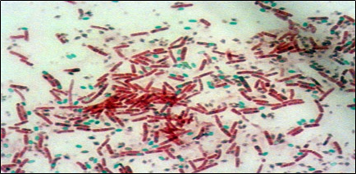
Observe the slide under oil-immersion microscope. From the microscopic field represent the size and position of the endospore. Write the colour of the spore and vegetative cell.
Result:
In B.cereus the endospore stain green and in vegetative cell stain red. Vegetative cell are rod shaped and S. aureus are spherical in shape.
Example:
Type
Example
Positive
Bacillus anthracis, C. botulinum, Clostridium perfringens
Metabolism is the collection of chemical reactions takes place to sustain life of an organism.
The main purposes of metabolism is to convert food to energy to run cellular activity; to convert food to building blocks for nucleic acids, lipids, protein and carbohydrates; and to remove metabolic waste.
The metabolic reactions which are enzyme catalysed are responsible for the growth and reproduction of organism, to maintain their structure and interact with environment.
Metabolic reaction is of two types one is catabolic reaction, means the breaking down of compounds and another is anabolic reaction- the building up of the compounds. The catabolic reaction liberates energy and the anabolic reaction uses energy.
Metabolic pathways include the steps through which one chemical is transformed into another and each step is facilitating by an enzyme.
Enzymes are the key component of the metabolic reaction; they act as catalyst- allows the reaction to proceed more rapidly.
Types:
Metabolic reaction is of two types:
Catabolism
Anabolism
Catabolism:
In catabolism the compound through the set of chemical reaction is broken down into simpler compound or molecules.
This is achieved by breaking down and oxidizing food molecules.
Catabolism is responsible to provide energy for working of the cell and component needed for the anabolic processes which build molecules.
The nature of these catabolic reactions based on the source of energy and carbon which is differ from organism to organism.
The chief metabolic processes in a cell are:
Glycolysis
Pentose-phosphate pathway
Entner-doudoroff pathway
Tricarboxylic acid cycle
Fermentation
Glyoxylate cycle
Lipid hydrolysis
Protein hydrolysis
Anabolism:
Anabolism is the set of constructive reactions which used energy released by the catabolic pathway to synthesize complex molecules.
The complex molecule construct cellular structure step by step, make up from small and simple precursor.
The biomolecules are necessary for the growth and reproduction, some biomolecule serve as the central metabolic intermediates.
Some organisms can synthesis all the necessary organic compound like autotrophs. They can be grown on simple media. On the other hand, the organisms which cannot synthesize organic compounds from atmosphere are known as fastidious organisms.
Following anabolic process takes place in organism:
Synthesis of glucose, lipids, amino acid and protein, nucleic acids
Synthesis of other growth factors like vitamins, hormones etc.
Metabolic process:
Glycolysis:
In the glycolysis process glucose and other sugar are partially oxidized to the smaller molecule i.e. pyruvate
Embden-Myerhof pathway, pentose phosphate pathway and Entner-Doudroff pathway are the three routes for the conversion of sugar into pyruvate.
It is anaerobic process in which organism obtain energy in the absence of oxygen, also called anerobic fermentation.
Tricarboxylic acid pathway:
Given by H. A. kerbs in 1973
Also known as citric acid cycle. Because citric acid is the first product of the kerb cycle which is as known as TCA cycle as the citric acid has three carboxylic group.
Glyoxyalte cycle:
It is anaplerotic reaction which means one product of a cycle is taken up by the other cycle
Oxaloacetate is taken from TCA cycle and used for carbon source from the amino acid synthesis.
Pentose phosphate pathway:
It is an alternative pathway for the sugar degradation.
Its main function is to generate power in the form of NADH in extramitochondrial cytoplasm and the second function is to convert hexoses into pentose for the synthesis of the nucleic acids. The third function is complete degradation of pentose.
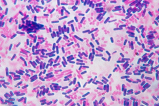
Gram staining is one of the most useful techniques used for the identification of bacterial population.
Developed by Hans Christian Gram a Danish Bacteriologist in 1884.
The procedure includes staining of bacteria and observation under microscope.
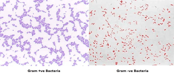
The organisms are differentiate on the basis of holding of stain, the organism called to be gram positive if it retain crystal violet after decolourization and appear purple while the gram negative bacteria after decolourisation appear pink to red in colour because of the safranin counter stain.
Principle:
The differences in gram positive and gram negative is due the difference in the cell wall composition of the bacteria.
Cell wall of a gram positive bacterium consist of a thick layer of peptidoglycan layer with numerous teichoic acid this complex structure resist decolourization.
Cristal violet (primary stain) penetrates cell wall and cell membrane after that iodine is used as moderant, crystal violet interacts with iodine and peptidoglycan layer and makes a complex called cv-I complex. Even after decolourizer is applied CV-I complex retains in the cell and making it to appear purple to dark blue.
In gram negative bacteria there is a thin peptidoglycan layer with loose cross linkage. The peptidoglycan layer is distributed loosely in inner and outer cell membrane. After the application of cristal violet and iodine on the cells, CV-I complex is not trapped in the peptidoglycan layer and decolourizer makes hole in the layer which allow to wash out the CV-I complex from cell and leaves the cell colourless. To make colourless cell visible a counter stain is used called safranin which gives pink to red colour.
Material required and Reagents:
Glass slide
Bunsen burner
Microbial strain (fresh 24 hours incubated)
Crystal violet
Gram’s Iodine
Safranin
70% ethyl alcohol
Dropper
Microscope
Procedure to follow:
Clean a microscope slide and make a smear of distilled water.
With the help of an inoculation loop pic a minute portion from the isolated colony and mix it with the smear on slide to make a thin smear.
Air-dry the slide at room temperature.
Ignite a Bunsen burner and heat fix the smear by heating the slide over the flame (not overheat).
Pour some drops of crystal violet over the smear and wait for 1 min
Slowly pour cold-water on the slide from the edges to wash out excess crystal violet .
Flood the slide with iodine wait for 1 minute and then wash if off.
Decolourize with the help of ethyl alcohol until colourless slide is appear.
Now add few drops of counter stain i.e. safranin to the smear and left for 30 second, then wash it off.
Dry the slide with bloating paper
Observe the slide under microscope at 40X or 100X.
Observation:
Examine properly the slide an identify the gram reaction, describes morphology and arrangement of the microbial cell
Results:
Bacteria which appeared blue colour under microscope are referred as gram negative and those cells which show pink colour are called gram negative .
Limitations:
Over decolourization may lead to identification of false gram negative cells and under decolourization lead to identification of false gram positive cells.
If the smear is too thick or viscous it may retain too much primary stain and makes identification difficult.
Examples:
Type
Example
Gram-Positive
Staphylococcus, Clostridium, Listeria, Bacillus
Gram-Negative
Pseudomonas aeruginosa, Shigella spp, Yersinia pestis, E. coli
Each copy of the DNA is pulled to the separate poles.
Synthesis of new cell wall begins
Once the new cell wall is synthesised fully it results in complete split of bacterium.
New daughter cells now have tightly coiled DNA, plasmids and ribosomes.
Types of binary fission
Example
Transverse
Paramecium
Oblique
Ceratium
Longitudinal
Euglena
Irregular
amoeba
Gene transfer:
Gene transfer means movement of genetic information in organisms.
There are two types of gene transfer method one is vertical in which gene is transferred from parents to offspring and another one is horizontal in which gene is transferred in between two organisms.
In prokaryotes vertical gene transfer is by the means of binary fission and horizontal gene transfer method consist of three process i.e. transformation, transduction and conjugation.
Transformation:
In 1928 Fred Griffith discovered this method of horizontal gene transfer.
In this process naked DNA molecule or fragment from surrounding environment is uptake by the recipient and incorporated in its chromosome.
It is of two types natural and artificial, natural transformation is very rare event and observed in both gram negative and gram positive bacteria.
Ability of bacteria to uptake DNA fragment and get transformed is known as competence.
Process of transformation:
Competent bacteria naturally pull DNA fragment into their cell from the environment.
These DNA fragment naturally released in the environment after a bacterial cell die.
Ds DNA once crosses the membrane in cytoplasm the 3’ end is leading.
The translocated strand interested in the chromosome of recipient bacteria by homologous recombination.
Now the recipient bacteria undergoes replication and the cells acquired new phenotype are said to be transformed.
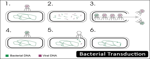
Transduction:
In transduction, DNA is transfer from donor bacteria to recipient bacteria by bacteriophage (functions as vector).
It was discovered by Lederberg and Zinder in 1951.
Bacteriophage due to high specificity of surface receptors has narrowest host range.
Transduction has one advantage over conjugation is that it doesn’t require physical contact of donor to recipient cell.
Transduction process is resistant to the DNase enzyme.
Steps:
The phage infects the host and inserts its phage DNA into the cytoplasm of the host.
During lytic cycle the phage DNA along with the bacterial chromosome is broken down into pieces
Bacterial chromosome packed into the viral capsid is released by the lysis of the bacterium.
Now the transducing phage with bacterial chromosome is ready to infect another bacterium in this way donor’s DNA enters into the cytoplasm of second bacterium.
Host recombinase recA is present in the cell due to which donor DNA recombines with homologous bacterial DNA and produces transductants.
Conjugation:
The process of transfer of plasmid or other transmissible DNA element from donor to recipient via sex pilus or conjugation tube.
Recipient of conjugation is known as transconjugants.
Is can transfer DNA regions of hundreds to thousands of kilobases and has board host range fro DNA transfer.
Occur in between many species of gram negative and gram positive bacteria even occurs between plants and bacteria.
Conjugation involves F plasmid is most common.
Steps:
F+ structure contains tra locus which has pilin gene with some regulatory proteins responsible for the formation of pili on surface.
Proteins present on pili attach to the F- cell surface and responsible for making contact between them but doesn’t transfer plasmid.
The traD enzyme on the base of the pili makes the membrane to fuse.
After the conjugation initiated the enzyme relaxes attached to the conjugative plasmid and make a nick at oriT.
The nicked strand is now transferred to the recipient cell
F+ cell carry such integrated F element is known as Hfr cell.
The F element of Hfr cell is replicated along with the bacterial chromosome and in this way transmitted from one to next generation.
Autoclave is a machine used in industrial and scientific process which require high pressure and high temperature
Autoclave provides a physical method for sterilization by killing bacteria, virus even spores by the means of steam under pressure.
It sterilizes the materials by heating them up at a particular temperature for a specific period of time.
It is considered as more effective method of sterilization based on moist heat sterilization,
Principle and working:
Autoclave work on the principle of moist heat which uses steam and pressure inside the chamber for sterilization.
The boiling point of water is 1000 C under atmospheric pressure (760 mm of Hg), high pressure in autoclave increases the boiling point of the water to achieve proper sterilization.
In addition high pressure facilitates the penetration of heat into the microbial cell and moisture helps in the coagulation of the protein cause death of the microbes.
Working temperature of autoclave is 1210 C at the pressure of 15 psi or 775 mm of Hg.
Steam comes in the contact with the surface of the material inside the chamber and kills the microbes by giving latent heat.
After completion of the sterilization the pressure is released from the whistle and the chamber is restored back to the ambient temperature.
Autoclave components/ parts:
Chamber:
It is the main part of an autoclave consist of inner chamber and outer jacket
Inner side of the chamber is made up of stainless steel or gunmetal and the outer side is made up of iron
A healthcare laboratory uses the autoclave which has an outer jacket filled with steam to reduce the time taken for the sterilization.
Size ranges from 100 L to 3000 L .
Lid:
It is the next important part of an autoclave. A lid is used to seal the chamber and create sterilize condition inside the autoclave.
Lid having rubber seal to make chamber airtight, screw clamps are also used.
It consists of various other components
Pressure gauge:
Pressure gauge is used to indicate the pressure created inside the chamber. It ensures the safety of the autoclave while operating.
Whistle/ pressure releasing unit:
It is present on the lid, work of the whistle is to control excess pressure by releasing it.
Safety valve:
It is important part because it is used in case when autoclave failed to work properly and continuously increasing pressure. It contains a thin layer of rubber inside it that burst itself to release pressure and avoid and damage.
Electric heater:
An electric heater in present underneath the chamber which is electrically operated and boils the water and produce steam inside the chamber.
Water level in the chamber is vital as if chamber doesn’t have sufficient water the heating element might be burnt and if there is too much water in chamber it could interact with trays and material kept inside it.
Waste water cooler:
Many autoclaves come with cooling system which is used to cool the hot effluent into the drainage.
Used to prevent any damage to the drainage pipe from hot water.
Procedure of running:
Before every operation autoclave should be checked for any item left from previous operation
Water level should be checked if needed add sufficient water.
All the material to be sterilized place inside the chamber.
Close the lid and tightened the screw and safety valves to ensure airtight condition and switch on the heater.
Once the water boils the chamber is allowed to release the mixture of air and water from the discharge tube to ensure only moisture inside the chamber. The complete displacement is observed once the bubbles cease to come out from the tube
Now let the steam allowed to reach the desirable pressure (15 lbs).
Once the pressure reached to its desired level the excess pressure is released by the whistle. After the first whistle the autoclave run for holding period i.e. 15 min.
After the completion of holding period the heater is switched off and leave autoclave to cool down until the pressure gauge indicate the pressure inside the chamber equals to atmospheric pressure.
Discharge pipe is now opened to allow air inside the chamber and lid is opened, sterilized material are taken out from the chamber.
Vaccine is an artificial biological preparation that contains antigens or mixture of antigens to acquired active immunity to the particular infectious disease.
Vaccine is prepared by the various different substances like disease causing microorganism or weakened or killed form of microbe, can be from one of the surface protein or its toxins.
Agent used in vaccine stimulates body’s immune system which recognise the antigen and destroy it and also encounter the agent if in future that antigen enters in the body.
Vaccine is prophylactic (prevent future infection by the pathogens) or therapeutic (fight against the disease already occurred).
The process of administration of vaccine is called vaccination; it is the most effective method to prevent infectious diseases.
The term vaccine and vaccination are coined by Edward Jenner. He described that how cowpox is to produce immunity against smallpox.
Vaccine stimulates T-cells and B- cells which further produces antibodies against the antigen.
First vaccine:
The first vaccine was introduced by Edward Jenner, used the cowpox virus to protect against smallpox in humans
Prior to this Asian physician used to give dried lesions from the diseased person to children. But by this process some individuals developed immunity while some develop disease.
Jenner introduced a safer way to counter this disease. He uses similar cowpox virus to confer immunity against smallpox (rare condition in which immunity of one virus protect against another virus).
Louis Pasteur in 1881 shows immunization against anthrax disease and four years later he develops vaccine of rabies.
Characteristics of vaccine:
Safe
Long term protection
Induced b and t cells
No or very few side effect
Low cost and biologically stable
Easy to use
Types of vaccine:
Live attenuated:
Is containing live organism which is weakened in the lab so that it cannot cause disease and activate the immune system against the antigen.
It is relatively easy to produce live attenuated vaccine for viruses then bacteria because bacteria have thousands of genes which is much harder to control.
Uses whole organism as vaccine which loses their pathogenicity but can induce immune response, they continuously multiply in human and provide immunity over the period of time.
Examples of live attenuated vaccine are mumps vaccine, measles vaccine, chickenpox, BCG, Sabin’s polio vaccine.
Killed or inactivated vaccine:
Microorganism causing diseases are killed by the means of chemicals, heat or radiation. These are more stable and safer than live vaccines reason is that the dead microorganism cannot mutate back to cause diseases.
They are the easiest preparations to use
Chemical which are used to kill microorganism are formaldehyde or beta-propiolactone, traditionally used chemical for virus is formalin.
The process should be observed carefully because excessive treatment can destroy immunogenicity and insufficient treatment leave infectious microbes to cause disease.
Like above it doesn’t uses whole organism, only the part which server as antigen and stimulate the immune system is used to prepare vaccine.
Composed of purified macromolecule derived form the pathogen known as subunit vaccine.
General forms of such vaccine are
Purified capsular polysaccharide vaccine: pathogenicity of some bacteria depend on their capsule and this capsule protect bacteria from binding to the antibody. In this way infants and younger children’s immune system cannot recognise and respond against them.Example: Hib vaccine, vaccine for Streptococcus pneumoniae
Recombinant microbial vaccine: various genes encoding surface antigen of viral, bacterial and protozoan pathogens were successfully cloned into the cells of a vector. This genetic material is in bacteria cause to represent other microbial gene on its surface i.e. harmless bacterium mimics the harmful microbe and provide immunity against it. Hepatitis B is the only recombinant vaccine at present.
Synthetic peptide: development of synthetic peptide vaccine depends on the immunogenic sites. They have many advantages like low cost production and relatively stable.it is not applicable for all viruses like polio virus.
Inactivated exotoxin:this vaccine is made for some bacteria that produce toxins or harmful chemical substances. This toxins or inactivated(toxoid) by formalin and serve as vaccine which produces the anti-toxoid antibodies and neutralizes the toxin. Example of such vaccine is diphtheria and tetanus vaccine.
DNA vaccine:
The DNA vaccine is the DNA sequence used as vaccine.
The sequence is responsible for the antigenic activity of the pathogen.
The gene of the microbe’s antigen is introduced in the body the host cell take up the DNA, DNA instruct to produce the antigen molecule which is represented by the cell on its surface, now the body’s own cells become the vaccine producing factory and stimulates the immune system.
Immune response is raised against the protein produces by the cell
Example: DNA vaccine against west nile virus, herpes and influenza virus.
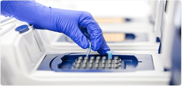
Polymerase chain reaction is a technique used to amplify the target DNA sequence from the source of DNA, it’s a rapid and a versatile in vitro methods
This method is introduced by Kary Mullis in 1985.
This method is able to amplify the specific sequence of the DNA from the heterogeneous collection of DNA molecules. For this selective amplification some prior information of the target DNA sequence is required.
Two primer synthesises by this sequence information get attached to the target DNA after denaturation (at the specific site). Primer initiates the DNA synthesis in the presence of heat stable DNA polymerase and DNA precursors (dnTPs).
Principle:
Technique based on enzymatic replication of DNA
Short segment of DNA is amplified by primers which are mediated by enzymes.
DNA polymerase synthesises complementary DNA from the template DNA. DNA polymerase adds nucleotide at 3’-OH group only so to attach DNA polymerase to the strand at specific site primers in required.
Components:
DNA template: segment of the interested DNA from the sample
DNA polymerase: a heat stable DNA polymerase which can withstand high temperature e.g. Taq polymerase.
Oiligonucleotide primers: short stretch of DNA complementary to the 3’ ends of sense and antisense strands.
Deoxyribonucleotide triphosphate: provide energy for polymerization and building blocks for the synthesis of DNA
Buffer system: provide optimum conditions for DNA denaturation and renaturation, magnesium and potassium are the key component. Important for polymerase activity and stability.
Steps of PCR:
It is a cyclic process because newly synthesized DNA strand serve as the template for further DNA synthesis. It consist three steps:
Denaturation:
It is the initial step in which the reaction mixture is heated to the temperature about 90-980 C that ensures the separation of the DNA strands known as DNA denaturation. Duration of denaturation step usually is 2 min at 94oC.
Annealing:
In the steps the mixture is cooled to the temperature that permits the attachment of the primer to the complementary strand of DNA.
Primer attached at the 3’-ends of the two strands.
Duration for annealing is 1 min at 40-600 C.
DNA synthesis:
The temperature is so adjusted that DNA polymerase synthesizes the complementary strand using the primers for attachment at 3’OH end
Primers extended towards each other so that the DNA segment lying in between is copied.
Duration of this step is usually 2 min at 720 C.
Types of PCR:
Nested PCR
Quantitative Real-Time PCR
RT-PCR
Inverse PCR
Anchored PCR
RACE
Touchdown PCR
RAPD
AFLP
Applications:
Forensic science
Genetic fingerprinting tool
Identification of criminal’s DNA collected from the crime site among the various people.
Tests for paternity
Research
Comparison of two organism’s genome for study
Gene mapping
Phylogenetic analysis of DNA from sources like fossils
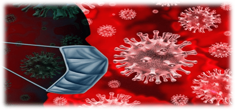
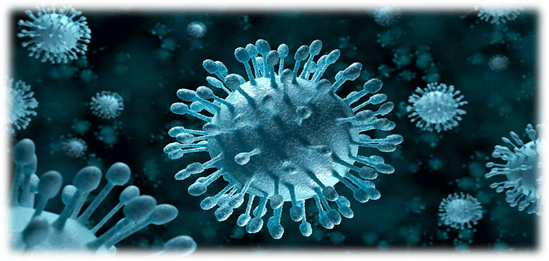
Coronaviruses are RNA viruses that cause diseases in mammals and birds derived from Latin word corona which means ‘crown’, named by June Alimedia and David Tyrrell who first observed human coronaviruses.
Coronavirus cause respiratory tract infection in human that range from mild to lethal, mild illness include common cold while lethal verities cause MERS(middle east respiratory syndrome), SARS(severe acute respiratory syndrome) and COVID-19.
Constitute subfamily orthocoronavirinae. The virus is enveloped and has a positive sense single-stranded RNA genome and helical symmetry nucleocapsid.
Coronaviruses are among the largest virus, size ranges between 26-32 kilobases.
History
Arthur Schalk and M. C. Hawn in 1931 described a new respiratory infection of chicken in North Dakota. The mortality rate was 40-90% in new born chicks.
Six years later Fred Beaudette and Charles Hudson successfully isolate and cultivated IBV (infectious bronchitis virus) which cause disease.
In 1960s human coronaviruses were discovered. E. C. Kendall, Malcom Byone, and David Tyrrell working at British Medical Research Council isolated a novel common cold virus B814 from a boy. In n1965 Tyrell and Byone cultivated the novel virus in organ culture of human embryonic trachea by serially passing through it, the virus cannot be cultivated by standard techniques used for rhinoviruses and other common cold viruses.
At the same time Dorothy Hamre and John Procknow at the University of Chicago from a medical student isolated a novel cold virus 229E grew in kidney tissue culture.
In 1967 June Almeida imaged this two novel strain by electron microscopy and said that the two strains were morphologically related by their club-like spikes.
Many human coronaviruese have been identified SARS (2003), MERS (2012) and SARS cov-2 (2019).
Structure:
Large, spherical particles with bulbous surface projection
The virus diameter is around 125nm, envelope diameter is 85nm and the spikes are 20nm long.
The envelope is made up of lipid bilayer in which the membrane, envelope and spike structural protein are anchored in the ratio of 1:20:300(E:S:M).
A single particle on an average has 74 surface spikes.
The spikes are homotrimers of S protein composed S1 and S2 subunit. S protein is the class 1 fusion protein responsible for receptor binding and membrane fusion with the host cell.
S1 subunit forms head and has RBD (receptor binding domain), S2 forms the stem which anchor the spikes in the viral envelope and enable fusion on protease activation
E and M contribute in envelope formation and maintaining the shape.
COVID-19
Novel coronavirus is a new strain which has not been identified in humans yet.
Coronaviruses are zoonotic which means they transfer from animal to human. Example SARS cov was transmitted from civet cats to human and MERS cov from dromedary camels to humans. Many coronaviruses strains are circulating in animals have not infected humans.
Transmission
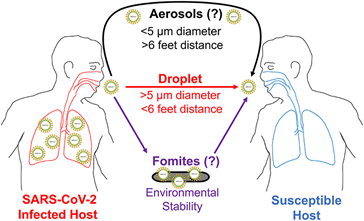
Infection carriers shed the viruses into environment while sneezing, coughing or sometimes speaking.
When these droplets come in contact with other person it gets inside the body through mouth, nose and eyes.
Symptoms
Most common symptoms:
Fever
Tiredness
Dry cough
Less common symptoms:
Diarrhoea
Aches and pains
Headache
Sore throat
Conjunctivitis
Loss of taste or smell
Serious symptoms:
Chest pain or pressure
Difficulty breathing or shortness of breath
Loss of speech or movement
Prevention and Control
It is better to prevent from any disease than cure
Prevention of coronaviruses is to avoid being exposed to it
The major source of transmission is transfer of droplets, this can be controlled by respiratory hygiene
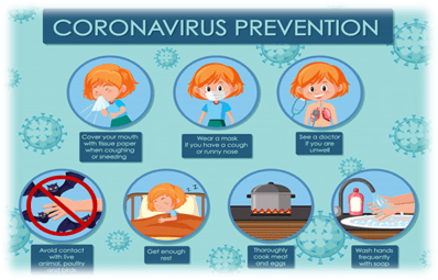
Following measures to be adopted to avoid transmission of these droplets
Always wash hand with soap and water for at least 20 second or if no dirty notice alcohol based rub can be used, especially after traveling or after sneezing, coughing and blowing nose
Avoid close contact with people who are sick or in contact with sick person because it may take few days to show symptoms and may be asymptomatic.
Wearing mask or cover moth with cloth when come contact with other people
Keeping a distance of at least 1 meter from other person
In any person doesn’t have cloth or mask covering their moth they should use tissue or under elbow while sneezing and frequently dispose tissue in the dustbin.
Regular cleaning and disinfection of the frequently touched surfaces i.e. Doorknobs, light switches, table, tapes
Precaution taken in hospitals for the patient and healthcare worker
Patient should be placed in a single room with proper ventilation if single rooms are not available the suspected patients should grouped together.
Health care staff should were proper medical mask, facial protection or eye protection to avoid contamination
In hospitals beds of the patient should be placed at least 1 meter apart
The equipment which are being shared among the patient should be disinfect after each use
Awareness should be generated among the patient, the general public families about the symptoms prevention and precautions
Virus is a parasite of sub-microscopic level on all the organisms. They infect all type of life forms like plants, animals, bacteria.
They are ambiguous in nature i.e. weather living or non-living, they are non-living in free-state but behave like living organism in a host.
In 1892 Dmitri Ivanovsky’s described about virus when he was working on bacterial pathogen infecting tobacco plants.
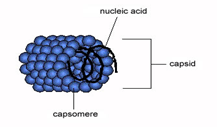
When virus invades a host organism it forced to replicate rapidly to produce thousands of copies of the virus. In contrast when virus is not inside a host or in the process of infecting it, virus exists in independent form known as ‘virions’ consists of a genetic material (RNA or DNA), a protein coat – capsid and the outer envelope of lipids.
Virus can be of various shapes ranging from helical to the icosahedral form.
Virus are much smaller than bacteria, size of the most virus that have been studied is between 20-300 nm.
They contain only one type of nucleic acid at a time either DNA or RNA but never both.
Virus are responsible for various infectious diseases in plant and animals rabies, AIDS (HIV), avian influenza, Ebola virus disease. Transmission of viral disease is through ‘vector’ (diseases bearing organism).
Structure:
Virus shows a wide variety of shape and size, a complete virus particle know as virions contain nucleic acids surrounded by the protein coat capsid, capsid is made up of small subunits of protein knows as capsomers and an outer layer called envelope made up of lipid which is derived from host cell membrane.
The main morphological structures are:
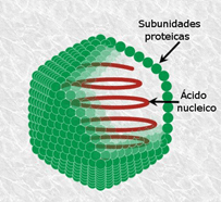
Helical
Composed of single type of capsomeres stacked around the central axis of helical structure having central cavity results in the formation of rod shape structure which may be short and rigid or long and flexible
Genetic material (typically ssRNA or ssDNA) bound to the protein helix by the interaction between the negatively charged nucleic acid and the positively charged capsomer protein.
Example: tobacco mosaic virus
Icosahedral
Most animal viruses exhibit icosahedral symmetry. The minimum no of triangular faces is three which gives rise to the 60 more capsomers. Rotavirus has more than 60 capsomers. Regular icosahedron is the optimum way to form closed shell symmetry with identical subunits.
Capsomers attaches with 5 other capsomers at the apices called pentons and on the triangular faces attaches with 6 capsomers called hexon.
Pentons and hexon may be of same protein or may be of other proteins.
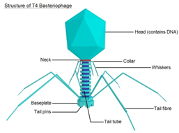
Prolate
Elongation of icosahedron along the fivefold axis, it is common in bacteriophage and composed of a cap with cylinder structure.
Envelope
In some species, the virus modify their cell membranes to envelope them self it may be either the outer membrane of the host or the internal nuclear membrane or endoplasmic reticulum making a outer lipid bilayer known as viral envelope.
The infectivity are depend on the envelope of the virus, the membrane i
Has protein coded by the viral and host genome and lipid membrane with any carbohydrates originate by the host.
Characteristics:
Non- cellular organism enclosed by a protective envelope
Virus attaches to their host by the help of spikes
Having nucleic acid (DNA and RNA) in the core which is surrounded by the protein coat
Considered as both living and non-living i.e. inactive when present outside the host and became active within the host cells
Virus uses host mechanism and enzymes to reproduce itself.
Classification :
On the basis of genetic material:
DNA virus:
DNA as genetic material
They attack on both humans and animals
Example: papillomavirus, parovirus and herpesvirus
RNA virus:
RNA as genetic material
Example: polio virus, ebola virus, hepatitis C virus
Their genetic material is RNA remains enclosed in the protein coat and infect plants
Example: potato virus, TMV, turnip yellow viruses
Bacteriophages:
Virus infect bacteria are known as bacteriophage, contain DNA as genetic material
On the basis of mode of transmission:
Transmitted through
Example
Respiratory route
Rhino virus, swine flu
Faeco-oral route
Polio virus, rota virus
Blood transfusion
HIV, hepatitis B virus
Sexual contact
Retro virus
Zoonotic virus
Alpha virus, flavi virus
Baltimore Classification:
In early 1970s Nobel prize winner David Baltimore developed the most commonly used virus classification system
Baltimore focus on how mRNA is produced during replication and classify virus into various groups
Group I: mRNA is produced as the same way as their cellular DNA by transcription, contain ssDNA as their genetic material
Group II: convert their ss genome dsDNA before transcription of mRNA occur, have ssDNA as genetic material
Group III: uses RNA dependent RNA polymerase to generate mRNA from the one of the strand used as template, genome is dsRNA.
Group IV: genetic material is ssRNA . Genomic RNA is with positive polarity means that is directly serve as mRNA
Multiple full length RNA strand with negative polarity are formed from the intermediate of dsRNA (replicative intermediates) made in the genomic copying process.
These serve as template form the production of positive polarity RNA
Group V: contain ssRNA with negative polarity; means that sequence is complementary to mRNA. Negative-strand is converted into mRna
Group VI: virus have two copies of genome i.e. ssRNA conveted using reverse transcriptase enzyme to dsDNA
dsDNA transported to the host’s nucleus in inserted in its genome viral DNA is produced by transcription with host DNA
group VII: genetic material is dsDNA which make ssRNA as intermediate acts as mRNA and also converted back to dsDNA by reverse transcriptase enzyme.
In Mendel’s crosses F1 and F2 from crosses show same results unaffected by whether it is a male or female both shows identical ratios.
But this rule is may not be applicable sometimes due to some exception produced by the sex linkage phenomenon.
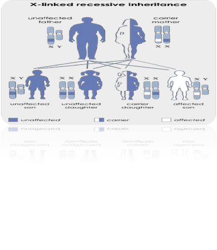
Sex linkage is the phenomenon of gene being located on X or Y chromosome.
Inheritance of trait is determined by this gene on sex chromosomes.
First known case of sex linkage is haemophilia in human beings. In haemophilia, affected person bleed profusely even from minor cuts because their blood doesn’t clot on exposure to air.
There are many more exa mple of sex linked inheritance but it is clearly explained by Morgan in 1910( white eye gene in drosophila).
Characteristics
Heterogametic sex has more no of individual showing recessive sex linked character than the homogametic sex.
Genes that control sex linked traits are not getting transferred from male parents to their male progeny directly.
Sex linked gene are transmitted from male parent to its entire daughter (daughter receives its half X chromosome from father) then daughter transmit this gene to half of its male progeny. As a result the gene transferred from male to female progeny and then to the half of male progeny. This transfer of male to female and back to the male is known as criss-cross inheritance.
Y chromosomes not carry sex linked genes. Heterogametic sex is present in hemizygous condition which lead to the expression of the recessive alleles of sex linked genes and in homogametic sex they have to be present in homozygous condition.
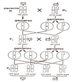
Inheritance of white eye in drosophila
Morgan in pedigree culture of normal dull red eye drosophila observed a single white eye male. He mate white eye male with red eye female and found F1 flies had all red eyes and in F2 3 red eyes and one white eye showing that white eye is due to a recessive gene.
But when he classified F2 flies on the basis of sex he found that all the female had red eyes and half of the male is red eyed and remaining half is white eyed. It seems as if the eye colour related to the sex of the flies.
After that Morgan mated white eyed female with red eyed male he observed that half of them were red eyed and other half were white eyed, in F2 generation the ratio of red eyed and white eyed is 1:1, which same as the F1.
Later Moran reasoned that X chromosome carry white eye gene of drosophila, and Y chromosome doesn’t carry any allele of this gene.
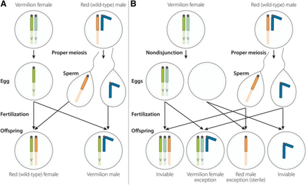
Nondisjunction of X chromosome
In 1916 Bridges, studying the inheritance of vermilion colour gene in drosophila. Observe that vermilion eye colour gene v (sex linked recessive gene) show the same pattern of inheritance as w
Cross between the vermilion females and red males, among the progeny of F1 some females were vermilion eyed and some males were red eyed. However, majority of females were red eyed and males were vermilion eyed.
Bridges explained these data and postulated that the X chromosome failed to separate during oogenesis of some oocytes of vermilion eyed females and both moved to the same pole and the opposite pole did not receive X chromosome such irregular distribution is known as nondisjunction.
Attached X chromosome in drosophila
Morgan in 1922, obtained all females with yellow body and all males with grey body when he mated yellow body female with grey body male drosophila
Yellow is recessive to the normal grey body located on X chromosome and determined by y.
Morgan reasoned that X chromosome nondisjunction is regularly takes place in oocytes and produced the following XvXv and O unusual eggs
Such highly regular disjunction only takes place when the two X chromosomes shared the same centromere and behave as a single chromosome this fused form is known as attached X chromosome.
Sex linkage in human and other organisms
In addition to drosophila sex linked inheritance is also known in mice, cat , insects, cattle , in man
Drosophila had more than 150 sex linked genes and over 200 genes in humans most of them cause genetic diseases. Example haemophilia ( failure in blood clotting ability), colour blindness ( inability to see one or more colour), optic atrophy (optic nerve degeneration), myopia(short-sightedness), etc.
For example like colour blindness it is a sex linked disorder occurs in 5-10% males and only 1% to the females. Human has there protein for red green and blue colour present on cone cells genes encoding for red and green light are located on x chromosome and gene for blue light is placed on autosome.
Partial sex linkage
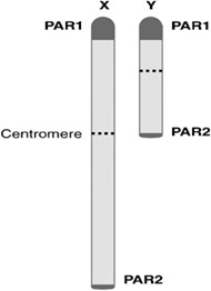
X and Y chromosome in human aur morphologically different. But during meiosis they pair in male cell
The paring in two telomeric region is called as pseudoautosomal regions
PAR1 major region of 2.6 Mb long, located at the tip of short arm of X and Y chromosomes, it is 70 times more than the normal recombination frequency. Example SHOX, WE7, Tramp
PAR2 minor region of 320 kb located at the tip of the long arms of X and Y chromosomes, crossing over is not so frequent. Example IL9R and SYBL1.
X chromosomes contain PAR1 and PAR2 but this genes don’t show inheritance pattern of sex linkage because Y Chromosome but they resemble of autosomal genes.it is known as partial sex linkage gene X chromosome show autosomal pattern