CARBOHYDRATES

BY- SREELAKSHMI (MSIWM012)

Carbohydrates are polyhydroxy aldehydes or ketones, or substances that produce such substances in hydrolysis. Most, but not all, carbohydrates have a positive effect (CH2O) n.

Stereo Isomerism:

     The presence of carbon asymmetric atoms allows the formation of isomers. Chemicals that have the same structure but differ only in localization are called stereoisomers or geometric Glucose isomers with 4 asymmetric carbon atoms with 2n (16) Isomers. n = unequal number of carbon atoms.

Epimers: a sugar that differs only in the configuration around a single carbon atom. E.g. D-glucose and D-Mannose 2. D-Glucose and D-Galactose

Diastereomers: One type of diastereomers (or geometric stereoisomers) differs in terms of “cis” and “trans”. In diastereomers, some chiral centres are similar and some are opposite. The molecule does not resemble a mirror image of its diastereomer.

D and L isomers:

Enantiomers: Enantiomers are mirror image molecules that cannot be elevated to each other. The suggestion suggests that two mirror molecules can be psychologically integrated into one object as they are integrated. Eg, D-glucose and L-glucose. When the OH group in the carbon atom adjacent to the terminal primary alcohol (carbon atom 5 in the right), sugar is a member of series D. On the left is a member of the L series. Most monosaccharaides are classified as D.

 Optical Isomerism: When a cooled light beam is transferred to a solution that reflects light performance, the part will be moved right or left depending on the type of combination present. The element that alternates the illuminated light to the right is said to be dextrorotatory and the plus (+) sign is used for designation. The rotation of the pole on the left (laevorotatory action) is marked with a minus sign (-). When an equal number of dextrorotatory and laevorotatory isomers are present, the resulting mixture has no optical functions, because the functions of each isomer overlap. That mixture is said to be a racemic mixture.

.

There are three categories of carbohydrates:

1. Monosaccharides

2. Oligosaccharides

3. Polysaccharides

Monosaccharides or simple sugars:

It contains one unit of polyhydroxy aldehyde or ketone. E.g.: Sugar, Fructose, Galactose.

Once the group is at the end of the carbon chain (i.e., in the aldehyde group) monosaccharide is an aldose. If the group can be in another position (in the ketone group) monosaccharide is ketoses. The simplest monosaccharaides are trios-carbon trioses including Glyceraldehyde aldotriose, and Dihydroxyacetone ketotriose.

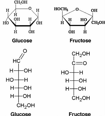
Glucose, a major source of energy for many living things. Glucose is present in both open chains and a ring form with rings forms when glucose is dissolved in water. It contains radical aldehyde as part of the structure. Group C = O in the aldehydic group has reduced concentrations and therefore lowers blood sugar. This is the most prominent monosaccharide in natural D-glucose, sometimes called dextrose. Fructose has a ketonic group as part of the structure. Group C = O in the group has reduced areas which is why it reduces sugar. Galactose is an aldohexose with reduced properties. One of the ingredients of Lactose.

Oligosaccharides: composed of short chains of monosaccharide units bound by glycosidic bonds. The most abundant are disaccharides, which have two monosaccharide units connected by glycosidic bonds. E.g., Maltose, Lactose, Sucrose.

• Maltose is also known as a sugar source made up of two sugar molecules. Maltose is formed by joining two alpha glucose molecules that meet the condensation reaction and form a glycosidic bond between molecules. It has reduced properties.

• Sucrose is a common household sugar or sugar cane and is composed of monosaccharides glucose and fructose bond. Sucrose does not contain an unknown free carbon atom.Anomeric carbons atoms of both monosaccharide units form a glycosidic bond. So sucrose sugar does not decrease. Non-reducing disaccharides are called glycosides.

Polysaccharides: Sugar polymers contain more than 20 or more units of monosaccharides called polysaccharides. E.g., Starch (Amylose, Amylopectin), Glycogen, Cellulose, Chitin. Polysaccharides are the main polymers of monosaccharaides .The polysaccharides may not be soluble or form colloidal suspensions. Starch is an alpha glucose polymer that is a mixture of two different polysaccharides.

AMYLOSE AND AMYLOPECTIN

AMYLOSE: They are long, unstable plates of sugar units. It is made up of a series of condensation reactions that include alpha glucose molecules that have been synthesized into an extended chain that forms many glycosidic bonds

AMYLOPECTIN – a very powerful polymer for glucose units. It contains an open series of alpha glucose units with branch points across all twelfth glucose. Branch points are formed when carbon 6 of the glucose molecule within an open chain forms a glycosidic bond with carbon 1 of the glucose molecule placed above the series

Glycogen

• Glycogen is often referred to as glycogen. The structure of Glycogen is almost identical to amylopectin but there are many branches in glycogen. Glucose is stored as glycogen in large mountains in both liver and bone tissue.

Cellulose:

• Cellulose is one of the most important structural polysaccharides because it is the major component of plant cell walls. Many identical chains of beta glucose units are formed and the whole chain contains hydrogen bonds between groups of OH adjacent chains.

Chitin:

• Chitin can be a polysaccharide that makes many invertebrate exoskeletons. N-acetyl glucosamine polymer in beta 1 to 4 glycosidic bonding. It is a key component of the insect and crustacean sac that protects and supports.

Necrosis

BY – SAI MANOGNA (MSIWM014)

Irreversible cell damage leads invariably to cell death as a result of interactions with noxious stimuli. Infectious agents, oxygen deprivation or hypoxia, and extreme environmental factors such as heat, radiation, or exposure to ultraviolet irradiation are all included in these noxious stimuli. The subsequent death is referred to as necrosis, commonly distinct from the other significant consequence of permanent damage, referred to as apoptosis cell death. Apoptosis is a cell death that is programmed or structured and may be physiological or pathological. Additional knowledge is beyond the scope of this chapter about this type of cell death. A pathological process is almost always associated with necrosis as a form of cell death. 

They show two main types of microscopes or macroscopic appearance as cells die from necrosis. The first is liquefactive necrosis, also referred to as colliquative necrosis, characterized by a partial or total breakdown of dead tissue and liquid, viscous mass transformation. The tissue and cellular profile degradation occur in liquefactive necrosis within hours. Coagulative necrosis, the other significant pattern, is distinguished, in contrast to liquefactive necrosis, by the preservation of typical necrotic tissue architecture for several days after cell death. 

The slimy, liquid-like essence of tissues undergoing liquefactive necrosis results in liquefaction. In part, this morphological appearance is due to the activities of hydrolytic enzymes that cause cellular organelles to dissolve in a cell undergoing necrosis. Liquefaction enzymes are derived either from bacterial hydrolytic enzymes or from lysosomal hydrolytic enzymes.

Types of necrosis :

Six types of necrosis are identified based on the morphological patterns associated with cell death.

a. Liquefactive necrosis

b. coagulative necrosis

c. Caseous Necrosis

d. Fat Necrosis

e. Gangrenous Necrosis

f. Fibrinoid necrosis

  1. Liquefactive necrosis :

The necrosis pattern that is seen with infections. The pattern is also seen in the brain following ischemic injury. The release of digestive enzymes and neutrophil constituents causes liquefaction in infections, although there is a poor understanding of the cause of liquefactive necrosis following ischemic injury in the brain.

Gross Appearance: Due to pus formation, the tissue is in a liquid state and sometimes creamy yellow. 

  • Coagulative necrosis :

Occurs in any organ in the body but the brain; this is the default necrosis trend associated with ischemia or hypoxia. 

Gross Appearance: tissue is solid, and for days after cell death, architecture is preserved. 

  • Caseous necrosis :

A rare form of tuberculosis-related cell death. 

Gross appearance: white, fluffy, cheesy-looking material (case-looking).

A granuloma is known as the entire structure formed in response to tuberculosis. 

  • Fat necrosis :

Acute inflammation affecting tissues with multiple adipocytes, such as the pancreas and breast tissue, causes fat necrosis. Digestive enzymes that break down lipids to produce free fatty acids are released by damaged cells. 

Gross Appearance: Whitish deposits as a consequence of calcium soap formation. 

  • Gangrenous Necrosis : 

Medical usage in the description of lower limb ischemic necrosis (sometimes upper limbs or digits). 

Gross appearance: varying degrees of putrefaction on black skin. 

  • Fibrinoid necrosis :

Vascular damage (autoimmunity, immune complex deposition, infections (viruses, spirochetes, rickettsiae)) is a pattern associated with this. 

Gross Appearance: Not necessarily grossly discernible. 


All of these reflect morphological patterns, grossly, and microscopically evident. Typically, fibrinoid necrosis is evident only microscopically. In the following paragraphs, we examine the characteristic gross and microscopic effects of liquefactive necrosis.

Difference between Apoptosis and Necrosis:

CharacteristicsApoptosisNecrosis
DefinitionProgrammed cell deathPremature cell death
Process Occurs through shrinkage of cytoplasm, followed by chromatin condensationOccurs through swelling of cytoplasm along with mitochondria followed by cell lysis
Cause Naturally occurring physiological processThe pathological process, caused by external agents such as toxins, trauma, and infections.
Membrane integrityPlasma membrane blebbing is observed without losing integrityThe membrane integrity is loosened.
ChromatinAggregation of chromatinNo structural changes in chromatin
OrganellesMitochondria become leaky often by forming membrane pores, while lysosomes kept their integrity. Organelles still function even after cell death.Lysosomes become leaky, while mitochondria kept their integrity. Organelles are disintegrated by swelling and do not function after cell death.
Vesicle formationMembrane-bound vesicles called apoptotic bodies fragments the cell into small bodiesNo vesicle formation. Complete cell lysis occurs and releases contents into the extracellular fluid.
RegulationTightly regulated by its activation pathway of enzymesUnregulated process
CaspaseCaspase dependent pathwayCaspase independent pathway
Energy requirementThe active process occurs at 40cThe inactive process does not occur at 40c
Digestion of DNANon-random mono and oligonucleosomal length fragmentation of DNA and show band pattern in Agarose gel electrophoresis.DNA in the cell is randomly digested and shows a smear in Agarose gel electrophoresis.
Timing of DNA digestionPrelytic DNA fragmentation.Postlytic DNA digestion.
OccurrenceThe localized process involves destroying individual cellsAffects contiguous cell groups
PhagocytosisEither by phagocytes or adjacent cellsBy phagocytes
SymptomsNeither inflammation nor tissue damageA significant inflammatory response is generated. May cause tissue damage
InfluenceOften beneficial, but abnormal activity may lead to diseases.Always harmful. If necrosis is untreated- it may be fatal.
FunctionInvolved in regulation of the number of cells in multicellular organisms. Involved in tissue damage and induction of the immune system, defending the body from pathogens as well.

CHROMOSOMAL ABNORMALITIES

BY- SREELAKSHMI (MSIWM012)

Chromosomal abnormalities are seen in many people. These are generally aneuploidy (presence of abnormal number of cells). it’s either autosomal or sex-chromosome. Translocation, duplication and deficiency in chromosome can also be the reason for this abnormality. It is found that certain disorders or diseases in man are associated with the abnormal number and nature of chromosome.

Abnormalities in Sex Chromosome

A normal male and female has XY and XX chromosome respectively.Klinefelter’s syndrome and Turner’s syndrome are an example for the anomalies in the number of sex chromosomes. Other important abnormal sex in man is the XYY male. They are usually tall and are more aggressive. Many cases it is observed they are mental retarded, but they are fertile.

Abnormalities of the Autosome

In some cases abnormal number of some of the autosomes or other autosomal aberrations. First autosomal anomaly that was described in man is Down’s syndrome. This abnormality is also called mongolism, as the features of such individuals resemble those of Mongolian races. This syndrome was first described by Seguin in 1844.The symptoms include: round face with small skull, short body, swollen tongue, eyelid fold resembling those of Mongolian’s and also mental retardation. In some cases congenital heart diseases is present in these individuals and mostly death occurs early in childhood. But some of them live upto young adult age with the low IQ.

Studies showed that Down’s syndrome is due to trisomy of the 21st chromosome. This trisomy is due to non-disjunction during meiosis. It may occur either during spermatogenesis or oogenesis. Sometimes the third chromosome in the set 21 may be attached to another autosome which is most commonly paired to .This is called translocation trisomy. Down’s syndrome can be related to the age of mother. Frequency of this syndrome increases with increase in mother’s age. In most cases, Down’s syndrome births occur in women conceiving after the age of 30.The trisomy’s of two slightly larger chromosome number 13 and number 18 which leads to early death.

13-Trisomy: It is observed less in number. Such people will have severe mental and physical deformities. The head and the eyes are small.in some cases eyes are absent. Such individuals have congenital heart disease. This disease is also called Patio’s syndrome. This syndrome can be also due to isochromosme of chromosome-13.

18-Trisomy: They show low set ears, simian crease, deformities of fingders, toes and feet, congenital heart disease and receding chain. Head will be flattened 90% of people die within one year.it is also called Edward’s syndrome.

Abnormalities of Chromosome Structure

Chromosomal abnormalities like deletions, translocations, ring formation etc. Are lethal even as heterozygotes, resulting in zygotic rules, still births or infant deaths. Sometimes infants with small chromosome deficiencies survive. However these infants die at an early age.

Cri-du-chat syndrome: It is due to the chromosome deficiency in the short arm of chromosome 5. This deficiency is designated as 5P. Infants with this syndrome cry like a cat mewing. Other features are small forehead, broad face with saddle nose, widely spaced eyes with epicanthic folds and physical and mental retardation.

Chronic Myelocytic Leukemia: This disease is caused when there is a deletion of chromosome 22. Exposure to X-rays can be a cause of this disease.

GENETIC DISEASES DUE TO DEFECTS IN THE CATABOLISM OF PHENYLALANINE

  1. Phenylketonuria (PKU): The conversion of phenylalanine into tyrosine is mediated by the enzyme phenylalanine hydroxylase. The absence of this enzyme causes a disorder in a man.It is called phenylketonuria. In people affected with this disorder,phenyalanine will not be converted tyrosine.Instead,phenyl-acetic acid(PAA) are formed.Phenylpyruvic acid causes damage to nervous system which leads to mental disorder.it is a serious disease and can be detected by simple blood and urine test.Affected infants can be given a diet that is low in phenylalanine.
  2. Alkaptonuria: This disease is caused due to the accumulation of homogentisic acid. Homogentisic acid is responsible for the oxidation of phenylalanine during catabolism. Absence of enzyme responsible for the oxidation of homogentisic acid, it gets accumulated and passes into the urine. When such urine is exposed to air, alcoptone gets oxidized and forms pigments. The absence of this oxidase enzyme is due to to air, alcaptone gets oxidase enzyme is due to and a recessive genes.Alkaptouria is not a serious gene.
  3. Albinism: The color of the skin and the hairs is due to the presence of a dark pigment called melanin. Individuals who do not have melanin are pale in colour.Such individuals are called albinos. This is due to a metabolic block in the conversation of tyrosine into melanin. This block is due to the absence of the enzyme catalyzing the conversion of tyrosine into DOPA (3, 4-dihydroxy phenylalanine).This enzyme is called tyrosine, Albinism may be due to deficiency in the processes that takes place after the formation of DOPA. 

Sickle-cell Anaemia

The anaemic condition of the person due to change in the blood cells is called sickle-cell anaemia.It is a chronic, haemolytic disease.In the past, patients died early due to infections or cardiac failure. The sickled red cells become trapped in the small blood vessels. This causes impaired blood circulation resulting in the damage of vital organs. The sickle cells are more fragile and hence haemolyse readily, so have a shorter life than the normal cells. They will suffer from severe anaemia.They are generally genetically transmitted. When two individuals who have sickle – cell trait marry 25% of the progeny will have sickle-cell anaemia, 50% will have sickle –cell trait (carriers) and 25% will be normal individuals.

AMINOCENTESIS

It is prenatal detection of inherited diseases. Human fetus lies in the amniotic cavity in the uterus of the mother. A fluid, amniotic fluid, surrounds the fetus and fills the amniotic cavity. Cells from the fetus are shed into this fluid. The amniotic fluid along with the fetal cells can be collected by introducing a syringe through the abdominal wall of the mother after the third month of pregnancy. This procedure is called amniocentesis. This amniotic fluid is further analyzed.

The fetal cells separate from the amniotic fluid are called amniotic cells. Identification of sex of the fetus helps to terminate pregnancy in people who have a history of sex-linked diseases such as hemophilia, color blindness. Turner’s syndrome and klinefelter’s syndrome can be detected by observing the number of Barr bodies.

Amniotic cells can be cultured in an appropriate medium for 2-6 weeks and can be used for chromosome analysis and biochemical studies. Chromosome analysis helps to determine the karyotype of the foetus .When any kind of an abnormality is found the mother is asked for abortion which is legal.

Disadvantage of Amniocentesis

It can be done only after 12-16 weeks after conception. Analysis of the culture will take another 2-6 weeks.Therefore, if any kind of abnormalities are found and the mother is asked for abortion it will be too late.Aminocentesis will give a mild shock to the fetus affecting its growth.

MICROBES IN FOOD SPOILAGE

BY- ABHISHEKA G.(MSIWM013)


INTRODUCTION:

 Food spoilage is defined as the process in which the destruction of food occurs then it becomes non edible to humans and it’s quality of edibility is decreased. It is also referred to be the changes in the visual, smell, and texture of the food that makes it unacceptable for consumption. The growth of microbes in food results in food spoilage. Spoilage of food occurs due to the action of microbial enzymes in the absence of viable cells. The microbial spoilage of food results in changes such as color, odor, texture, the formation of slime, accumulation of gas, and release of liquids.

 The microbes like Bacteria, yeasts, and molds causes of food spoilage in high amount. These microbes synthesis different enzymes to decompose the various food items. Molds are the major causes of spoilage of foods with reduced water activity like dry cereals and cereal products. Bacteria spoil foods with relatively high water activity such as Milk and its products.

RELATION BETWEEN WATER ACTIVITY AND FOOD SPOILAGE MICROBES:

1.Water activity has an important role in food preservation. Each microbe has a critical water activity, below this critical water activity, the growth of the microbes do not occur.

2.For example, pathogenic microbes cannot grow at water activity below 0.86, Yeasts and molds are tolerant to water activity and they grow at or below water activity 0.62. Hence water activity is important in foods and it is a major factor in food spoilage and food safety.

3. Decrease in water activity retards the growth of microorganisms by slowing down enzyme-catalyzed reactions and retards nonenzymatic browning.

WATER ACTIVITY OF SOME OF THE FOODS:

Fruits and Vegetables – 0.97 to 1.

Meats- 0.95-1.

Cheese – 0.68 – 1.00

Jams and Jellies- 0.75 – 0.94

Honey – 0.54 – 0.75

Noodles – 0.50

Dried Milk – 0.20

MINIMUM WATER ACTIVITY REQUIRED FOR THE GROWTH OF MICROBES IN FOOD

Clostridium botulinum – 0.95

Bacillus cereus – 0.95

Pseudomonas aeruginosa- 0.95

Salmonella species – 0.95

Staphylococcus aureus- 0.90

Candida species- 0.90

Saccharomyces cerevisiae- 0.90

Staphylococcus aureus – 0.86

Penicillium species – 0.82

Most spoilage yeast – 0.88

Most spoilage molds – 0.80

SOURCES OF MICROBES IN FOOD:

  1. Soil and water
  2. Plant and plant products
  3. Food utensils
  4. The Intestinal tract of Humans and Animals
  5. Air and dust
  6. Food handlers.

PROCESS OF FOOD SPOILAGE BY MICROBES: Microbes like bacteria, molds attack the food items. Then preserved food items undergo degradation. As a result of degradation, food items result in changes such as physical changes like the texture of food, visual, smell, and quality of the food and loss of taste, etc. Later the microbes which attacked the food items releases the necessary enzymes required for the degradation of spoiled food items. Then enzymatic action brings about the degradation process.

FOODS AFFECTED BY VARIOUS GROUPS OF MICROBES:

  1. Anaerobic or facultatively anaerobic: These groups of microbes are most likely to grow in canned foods.
  2. Microaerophilic bacteria: These are most likely to grow in vaccum packed foods since they have low oxygen tension.
  3. Aerobic bacteria: They grow on the surface of the raw meat.
  4. Aerobic molds: Grow on insufficiently dried or salted products.

FACTORS AFFECTING THE GROWTH OF MICROBES IN FOOD:

  1. pH: Most of the bacteria grow best at neutral or slightly alkaline nature of foods, the pH ranging from 6.8 to 7.5. Bacteria like Salmonella grow at a pH range of 4.5 to 9.0. Molds grow between 1.5 to 11.0, whereas yeasts grow between 1.5 to 8.5.
  2. Moisture content: The effect of moisture is measured in terms of water activity. The water activity of a food is defined as the amount of free water present in the food medium. If there is a lack of water the microorganisms do not grow in the food medium.
  3. Nutrient contents in the food: Microbes require proteins, carbohydrates, lipids, water, energy, nitrogen, Sulphur, phosphorous, vitamins, and minerals for their growth. Various foods have specific nutrients which support the growth of microbes in the food. Foods like meat, milk, and eggs contain a large number of nutrients required for the growth of microbes. Hence these foods are more susceptible to microbial spoilage.
  4. Anti-microbial substances: The antimicrobial substances like lactenin, anti-coliform factors in milk, and lysozyme in eggs prevents the growth of microbes.
  5. Temperature: Microbes require optimum temperature for their growth. Psychrophilic bacteria causes spoilage of food at low temperature conditions. Mesophilic bacteria can grow between 5*c to 40*c. Thermophilic bacteria grow above 45*C temperaturze.

APOPTOSIS

BY: SAI MANOGNA (MSIWMO14)

Apoptosis or Programmed cell death. It is an induced and ordered mechanism in which the cell is actively involved in bringing about its demise. This is a crucial factor in the homeostatic control of many cell populations, including hematopoietic ones. 

1. Cells undergoing programmed cell death also show distinctive morphological changes, collectively called apoptosis.

2. These changes include a marked decrease in cell volume, cytoskeleton alteration resulting in membrane blebbing, chromatin condensation, and DNA degradation into smaller fragments.

3. Following these morphological changes, an apoptotic cell sheds tiny apoptotic bodies containing intact organs. 

Macrophages rapidly phagocytose apoptotic bodies and cells in advanced apoptosis. This ensures that their intracellular content is not released into the surrounding tissue, including proteolytic and other lytic enzymes, cationic proteins, and oxidizing molecules. Apoptosis causes no local inflammatory response. Apoptosis varies markedly from necrosis, cell death associated with an injury. In necrosis, the wounded cell swells and bursts, releasing its contents and likely causing an inflammatory response. 

Each hematopoiesis-produced leukocyte has a characteristic lifetime and then dies by programmed cell death. For example, there are around 5 ×10¹⁰ neutrophils in the circulation in adults. These cells only last a few days before programmed cell death is triggered. Together with constant neutrophil development, this death maintains a stable number of these cells. If programmed cell death fails, leukemia can develop. Also, programmed cell death plays a part in holding decent numbers of hematopoietic progenitor cells. For example, in removing colony-stimulating factors, progenitor cells undergo apoptosis. Beyond hematopoiesis, apoptosis is critical in tolerance and killing target cells by cytotoxic T cells or natural killer cells.

Regulation of Activated B-cell numbers by Apoptosis :

1. Bcl-2 levels were found to play an essential role in controlling the expected lifespan of different hematopoietic cell lines, including lymphocytes.

2. An average adult has around 5 L of blood with about 2000 lymphocytes / mm3 for around 1010 lymphocytes.

3. During acute infection, the lymphocyte count increases 4- to 15-fold, resulting in a total lymphocyte count of 40 ×50 ×109.

4. Since the immune system cannot withstand such a massive increase in cell numbers for a prolonged time, the system needs to remove excess activated lymphocytes after the antigenic threat has passed.

5. Activated lymphocytes express lower levels of Bcl-2 and are thus more vulnerable to apoptotic death induction than naive lymphocytes or memory cells.

6. However, if antigen activates the lymphocytes, then the signals obtained during activation block the apoptotic signal. As antigen levels subside, block activation and lymphocytes begin to die from apoptosis.

Several gene expressions accompany leukocyte apoptosis and other cell types. Some proteins specified by these genes are apoptosis induced; others are essential during apoptosis, while others inhibit apoptosis. For example, radiation can cause apoptosis in thymocytes, but only if the protein p53 is present; Fas signals cause many cell deaths, a molecule found on the surface of several cells, and proteases known as caspases are involved in a cascade of reactions that lead to apoptosis. By contrast, the family members of the bcl-2 (B-cell lymphoma 2) gene, bcl-2, and bcl-XL encode protein products that inhibit apoptosis. Interestingly, in studies involving non-cell death but an uncontrolled proliferation of B cells in the form of cancer called B-lymphoma. bcl-2 was recognized as the first member of this gene family. The bcl-2 gene was at the break-point of chromosomal translocation in human B-cell lymphoma. The translocation transferred the bcl-2 gene into the immunoglobulin heavy-chain locus, resulting in transcriptional activation of the bcl-2 gene and lymphoma cells overproduction of the encoded Bcl-2 protein. The resulting high Bcl-2 levels are thought to help transform lymphoid cells into cancer cells by inhibiting signals that would typically induce apoptotic cell death. 

Functions of genes and their role in apoptosis :

GenesFunctionRole in Apoptosis
bcl-2Prevents apoptosisInhibits
baxOpposes bcl-2Promotes
bcl-XL (bcl-Long)Prevents apoptosisInhibits
bcl-XS (bcl-Short)Opposes bcl-XLPromotes
CaspaseProteasePromotes
fasInduces apoptosisInitiates

SALMONELLA

BY: SREELAKSHMI (MSIWM012)

Salmonellas is an infectious disease caused by bacteria. It is also known as salmonellosis. Causes Enteric fever, Gastroenteritis, Septicaemia etc. Salmonella typhi rank Typhoid. Salmonella is two groups that include a group that includes the enteric flu that contains only typhoid & Paratyphoid bacilli or major human parasites and the other group includes a group that causes food poisoning but can infect people who cause stomach infections.

Causes: Often the cause of contaminated food or water. Food delivery can enter

• Raw meat, poultry and seafood: Sewage enters immature meat and poultry causes contamination and disease. The transport of marine fish can be caused by water pollution in the agricultural environment.

• Raw Eggs: some infected chickens produce salmonella-containing eggs before the shell is formed. Raw eggs are used as versions of mayonnaise and hollandaise sauce.

• Fruits and vegetables: New products like imported varieties, can be watered or washed during salmonella contamination. Contamination may also occur in the kitchen, where juice from raw meat and poultry comes in contact with uncooked food.

The Food and Drug Administration claims that some outbreaks of salmonella have been caused by spice contamination. Food can also be contaminated if it is prepared by people who do not wash their hands properly after using the toilet or changing diapers. Infection can occur if you touch anything contaminated including pets, birds and reptiles and put your fingers in your mouth.

Salmonella are negative gram sticks. With the exception of S. gallinarum pullorum they travel with peritrichate flagella. Salmonellae is an aerobic plant that grows in light media at a pH between 6-8 and a temperature of 15-41 ° C. They can be killed at 55oC for one hour or 60oC for 15 minutes. Boiling or dehydrating and dehydrating helps to eliminate bacilli. In dirty water it can live for weeks and freeze for months.

Salmonella infection is not a life-threatening disease. In some cases, especially when infants and young children, elderly adults, transplants, pregnant women, and people with weakened immune systems find this infection can be difficult.

It can be dangerous. It causes dehydration as you get diarrhoea and do not take a sufficient amount of water to compensate for water loss. Warning signs include reduced urine, dry mouth, sunken eyes, and decreased production of tears.

When salmonella infection enters the bloodstream, it can cause tissue damage throughout the body. When it affects the tissues around your brain and spinal cord it can lead to meningitis. If you have a heart attack or valve, it could be endocarditis.

 Infections in the bones or bone marrow can cause osteomyelitis. People who have been infected with salmonella are at greater risk of developing active arthritis. Also known as Reiter’s syndrome.

Active rheumatoid arthritis is the cause: Eye irritation, painful urination, painful joints Prevention can be done with care to avoid spreading the virus to others. Immunizations are really important when preparing food, caring for infants, the elderly and people with weakened immune systems. Washing hands thoroughly can help prevent the transmission of salmonella virus to your mouth from any food you prepare.

Salmonellae causes the following clinical disease in humans

 Enteric fever

It is also called typhoid fever. It is usually caused by Salmonella enteric serotype typhi and S. enteric serotypes paratyphi A, B, and C.

 They are phagocytosed by polymorphs and macrophages. They are able to withstand intercellular killings and multiply within cells. They enter the lymph nodes where they multiply .They enter the bloodstream through the thoracic tunnel causing bacteraemia.

 Bacillus is more abundant in the bladder as bile is a good culture. It is then continuously excreted in the intestine where it implants Peyer’s Patches and lymphoid follicles on the ileum, which is ulcerative and can lead to intestinal insufficiency & bleeding as a problem.

 The incubation period is usually 7-14 days but may occur in 3-56 days. Bacteraemia occurs early in the disease and blood pressure is very good in the first week of the flu. The diagnosis is collected about 5-10ml of blood and injected into a traditional bottle containing 50-100ml of 0.5 per cent of bile broth.

The sample was incubated overnight at 37oC. After that bile broth is a subculture in MacConkey agar, pale lactose fermenting colonies from this method were selected for chemical experiments. Salmonellae is usually motile, indole and urease non-fertile glucose and ferment. Another option is the display of typhoid bacilli in the blood or urine.

If Salmonellae is not found in the original small cultures, the smaller cultures should be repeated every other day until growth is achieved. If possible, if Salmonellae cannot be detected in the first subculture from the broth, subcultures should be repeated daily until growth is achieved.

WESTERN BLOTTING:

BY: SAI MANOGNA (MSIWMO14)

Western blotting, also known as immunoblotting. This method is used for detecting proteins and post-translational protein changes, using antibody-based samples to extract precise protein information from complex samples.

It’s a standard approach in molecular biology, biochemistry, and cell biology with several applications. It can provide semi-quantitative or quantitative protein data in simple or complex biological samples.

Western Blot is also used to separate and identify proteins. In this process, a protein mixture is separated by gel electrophoresis based on molecular weight. These observations are then transferred to a membrane that produces a band for each protein. The membrane is then incubated with protein-specific antibody labels of interest.

The unbound antibody is washed away, leaving the protein of interest with only the bound antibody. By developing the film, the bound antibodies are then detected. Since the antibodies only bind to the protein of interest, there should be only one band visible. The band’s thickness corresponds to the amount of protein present in the sample.

Sample Preparation :

1. Cell lysates are the most common type of sample used in the western blot technique. In the cell cytosol, protein extraction aims to gather all the proteins. This can be achieved with protease inhibitors at a cold temperature to avoid protein denaturing.

2. Using a spectrophotometer, protein concentration is also measured. Using this concentration, the relationship between concentration, density, and volume allows the density of the protein-loaded into each well to be measured.

3. After deciding the necessary sample amount, it is diluted into a glycerol-containing loading buffer so that the samples quickly fall into the gel wells.

4. There is also a monitoring dye (bromophenol blue) in the buffer, allowing the observer to see how far the separation has advanced.

5. After being diluted into a loading buffer, the sample is heated to denature the higher-order structure while maintaining sulfide bridges. High structure denaturing ensures that the negative charge of amino acids is not neutralized, allowing the protein to shift in an electric field.

6. Having positive and negative controls for the sample is also very relevant.

7. A known target protein source, such as a distilled protein or a control lysate, is used for positive control. This helps to validate the protein’s identity and the antibody’s activity.

Gel Electrophoresis :

There are two different kinds of agarose gels used by western Blot: stacking and separating gel.

Stacking gel: The stacking gel is slightly acidic (pH 6.8), which is present at the top position and has a lower concentration of acrylamide, making a porous gel that poorly separates protein, but allows thin, sharply formed bands on the gel.

Separating gel: The separating gel is basic (pH 8.8), also known as resolving gel, which has a higher polyacrylamide content, making the pores of the gel narrower. Therefore, in this gel, the protein is more differentiated by its size, since the smaller proteins migrate more quickly and thus faster than the larger proteins.

The proteins have a negative charge when mounted on the gel since they have been denatured by heating, and when a voltage is applied, they will migrate towards the positive electrode. Using the buffer solution, gels are typically created by pouring them between two glass or plastic plates. Samples are loaded with a marker, and a sample buffer is loaded into the empty wells. The gel is then attached and allowed to run on the power supply. If the voltage is high, it can overheat and distort the bands.

Blotting: After the separation of protein samples, It is then transferred to a membrane.

Nitrocellulose membrane: Two membrane forms exist: nitrocellulose and PVDF. Nitrocellulose is used because of its high protein affinity and retention capacities. It is brittle, however, and does not permit the membrane to be used for reprobation. PVDF membranes provide better mechanical support in this regard and allow reprobation and storage of the Blot. In the PVDF membranes, however, the background is higher; washing is therefore essential.

1. Using an electric field directed perpendicular to the gel surface, the transition is performed, which allows the proteins bands formed to travel out of the gel and onto the membrane.

2. The membrane is embedded into a sandwich between the gel surface and the positive electrode.

3. To cover the gel and blotting membrane, a fiber pad at both ends and a paper towel are used as a sandwich.

Two aspects are essential here:

a. the close contact between the gel and the membrane to ensure a clear image

b. the location between the gel and the positive electrode of the membrane.

For a successful transfer, the membrane has to be placed so that the negatively charged proteins move from the gel to the membrane. This transfer form is called an electrophoretic transfer, which can be achieved in semi-dry or wet conditions. Wet conditions are typically more stable, as the gel is less likely to dry out, and larger proteins are preferred.

Washing, blocking, and antibody incubation :

Blocking is a very significant step in western blotting, as it prevents the non-specific binding of antibodies to the membrane. To minimize the background, blocking is often made with 5 percent BSA or non-fat dried milk diluted in TBST.

Sometimes, non-fat dried milk is preferred as it is readily available and inexpensive. Even milk proteins are not compatible with all detection markers, so the required blocking solution must be selected with caution.

Example- , BSA blocking solutions are preferred to be milk containing casein, a phosphoprotein, and biotin. Incubating the primary antibody with BSA is also a successful idea since it is generally required in higher quantities than the secondary antibody. If the Blot does not provide satisfactory results, placing it in a BSA solution allows the antibody to be reused.

The antibody concentration depends on the sample used. In a wash buffer, such as PBS (Phosphate buffer saline) or TBST (Tris Buffer Saline with Tween 20), the antibody can be diluted. As it minimizes background and removes unbound antibodies, washing is essential. For a very long time, the membrane should not be left to wash, as the signal can also be diminished.

Using the labeled antibody, the membrane is then detected, usually with an enzyme such as horseradish peroxidase (HRP), which is then detected by the signal as it creates corresponding to the target protein’s location. This signal is recorded in a film that is usually made in a dark space.

Quantification :

It is essential to know that data generated with a western blot is usually called semi-quantitative. This is because it offers a relative comparison, though not an absolute quantity calculation, of protein levels.

There are two causes for this; first, there are differences between the samples in separate lanes in the loading and transfer rates that are different on independent blots. Until a more reliable comparison can be made, these variations would need to be standardized. Second, the detection-generated signal is not linear across the sample concentration spectrum. Therefore, because the produced signal is not linear, the concentration model should not be used.

Fig: Sequential stages of the Western blot process.

REFRENCES:

Photo courtesy of; http://onlinelibrary.wiley.com/doi/10.1111/sms.12702/full

DNA REPLICATION: TRANSCRIPTION & TRANSLATION

BY: SAI MANOGNA (MSIWM014)

Every time a cell divides, DNA breaks each of its double strands into two single strands. Each of these single strands functions as a template for a new complementary DNA strand. Each new cell, as a result, has its complete genome. This method is known as the replication of DNA. Replication is regulated by the pairing of template chain bases with incoming deoxynucleoside triphosphates and is driven by DNA polymerase enzymes. It is a complicated process involving an array of enzymes, particularly in eukaryotes.

1. DNA biosynthesis begins in the direction of 5′- to 3′. This makes it difficult for both strands to be simultaneously synthesized by DNA polymerases. It is first necessary to unwind a portion of the double helix, and helicase enzymes mediate this.

2. The leading strand is continuously synthesized, but in short bursts of about 1000 bases, the opposite strand is copied as the lagging strand template becomes available. The resulting short strands are called Okazaki fragments.

3. There are at least three distinct DNA polymerases in bacteria: Pol I, Pol II, and Pol III; Pol III is primarily involved in the elongation of the chain.

4. However, DNA polymerases may not initiate de novo DNA synthesis, but require a short primer with a free 3′-hydroxyl group. This is provided by an RNA polymerase (also called DNA primase) in the lagging strand that can use the DNA template and synthesize about 20 bases in length for a short piece of RNA.

5. Pol III can then take over one of the short RNA fragments previously synthesized. Pol I take over at this stage, using it’s 5′- to 3′-exonuclease activity to digest the RNA and fill the DNA gap until it reaches a continuous DNA stretch.

6. This leaves a gap between the newly synthesized DNA 3′-end and the DNA 5′-end previously synthesized by Pol III.

7. DNA ligase, an enzyme that creates a covalent bond between a 5′-phosphate and a 3′-hydroxyl group, aims to fill the gap.

Transcription:

The mechanism of producing an RNA copy of a gene sequence is transcription. This version, called a molecule of messenger RNA (mRNA), leaves the cell’s nucleus and enters the cytoplasm, guiding the protein synthesis that encodes.

One of the fundamental processes that happen to our genome is transcription. It’s the mechanism by which DNA is converted into RNA. And you may have heard of the Central dogma of Molecular biology in which DNA is transcribed to RNA and translated to protein. Well, the first part of moving from DNA to RNA relates to transcription. And in unique locations, we transcribe DNA to RNA.   But there are many other transcribed RNAs, including transfer RNAs and ribosomal RNAs, which do other genomic functions.

Formation Of pre-messenger RNA:

Initiation: There are similarities between the transcription machinery and that of DNA replication. As with DNA replication, before transcription can occur, a partial unwinding of the double helix must happen, and it is the RNA polymerase enzymes that catalyze this process.

Elongation: Unlike DNA replication, only one strand is transcribed. The strand containing the gene is called the sense strand, while the antisense strand is the complementary strand. A copy of the sense strand is the mRNA produced in transcription but transcribed as the antisense strand.

Termination: Ribonucleoside triphosphates (NTPs), with Watson-Crick base pairing (A pairs with U), align along the antisense DNA strand. For the formation of a pre-m-RNA molecule that is complementary to a region of the antisense DNA strand, RNA polymerase binds the ribonucleotides together. Transcription stops when a triplet of bases read as a “stop” signal enters the RNA polymerase enzyme.

RNA Splicing: Gene expression is regulated at several different stages during transcription and translation to ensure that the right products are produced. The gene includes different sequences in eukaryotes that do not code for protein. The transcription of DNA generates pre-mRNA in these cells. These pre-mRNA transcripts often contain regions known as introns, which are interfering sequences removed by the splicing process before translation. Exons are called the areas of RNA that code for protein. In a process called alternative splicing, splicing can be regulated so that various mRNAs can contain or lack exons. Alternative splicing makes it possible to create more than one protein from a gene. It is a significant regulatory step in deciding which functional proteins are produced from the expression of genes.

Alternative splicing:

Alternative splicing occurs during gene expression and enables multiple proteins (protein isoforms) generated from a single gene coding process. Due to the distinct forms in which an exon can be exempted from or included in the messenger RNA, alternate splicing can occur. It can also occur if parts of an exon are excluded or included, or if introns are included. For example, if four exons (1,2,3 and 4) are present in a pre-mRNA, they can be spliced and translated into many different combinations. It is possible to translate Exons 1, 2, and 3 together or translate Exons 1, 3, and 4 together.

By binding regulatory proteins (trans-acting proteins that contain the genes) to cis-acting sites located on the pre-RNA, the pattern of splicing and production of alternative-spliced messenger RNA is regulated. Splicing activators (that encourage specific splicing sites) and splicing repressors (that minimize the use of particular sites) are among some of these regulatory proteins.

Heterogeneous Nuclear Ribonucleoprotein (hnRNP) and Polypyrimidine Tract binding protein (PTB) include some common splicing repressors. The proteins translated from; spliced messenger RNAs, alternatively differ in their amino acid sequence, resulting in the protein’s altered function. This is one of the reasons, the human genome can encode a broad range of proteins. A typical process in eukaryotes is alternative splicing; most of the multi-exonic genes in humans are alternatively spliced. Unfortunately, the explanation that there are many hereditary diseases and disorders is often irregular differences in splicing.

Spliceosome:

Messenger RNA splicing is achieved and catalyzed by a macro-molecule complex called the spliceosome. The ligation and cleavage regions are determined by several subunits of the spliceosome, including branch sites and splice sites of 5′ and 3′. Interactions between these sub-units and small nuclear ribonucleoproteins (snRNP) found in spliceosome produce a complex that helps decide which introns to leave out, exons that hold together to bind together. After the introns are cleaved and detached, a phosphodiester bond binds the exons together.

Reverse Transcription: Reverse transcription is a technique used to create a complementary DNA strand from RNA. This mechanism is based on a retroviral mechanism whereby reverse-transcriptase enzymes can reverse transcribe RNA into DNA. This is incredibly helpful when scientists only have tissue and want to study gene sequences. Researchers will separate mRNA from the tissue; in this case, use reverse transcription to generate cDNA.

Translation:

The translation is the process in which proteins are generated using the information carried in mRNA molecules. The nucleotide sequence in the mRNA molecule provides the code for an essential amino acid sequence to create a protein. A protein is made from many amino acids, similar to how RNA is constructed from many nucleotides. A ‘polypeptide chain’ is considered a chain of amino acids, and a polypeptide chain bends and folds on itself to form a protein.

The RNA strand information is translated from RNA’s language into the language of polypeptides during translation, i.e., the nucleotide sequence is translated into an amino acid sequence.

Initiation:

The smaller and larger subunits of the ribosome bind to the mRNA transcript at its binding site during the translation, in the initiation stage.

When the starting codon AUG is recognized by the tRNA, the process of protein construction begins.

Elongation:

The mRNA triplet codon is “read” during the translation elongation process, and the tRNA is added to the complementary amino acid. Ribosomal RNA catalyzes the whole reaction.

Termination:

If the termination codon is reached, the polypeptide chain synthesis ends with peptidyl tRNA. Here, the whole process depends on the RNA polymerase’s involvement in the transcription, although no polymerase is involved in the translation. Interestingly, transcription is a method of encoding information in mRNA (messenger RNA), while translation is a decoding method.

In eukaryotes transcription occurs in the nucleus, while translation in the cytoplasm. However, the entire process occurs only in the cytoplasm in the prokaryotes.

Rifampicin antibiotics inhibit transcription, while translation is hindered by puromycin and anisomycin. The final transcription product consists of Adenine, Guanine, cytosine, and Uracil messenger RNA. At the leading site, it has the initial codon and, in the end, the termination codon. Due to a long adenine chain, the 3 ‘end of the mRNA is called a poly-A tail. The final translation result is a long amino acid chain, a fundamental building block of a protein called the polypeptide chain. The first amino acid is methionine in the amino acid chain. (Although, in most cases, it is removed).

Transfer RNA (tRNA):

1. Ribonucleic acid transfer (tRNA) is a type of RNA molecule that helps decode a sequence of messenger RNA (mRNA) into a protein.

2. During translation, tRNAs act at specific sites in the ribosome, which is a mechanism that synthesizes a protein from an mRNA molecule.

3. Proteins are formed from smaller units called amino acids, which are specified by codons called three-nucleotide mRNA sequences.

4. A codon represents a specific amino acid, and a particular tRNA is known for each codon.

5. With three hairpin loops forming a three-leafed clover, the tRNA molecule has a distinctive folded structure.

6. A series called the anticodon includes one of these hairpin loops, which can identify and decode an mRNA codon. Each tRNA has attached its corresponding amino acid to its end.

7. The tRNA transfers the necessary amino acid to the end of the increased amino acid chain when a tRNA recognizes and binds to its respective codon in the ribosome.

8. Then, the mRNA molecule begins to decipher the tRNAs and ribosomes until the whole sequence is converted into a protein.

Genetic Code: In the early 1960s, American biochemists Marshall W. Nirenberg, Robert W. Holley, and Har Gobind Khorana completed the deciphering of the genetic code. Genetic code is the term we use for the context in which the four DNA bases — A, C, G, and Ts — are linked together to be read and converted into a protein by the cellular machinery, the ribosome. Each of the three nucleotides in a row counts as a triplet in genetic code and codes for a single amino acid. So, each of the three sequences codes for an amino acid. And often, proteins are made up of hundreds of amino acids. Thus, the code that would make one protein could contain hundreds, sometimes even thousands, of triplets.

BLOTTING TECHNIQUES

BY: SREELAKSHMI (MSIWM012)

Blotting is that the technique used for the transferring of nucleic acids or proteins by immobilizing them onto a solid support generally nylon or nitrocellulose membranes. Blotting of macromolecule is that the central technique for hybridization studies. Macromolecule labeling and hybridization on membranes have formed the thought for a selection of experimental techniques involving understanding of phenomenon, organization, etc. Identifying and measuring specific proteins in complex biological mixtures, like blood, have long been important goals in scientific and diagnostic practice. More recently the identification of abnormal genes in genomic DNA has become increasingly important in clinical research and guidance. Blotting techniques are done to identify unique proteins and macromolecule sequences. They have been developed to be highly specific and sensitive and became important tools in both biology and clinical research. Generally principle behind  the blotting methods are fairly simple and typically contains four separate steps: electrophoretic separation of protein or of macromolecule fragments within the sample; transfer to and immobilization on paper support; binding of analytical probe to concentrate on molecule on paper; and visualization of bound probe. Molecules during a sample are first separated by electrophoresis then transferred on to an easily handled support medium or membrane. This immobilizes the protein or DNA fragments and they provides a faithful replica of the first separation, they also facilitates subsequent biochemical analysis. After being transferred to the support medium the immobilized protein or macromolecule fragment is localized by the utilization of probes, like antibodies or DNA, that specifically bind to the molecule of interest. Finally, the position of the probe which is suppose bound to the immobilized target molecule is visualized usually by autoradiography.

SOUTHERN BLOTTING TECHNIQUE

Southern Blot is the analytical technique which is utilized in biology, immunogenetics and other molecular methods to detect or identify DNA of interest from a mixture of DNA sample or a specific base sequence within a strand of DNA.The technique was in 1975 by a biologist E.M.Southern for analyzing the related genes during a DNA fragment and thus named as Southern blotting in his honor.

Principle of southern blotting

The process involves the transfer of electrophoresis-separated DNA fragments to a carrier membrane which is usually nitrocellulose and thus the next detection of the target DNA fragment by probe hybridization. Hybridization refers to the tactic of forming a double-stranded DNA molecule between a single-stranded DNA probe and a single-stranded target DNA. Since the probe and target DNA are complementary to each other, the reaction is restricted which aids within the detection of the precise DNA fragment.

PROCEDURE:

The gene of interest is isolated from the DNA employing a restriction endonuclease .by the action of restriction endonuclease the DNA gets into small fragments during which the specified fragment is additionally present. In order to seek out the proper the fragment agarose gel electrophoresis is performed. Supported the relative molecular mass of strands it get separated during electrophoresis. As we’ve used the double stranded DNA The separated strands also are double stranded it’s converted to single strand the gel is treated with mild alkali like NaOH .It helps in breaking the chemical bond between the double stranded DNA the obtained single stranded DNA is transferred to membrane with the assistance of a transfer solution .IN a tray transfer solution is kept, a glass is kept for the support of gel. On top a nitrocellulose membrane is employed alongside some papers .Above all a weight is kept. Due to the capillarity transfer solution transfers the strands to membrane. To fix the DNA on membrane heat treatment or UV rays are provided. Now the membrane is incubated in a probe solution. Probes are short oligonucleotide sequence used for the detection of molecules. Probes contains radiolabeled molecules which are complementary to the gene of interest. The probes get attached to the DNA strands. This strands are often visualized as using autoradiography to understand whether the gene of interest got transferred to the membrane or not. Southern blotting is additionally called as macromolecule hybridization. As the probe which may be a single strand and therefore the single stranded DNA gets hybridized.

APPLICATIONS:

• Identifying specific DNA during a DNA sample.

• Making of RFLP (Restriction Fragment Length Polymorphism) maps

• Detection of mutations, deletions or gene rearrangements in DNA

• For criminal identification and DNA fingerprinting (VNTR)

• Detection and identification of Trans genes for transgenic individual

• Mapping of restriction sites

• For diagnosis of infectious diseases

• Prognosis of cancer and diagnostic procedure of genetic diseases

• Determination of the relative molecular mass of a fragment and to live relative amounts in several samples.

IMMUNOLOGY

BY- SREELAKSHMI (MSIWM012)

Immunology is the branch of science dealing with the study of immunity. Louis Pasteur is considered as the Father of Immunology.

HISTORY OF IMMUNOLGY

 Immunology started from the observation of people who recovered from certain infectious diseases and who never got infected with the same.

  • The earliest written evidence on immunology is by Thucydides during 430 BC .He was describing about a plague in Athens where he mentioned that people who recovered from plague could only nurse the sick because they won’t get the disease again.
  • The first recorded attempt was by Chinese and turks in the 15 century. Dried crusts of from smallpox pustules was inhaled through nostrils or inserted into cuts in the skin. They used in this technique called variolation  to prevent the deadly and fatal smallpox.
  • Variolation technique was later improved by Edward Jenner in 1718.
  • Next major advancement was that success of Louis Pasteur in growing bacterium responsible for fowl cholera in chicken. After completing, he concluded that ageing weekend the virulence of pathogen. He called the attenuated strain as vaccine .he named it so in honor of Jenner’s technique of cowpox inoculation.
  • Next decade various researchers demonstrated that an active component from the serum of immune animals are capable neutralizing toxins, precipitating toxins and occlude rich bacteria. They were termed as angio toxin precipitating and agglutinating respectively. Gamma –globulin present in serum is responsible for this activities. This active molecule is called as antibody.

DIFFERENT TYPES OF IMMUNE RESPONSE

  1. Inherent Immunity

It’s a first line of defense mechanism and non-specific. Inherent immunity include physical barriers (e.g., skin, saliva etc.)and cells (e.g. Macrophages, neutrophils, basophils, mast cells etc.).It is active for first few days during infection period.

  • Adaptive Immunity

It is the second line of defense. It responds to anything that is foreign and also remembers it.It involves antibodies and lymphocytes. Active and passive immunity comes under Acquired immunity

ANTIGEN

It can be any substance that can be recognized by immunoglobulin receptor of B-cells or by the T-cell receptor when complexes with MHC. Antigens include toxins, bacteria, foreign blood and the cells of transplanted organs.

   TYPES OF ANTIGENS

  • Exogenous Antigen: Antigens that have entered the body from outside either by inhalation, ingestion or injection .Immune response to these antigens is often sub-clinical. Some Exogenous Antigen later become endogenous Antigens.
  • Endogenous Antigens: They are generated within an individual normal cells as a result of cell metabolism. Endogenous antigens include xenogenic, autologous and idiotypic antigens.
  • Tumour Antigens: They are present on the surface of tumor cells. They can sometimes be presented only by tumour cells and never by the normal ones due to some tumour specific mutations, such antigens are called Tumour specific Antigens (TSAs).Commonly these antigens are presented by both tumour cells and normal cells, and they are called Tumour Associated Antigens.

       ANTIBODIES (IMMUNOGLOBULINS)


They are group of glycoproteins which are present in the serum and tissue fluids of all mammals. They are produced by the immunocompetent B-cells called as plasma cells .Some of these antigen-binding proteins are carried on the surface of B-cells, where they act as receptors for specific antigens and thus, confers antigenic specificity on B-cells.

Structure of Antibodies: It is Y-shaped in appearance whose arms can swing at an angle of 180 degree. It consist of two identical light chains and heavy chains which are linked by disulphide bonds and non-covalent interactions such as hydrogen bonds, salt bridges and hydrophobic bonds in the form of heterodimer.

TYPES OF ANTIBODIES

  • Immunoglobulin G (IgG): It is a major immunoglobulin present in serum. It is the major Ig produced during the secondary response. It is the only Ig which can cross placenta. It also helps in the activation of classical compliment pathway.
  • Immunoglobulin A (IgM): It accounts for approximately 5%-10% of the total serum immunoglobulin with an average serum concentration of 1.5 mg/mL is the first immunoglobulin to be synthesized by the newborn. It is confined to the intravascular pool only. IgM are capable of agglutinating the antigen as well as it can neutralize the viral particles.IgM is also more efficient activator of the classical complement pathway.
  • Immunoglobulin a (IgA): It constitutes only 10%-15% of the total immunoglobulin in serum. It served as a first line of defense against the microbial invasion at the mucosal surfaces. Secretory IgA present in breast milk protect the newborn against infection during the first month of life.
  • Immunoglobulin E (IgE): It’s present in extremely low in serum. It mediate the immediate hypersensitivity reactions or allergic reactions. On the exposure of allergen, IgE will be produced which binds to Fc receptors present on the membranes of blood basophils and tissue mast cells. It also plays a major role in parasitic infections
  • Immunoglobulin D (IgD): It constitutes only 0.2% of the total immunoglobulin in serum. It is expressed by mature B-cells on its surface together with IgM.

SCOPE OF IMMUNOLOGY

Immunology is a diverse and growing discipline. It plays an important role in the development vaccines. Immunology is associated with the treatment of allergy and asthma. It plays a major role in the disciplines of medicine especially for organ transplantation, oncology, virology, bacteriology. Immunoinformatics is special stream which link immunology and bioinformatics.Majorily for vaccine design.