Radioimmunoassay (RIA)

BY- SAI MANOGNA (MSIWM014)

Radioimmunoassay (RIA):

Radioimmunoassay (RIA) is one of the most responsive antigen or antibody detection techniques. In 1960, this procedure was first developed by two endocrinologists A. Berson and Rosalyn Yalow, to evaluate levels in diabetics of insulin-anti-insulin complexes. While their technique addressed some skepticism, at concentrations of 0.001 micrograms per millilitre or less, it soon proved its usefulness for testing hormones, serum proteins, medicines, and vitamins. The importance of the technique was recognised in 1977, some years after Berson ‘s death, by the granting of the Nobel Prize to Yalow.

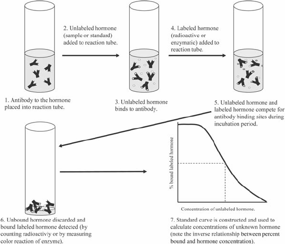
Principle : RIA’s technique involves the competitive binding to a high-affinity antibody of radiolabeled antigen and unlabeled antigen. The labeled antigen is mixed with an antibody at a concentration that saturates the antigen binding sites of the antibody.. Then, in increasingly greater quantities, test samples of unlabeled antigen of unknown concentration are added. The antibody does not differentiate between labelled and unlabeled antigen, so the two kinds of antigen compete against the antibody for available binding sites. If the concentration of unlabeled antigen increases, it will displace more labelled antigen from the binding sites. In order to assess the amount of antigen present in the test sample, the decrease in the quantity of radiolabeled antigen bound to a particular antibody in the presence of the test sample is measured.

Procedure :

A gamma-emitting isotope such as 125I is commonly labelled with the antigen, but beta-emitting isotopes such as tritium (3H) are often used as labels. The radiolabeled antigen is part of the assay mixture; a complex mixture, such as serum or other body fluids, containing the unlabeled antigen (test sample).

1. The first step in setting up an RIA is to decide the amount of antibody required in the assay mixture to bind 50 percent to 70 percent of a fixed amount of radioactive antigen.

2. Unlabeled antigen applied to the sample mixture would also compete for the limited supply of antibodies with radiolabeled antigen. (Even a small amount of unlabeled antigen added to the labelled antigen and antibody assay mixture will cause the amount of radioactive antigen bound to decrease, and this decrease will be proportional to the amount of unlabeled antigen added).

3. The Ag-Ab complex is precipitated to distinguish it from free antigen (antigen not bound to Ab) to assess the quantity of labelled antigen bound, and the radioactivity in the precipitate is calculated.

4. Using unlabeled antigen samples of a known concentration (in place of the test sample), a standard curve can be produced and the amount of antigen in the test mixture can be accurately calculated from this map.

For the separation of the bound antigen from the free antigen in RIA, several methods have been established. One strategy involves precipitating the complex of Ag-Ab with a secondary antiserum anti-isotype.

For example, if rabbit IgG antibodies are included in the Ag-Ab complex, then goat anti-rabbit IgG will bind to rabbit IgG and precipitate the complex.

Another technique makes use of the fact that Staphylococcus aureus protein A has a high IgG affinity. If the Ag-Ab complex produces an IgG antibody, it can be combined with formalin-killed S to precipitate the complex. The amount of free labeled antigen remaining in the supernatant can be determined in a radiation counter after removal of the complex by one of these methods; subtracting this value from the total amount of labelled antigen added yields the amount of labeled antigen bound.

Advantages:

  1. Used to detect very small amounts of serum antigens and antibodies.
  2. Used to quantify hormones, pharmaceutical products, HBsAg and other viral antigens.
  3. Analysis of nanomolar and picomolar concentrations in biological fluids.

Disadvantages:

  1. Cost effective
  2. Radio-labeled compounds have short shelf life
  3. The concerns surrounding the handling of radioactive (nuclear) wastes.

ELISA

BY: SAI MANOGNA (MSIWM014)

ELISA stands for Enzyme Linked Immunosorbent Antigen assay. ELISA is an ANTIGEN-ANTIBODY response.
In 1971, ELISA was founded. By Peter Perlmann and Eva Engvall at Stockholm university in Sweden. This is a plate-based assay technique used to detect and quantify substances such as peptides, proteins, antibodies and hormones. An antibody conjugated enzyme interacts with a colorless substrate to create a colored substance. This substrate is considered as a chromogenic substrate. A variety of enzymes has been used in ELISA, such as alkaline phosphatase, beta galactosidase and horse-radish peroxidase. Some substrates such as ortho-phenylenediamine dihydrochloride (for peroxidase), p-nitrophenyl phosphate (for alkaline phosphatase), which are hydrolysed by the enzyme referred to above, are used to generate a coloured end product.

In ELISA, an antigen must be immobilised on a solid surface and complicated with an antibody associated with an enzyme. Detection is achieved through an incubation with a substrate to create a measurable product by measuring the conjugated enzymes activity. A highly specific antimicrobial interaction is the most important aspect of the detection strategy.

Principle : Usually an ELISA test is performed in a multi-well plate of 96 or 384 well plates. The multi-well plate provides the stable surface for the antigen to stabilise. The immobilisation of the analyte makes it possible to isolate the antigen from the other components in the sample. This function makes ELISA one of the easiest experiments to conduct simultaneously in the multiple samples.

Types of ELISA :

ELISA assays can be found in various formats, based on the binding interactions between antigen-antibody.

They are :

  1. Direct ELISA
  2. Indirect ELISA
  3. Sandwich ELISA
  4. Competitive ELISA

  • Direct Elisa : the antigen is immobilised and detected on the surface of the multi-well plate with an antigen specific antibody, directly conjugated to HRP or other detection molecules.

  • Indirect ELISA :  With an indirect ELISA, an antibody can be detected or quantitatively determined.

    1. An antigen-coated microtiter well is applied to the serum or any other biological sample containing primary antibody (Ab1) and allowed to react with antigen attached to the well.
    2. The presence of an antibody bound to the antigen is identified after washing away any free Ab1 by adding an enzyme-conjugated secondary anti-isotype antibody (Ab2), which binds to the primary antibody.
    3. Any free Ab2 antibody is then washed away, and an enzyme substrate is added.
    4. Specialized spectrophotometric plate readers calculate the amount of colored reaction product that forms and can measure the absorbance of all the wells of a 96-well plate in seconds.
    4. This method of choice is for detecting the presence of serum antibodies against Human immunodeficiency virus (HIV), the causative agent of AIDS.


  • Reaction combinant envelope and core HIV proteins are adsorbed as solid-phase antigens to microtiter wells in this assay. HIV infected individuals can develop serum antibodies to these viral proteins in the epitopes. HIV serum antibodies will usually be detected by indirect ELISA within 6 weeks of infection.
  • Sandwich ELISA :  The antigen can be detected or measured using sandwich ELISA

    1. In this procedure, the antibody is immobilised on a microtiter well (rather than the antigen).
    2. An antigen containing a sample is added and allowed to react antibody being immobilised.
    3. A second enzyme linked antibody specific to different epitopes on the antigen is added after the well is washed and allowed to react with the bound antigen.
    4. After washing, substrate is added after any free second antibody is removed and the coloured reaction product is detected.
  • Competitive ELISA : Competitive ELISA is another variation for measuring the amounts of antigen in the sample.

    1. In this procedure, antibodies are first incubated in a solution with an antigen-containing sample.
    2. To an antigen-coated microtiter well, the antigen-antibody mixture is then added.
    3. The more antigen present in the sample, the less free antibody will be available for binding to the well coated with antigen
    4. The addition of an enzyme-conjugated secondary antibody (Ab2) unique to the primary antibody isotype can be used to calculate the quantity of primary antibody bound to the well. (as well as in Indirect ELISA).
    5. Nevertheless, in the competitive assay, the higher the concentration antigen, the lower the absorbance i.e present in the original sample.

Chemiluminescence : During certain chemical reactions, the measurement of light emitted by the chemiluminescence provides a convenient and highly sensitive alternative to absorption measurements in ELISA that use chemiluminescence. For Example, the oxidation of the luminol compound by H2O2 and the horse-radish peroxidase (HRP) enzyme produces light.

Enhanced sensitivity is the value of chemiluminescence assays over chromogenic ones. In general, by switching from a chromogenic to luxogenic substrate and with the addition of enhancing agents, more than 200 times, the detection limit can be increased at least tenfold. In fact as few as 5moles (5 attomoles) of the target antigen have been detected under ideal conditions.

ELISPOT Assay :  ELISPOT Assay is the modification of the ELISA assay.  This assay makes it possible to quantitatively determine the number of cells present in a population that contain antigen-specific antibodies or an antigen for which a specific antibody is present.

1. In this method, the plates are coated with the antigen recognised by the antibody of interest or with the antigen-specific antibody which is being tested for production. These specific antigen and antibody are known as capture molecules.

2. The coated plates are then supplemented with a suspension of the cell population under examination and incubated.

3. The cells settled on the plate surface. The secreted molecules that react with the captured molecules, will create a ring of antigen-antibody complexes around each cell that produces molecules of their interest.

4. The plate is then washed, added and allowed to bind an enzyme-linked antibody specific to the secreted antigen or specific to the species ( eg; goat anti-rabbit) of the secreted antibody.

5. Subsequent production of the assay shows the location of the each antigen or antibody producing cell as a point of colour or light by introducing an appropriate chromogenic or chemiluminescence-producing substrate.

Advantages and Disadvantages :

ELISAAdvantagesDisadvantages
Direct ELISA– Saves time and reagents (Short protocol) – No cross reactivity from secondary antibodies– In sample all proteins bind to the surface – Primary antibody must be labelled individually. – No signal amplification – Low flexibility
Indirect ELISA– Has signal amplification – High flexibility– Long protocol compared to direct ELISA – Potential cross reactivity from secondary antibody.
Sandwich ELISA– High specificity – Suitable for complex samples. – High flexibility and sensitivity– Finding two antibodies to the same target that identify different epitopes and function well together even at difficult times.
Competitive ELISA– Suitable for small antigens.– Depends on base ELISA selected.

APPLICATIONS OF RECOMBINANT DNA TECHNOLOGY

         BY: ABHISHEKA (MSIWM013)

The recombinant DNA technology has diversified applications in the field of Medicine, Industries, Agriculture, etc.

1.Medicine: In the field of Medicine recombinant DNA technology is one of the important milestones.

a) Production of vaccines: The RDNA technology is employed to produce vaccines by modifying cells and viruses to produce vaccines like the Influenza vaccine, Hepatitis B vaccine, etc. RDNA technique helps scientists to develop vaccines through cloning the gene used for protective antigen protein.

b) Production of human proteins and Hormones: The RDNA technology is used to produce hormones like Insulin, hGH, INF alpha, INF beta, and INF gama, etc. Recombinant DNA technology helped scientists to develop human insulin by using bacteria as a host cell. Now it is even available in the market.

c) Production of HCH: Now a days scientists have developed many growth hormones using rDNA technology, dwarfism is treated with this hormone.

d) Treating infectious diseases: RDNA technology has helped to develop many tests to diagnose diseases like Tuberculosis, Cancer, AIDS, etc. In the diagnosis process, certain pathogens are isolated, identified and then the kits to diagnose are produced when the genome of the specific pathogen is known to kill it or block its pathogenic activity.

e) Production of Interferons: Interferons are virus-induced proteins produced by virus-infected cells. Now, these interferons are produced using rDNA technology. These interferons are used for HCV infection.

f) Diagnosis of diseases: The RDNA technology has provided many tools for doctors to diagnose diseases like food poisoning salmonella, pus-forming staphylococcus, hepatitis virus, and HIV.

2. Agriculture: In the field of Agriculture the rDNA technology plays an important role to improve nutritional value and yield.

Example:

  1. Tomatoes are allowed to ripen on the vine and shelf life of the tomatoes is increased.
  2. BGH hormone produced through rDNA technology allows cattle to gain weight more rapidly and also to produce meat with lower fat content and produce 10% more milk.
  3. Crops sprayed with genetically modified bacteria can tolerate mild freezes.
  4. Organic farmers use Bt toxin to reduce insect damage to crops. This gene for Bt toxin is inserted into various crop plants using rDNA technology.
  5. Gene for beta carotene is inserted into rice crops through rDNA technology.
  6. Production of novel plants with high yielding and pest resistant abilities.
  7. The bacterial genes help in nitrogen fixation can be transferred to cereal crops like wheat, rice, maize, barley, etc.

3. Industry:  RDNA technology has numerous industrial applications.

a) Fermentation: The recombinant technology is used to improve strains of microbes which play important role in fermentation.

b) Production of chemical compounds and enzymes which are commercially and industrially important.

c) Developing more efficient strains of microorganisms.

4.Biotechnology:

a) Gene therapy: This RDNA technology helps scientists to replace the defective genes in the genome of the organism. It helps to treat genetic diseases by replacing defective genes in place by normal genes.

b) Creating transgenic animal: Gene of our interest is manipulated and prepared in the laboratory, then transgene is inserted into the selected cattle. Then the transgenic animals are produced.

Example: Transgenic cattle, transgenic chicken, Dolion, Lozebra.

c) Production of monoclonal antibodies and genetically modified plants and animals.

d) Bioremediation: Production of genetically modified bacteria to promote bioremediation.

  The use of RDNA technology in the present era is one of the important major advancements in the world.

CRISPR GENOME EDITING

BY: SREELAKSHMI (MSIWM012)

Clustered Regularly Interspaced Short Palindromic Repeats (CRISPR), are the hallmark of a bacterial defines system that forms the basis for CRISPR-Cas9 genome editing technology. In the field of genome engineering, the term “CRISPR” or “CRISPR-Cas9” is often used  to refer to the various CRISPR-Cas9 and -CPF1 and other systems that can be edited  to target specific stretches of genetic code and to edit DNA at precise locations and also  for other purposes, such as for new diagnostic tools. With these systems, researchers can permanently modify genes in living cells and organisms. In the future, it may be possible to correct mutations at precise locations in the human genome in order to treat genetic causes of disease. Other systems now available includes systems such as CRISPR-Cas13 which has the ability to target RNA and provide alternate ways for use and with unique characteristics that have been leveraged for sensitive diagnostic tools, such as SHERLOCK. Francis Mojica who is a microbiologist discovered Crisper.In2017 gene editing experiment on human embryo for the correctness of heart condition was successful. It was done to edit a gene MYBPC3 which caused hypertrophic cardiomyopathy(HCM),which affects 1 in 500.It has no cure and it causes cardiac arrest which leads to sudden death.

HISTORY OF CRISPRs

CRISPRs were first discovered in archaic and later in bacteria’s by Francisco Mojica, a scientist at the University of Alicante in Spain. He stated that CRISPRs serve as part of the bacterial immune system, defending against invading viruses. It consists of repeating sequences of genetic code which was interrupted using spacer sequences which are the remnants of genetic code from past invaders. Mojica’s theory was experimentally demonstrated in 2007 by a team of scientists led by Philippe Horvath. In January 2013, the Zhang lab published the first method to engineer CRISPR to edit the genome in mouse and human cells. Later many of the scientists and teams contributed to the understanding and development of the CRISPR system from the initial discovery to the first demonstrations of CRISPR-mediated genome editing. Feng Zhang and his team have trained thousands of researchers in the use of CRISPR genome editing technology and by sharing more than 40,000 CRISPR components with academic laboratories around the world.

 WORKING OF CRISPR

CRISPR mainly uses spacer sequences which are transcribed into short RNA sequences capable of guiding the system to matching sequences of DNA. When the target DNA is found, Cas9 which is one of the enzymes produced by the CRISPR system binds to the DNA and cuts it, shutting the targeted gene off. Modified versions of Cas9 is used by the researchers which can activate gene expression instead of cutting the DNA. These techniques allow researchers to study the gene’s function.

Research also tells that CRISPR-Cas9 can be used to target and modify typos in the three-billion-letter sequence of the human genome which can be helpful in treating genetic disease.

 IMPORTANCE OF CRISPR-Cas9 COMPARED TO OTHER GENOME EDITING TOOLS

CRISPR-Cas9 is proved to be an and efficient and tailored alternative to other existing genome editing tools. CRISPR-Cas9 system itself is capable of cutting DNA strands. They do not need to be paired with separate cleaving enzymes as other genome editing tools need. They can also be easily matched with tailored “guide” RNA sequences designed to lead them to their DNA targets. They are generally called as gRNA. Thousands of such gRNA sequences are already created and are available to the research community for studies. CRISPR-Cas9 can also be used to target multiple genes simultaneously, which is another advantage while comparing with other gene-editing tools that.

 DIFFERENCE BETWEEN CRISPR-Cpf1 AND CRISPR-Cas9

CRISPR-Cpf1 differs drastically from CRISPR-Cas9 while looking through the angle of research.Cas9 which is a DNA cutting enzyme in its natural form forms a complex which consist of two RNAs which are required for cutting activity. The Cpf1 is a simple system that   requires only a single RNA. The Cpf1 enzyme is a smaller than the standard SpCas9 which makes it easy to deliver into cells and tissues.Cpf1 cuts the DNA in different manner than Cas9.  The Cas9 complex cuts the DNA into both strands at the same place by leaving the blunt ends that often undergoes mutations while they are rejoining. Whereas Cpf1 complex cuts in the two strands which are offset and leaves short overhangs near the exposed ends. This is done to get help with precise insertion which allows the researchers to integrate a piece of DNA more efficiently and accurately.Cpf1 cuts far away from the recognition site which indicates that even if the targeted gene becomes mutated at the cut site it can still be cut again which allows multiple opportunities for correct editing to be done. The Cpf1 system provides flexibility in choosing the target sites. Similar to Cas9, Cpf1 complex must first get attached to a short sequence known as a PAM, and targets must be chosen which are adjacent to naturally occurring PAM sequences. The Cpf1 complex recognizes very different PAM sequences than those of Cas9. This could be an advantage while targeting, for example the malaria parasite genome and even the human genome.

 Scientific uses of CRISPR 

CRISPR genome editing allows the scientists to quickly create cell and animal models, which are used by researchers to accelerate their research into diseases such as cancer and mental illness. In addition, CRISPR is now being developed as a rapid diagnostic method.

MITOCHONDRIA

BY – SREELAKSHMI (MSIWM012)

MITOCHONDRIA

Mitochondria is majorly known as site of metabolism. It was discovered by collier in the flight muscles of insect. Term Fila was given by Fleming and term bio plast was given by Altmen .The term mitochondria was given by Benda.

 It is also called semi-autonomous organelle due its ability to perform certain activities. It is an important organelle in eukaryotes that produce adenosine triphosphate (ATP) which is the energy molecule for the cell. It is also called as the power house of cell. It is believed that mitochondria arises from free-living bacteria which were incorporated into cells.

Generally one mitochondria/cell is observed in unicellular organisms whereas 5 lakhs can be found in the flight muscle cells of insect which is the maximum observed till the date.

Structure of Mitochondria

  • Most common shape of mitochondria is disc or oval shape.
  • It have two membranes, an inner and outer membrane, which are made up of phospholipid layers.
  • Outer membrane has less proteins and they are permeable
  • Inner membrane is made up of several folds cristae, which increases the surface area. It also holds many proteins which supports the electron transport chain. It is semi-permeable. Many chemical reactions also take place in inner membrane. The increased surface area enhances the chemical reactions.
  • The inner area of mitochondria which is covered by inner membrane is called matrix. It is reach in enzymes foe cellular respiration and divalent ions like magnesium, ferric ions which are activators of enzyme present in matrix. It contains about 2/3 of total proteins. It is where the ATP production takes place. Space between inner and outer membrane is called perimitochondrial space.
  • Mitochondria have their own generic material and they produce their own RNA and protein. It also contains ribosomes.DNA present in mitochondria is reach in C and G nucleotides which results in the increase of denaturation temperature.
  • Mitochondria contains70s ribosomes .Generally 70s ribosomes are present in prokaryotes and this is the reason why Altman proposed a theory that eukaryotic mitochondria are either prokaryotic in origin or symbiotic.
  • But ribosomes in mammalian cells are 55s type. It has a larger subunit of 38s and smaller subunit of 28s.
  • It contains some nob like structures on cristae and this particles are called oxisomes.

FUNCTIONS OF MITOCHONDRIA

  • Most important function of mitochondria is to produce ATP.The simpler molecules are sent to mitochondria to be converted to ATP molecules. This process is called oxidative phosphorylation.
  • It performs major functions like like oxidation, dehydration, oxidative phosphorylation.
  • It also produces heat and ions of calcium or phosphate.
  • It also helps to maintain proper concentration of calcium ions within the cell compartments.
  •  Helps in building certain parts of blood and hormones
  • Mitochondria play an important role in apoptosis.

MITOCHONDRIAL DISEASE

Every year 1000 to 4000 children are born with mitochondrial disease in US. It is really difficult diagnosed.

In many cases, it is an inherited disease, it can also be due to environmental factors. Mutation in any one of the genes present in mitochondria can lead to mitochondrial disease. The mutation becomes so dangerous that it causes the proteins not to function properly.

This causes the mitochondria not to work properly which results in the decrease in the production of energy. Decrease in energy production can lead to any organ failure or even multiple organ failure.

There is no specific treatment for mitochondrial disease .It’s not having particular symptom or screening method which is the cause for misdiagnosis.

Symptoms can also include poor growth, developmental delays and muscle weakness. Mitochondrial diseases are generally transmitted by mother. Transmission of mitochondrial DNA mutation from mother can be reduced by mitochondrial replacement technique.

It is the replacement of mitochondria one or more cells. This technique uses healthy mitochondria of the donor’s egg. It is an in vito-fertilisatyion on which the mutated mitochondrial genes from mother’s cells is replaced by a third party.

The most common method in mitochondrial donation are maternal spindle fiber transfer using unfertilized egg and pronuclear transfer using fertilized egg. In maternal spindle fiber healthy nucleus of the mother is removed and from the donor egg is taken out without nucleus.

The healthy nucleus of the mother is now transferred to the donor egg and is fertilized. Pronuclear transfer involves the transfer of pronuclei from one zygote to another. It requires the fertilizes egg of donor also.

The healthy nucleus from the fertilized egg of parental couple is taken and the nucleus from donor egg is removed. Now the healthy nucleus is fused with the egg of donor. Mitochondrial disease include

  • Leigh Syndrome: It is a severe neuro disorder which is usually seen children. This leads to the eventual loss of mental and movement abilities. It results in patient’s death within 2-3 years due to respiratory failure. It can be due to the mutation or due to the deficiencies of pyruvate dehydrogenase enzyme. The symptoms also include weakness, muscle spasms. Lack of muscle ton, tremors which are symptoms of various other diseases also .Hence, it is really difficult to diagnose the disease.

FLOW CYTOMETRY

BY- SAI MANOGNA (MSIWM014)

Flow Cytometry : Flow cytometry is an efficient technique, because it enables the physical and chemical properties of cells up to thousands of particles per second in a heterogenous population. Multiple parameters of single cells can be analyzed simultaneously using this approach. This makes it a quick and quantitative technique for the analysis and purification of suspended cells. We can decide the phenotype and work using flow cytometry and even sort live cells.

It is primarily used to measure the fluorescence intensity provided by the protein- detecting fluorescent labelled antibodies, or ligands that bind to specific cell associated molecules such as DNA-binding propidium iodide. The staining process involves creating a single cell suspension from samples of cell culture or tissue. Cells are then incubated and tested on the flow cytometer in tubes or microtiter plates with unlabelled or fluorochrome- labelled antibodies.

Principle : Flow cytometry is used when a large number of different cell types need to be profiled in a population. On the basis of variations in size and morphology, the cells are separated. In addition to recognizing and segregating different subpopulations, fluorescently-tagged antibodies targeting the particular antigens on the cell surface may be used.

Components in flow cytometer :

  1. A flow cell
  2. A measuring system
  3. A detector
  4. An amplification system
  5. Computer analysis of the signals.
  1. A flow cell : this has a liquid stream containing the cells so they pass only single cells through the beam for sensing.
  2. Measuring system : This uses the measurement of conductivity and optical system lamps such as mercury and xenon, high power water cooled lasers such as argon and krypton, diode lasers such as blue, green. Red, violet resulting in light signals.
  3. Detector : The detector converts the measurement of forward scattered light and side scattered light as well as dye specific fluorescence signals into digital signals which further can be processed by computer.

Flow Cytometric Analysis :

 A.  Functional analysis : This method can determine the biological activity of cells including reactive oxygen species production, changes to the mitochondrial membranes during apoptosis, rates of phagocytosis in labelled bacteria, indigenous calcium content and changes in metal content during drug reaction, etc.

B.  Cell cycle analysis : The amount of DNA present in each phase of cell cycle varies. The fluorescent dyes which bind to DNA or monoclonal antibodies which can detect antigen expression, can evaluate this variation in the content of DNA. This approach can also be used to quantify other factors including cell pigment content such as chlorophyll, DNA copy count variance, intracellular antigen, enzyme activity, oxidative bursts, glutathione and cell adherence.

C.  Apoptosis and Necrosis Assessment : Apoptosis or programmed cell death is followed by typical changes in morphology of cells, structure loss, cell detachment, cytoplasm condensation, cell shrinkage, cell residue phagocytosis and nuclear envelope changes. Oncosis is a necrotic occurence in which a cell tends to swell instead of to decrease in its size. The plasma membrane breaks down and proteolytic enzymes are released, which can also damage the surrounding tissue. These changes can be analysed by flow cytometry in plasma membrane and cell type.

D. Determination of cell viability : This approach can also be used to test cell viability following addition of pathogens or drugs. Any defects in the integrity of a cell membrane can be assessed with the usage of dyes that can penetrate the cell membrane. Fluorescent samples like bis-oxonol will bind to proteins on the cell membrane, so that different stages of necrosis are established.

Working of Flow cytometry :

During the flow cytometry, a sheath fluid concentrates the cell suspension hydrodynamically through a small nozzle, so that only one cell can pass the laser at a time. A detector is positioned in front of the laser beam to capture the forward scattered light from the cells, while multiple detectors are also mounted on to the sides to determine the amount and intensity of scattered light in each direction.

As of now we understood that there are two ways in which the light signals can be analysed;

They are:

  1. Scattering
  2. Fluorescence emission
  1. Scattering :

    i. Forward scattering : forward scattering refers to the light refracted by a cell which is moving in the same direction as it was initially moving. The proportion of light scattered i.e, forward scattered determines the cell size, where larger particles emit more forward scattered light than the smaller particles, and also larger cells will have a stronger forward scatter signal.

    ii. Side scattering : Side scattering refers to the refracted light that is orthogonal to the light path direction. This provides information about granularity in which highly granular cells emit more light than cells with low granularity.

    For instance, cells with large granules, emit high forward scattered and high side scattered light. Monocytes that show low granularity emits high forward scattered light but low side scattered light. Therefore, based on the forward and side scattered light proportions, different types of populations can be differentiated.


Forward scattered light is directly proportional to cell size and refractive index.
Side scattered light depends on the shape and granularity of the cells.


  1. Fluorescence emission :  apart from the forward and side scattered light, various cell types may also be segregated by fluorescent molecules. Fluorescent light may be emitted by fluorescent molecules after excitation by a compatible wavelength laser. Fluorescent light may originate from naturally occurring fluorescence materials in the cell such as NADPH and FAD; (this mechanism is termed as autofluorescence), or may originate from the fluorescent dyes or fluorescence-tagged antibodies that have been used to label a specific structure of the cell.

Data Analysis : Each cell passing through the ;laser light is detected as a separate event. A distinct channel is often assigned to various forms of scattered light i.e, forward-scattered, side-scattered and fluorescence emission wavelengths. The data is separately plotted for each of these occurrences and can be interpreted by two methods: Histograms and Dot-plots.

Histograms :

  • Fast to read and easy to understand
  • Most useful when only one parameter is important
  • Representation includes intensity of single channel on x-axis and number of detected events on y-axis.
  • Multiple overlaid histograms : used to compare single parameters from two different sample populations.

Dot-plots :

  • Most useful for multi-parametric data
  • Can be 2-D or 3-D
  • Each distinct event is represented as a single dot and intensity of each channel is represented on its own axis.
  • More complex.

Uses of Flow cytometry:

  1. Cell counting
  2. Cell sorting
  3. Determining cell characteristics and function
  4. Detecting microorganisms
  5. Protein engineering detection
  6. Biomarker detection
  7. Diagnosis of health disorders such as blood cancers

RECOMBINANT DNA (RDNA) TECHNOLOGY

                   BY- ABHISHEKA G.(MSIWM013)

INTRODUCTION:

1.Recombinant DNA or RDNA technology is defined as the procedure of joining DNA molecules of two different species together and inserted into the host organism to produce a variety of new genetic combinations. This is also known as Genetic engineering.

2. The DNA fragments are selected from two different species and combined. This technique was developed by two scientists namely Boyer and Cohen in 1973.

3. The DNA molecule which is inserted into another DNA molecule is called a VECTOR. The recombinant vector is then introduced into a host cell where it replicates itself, and the new gene is produced. This is the basic principle behind Recombinant DNA technology.

TOOLS OF THE RECOMBINANT DNA TECHNOLOGY:

  1. Restriction endonucleases: These are used to cut DNA molecules at specific sequences into many smaller DNA fragments.
  2. Plasmids: These are extrachromosomal circular DNA present in the bacteria, which can replicate independently. During cloning, these plasmids carry drug resistance genes that are used for selection. Foreign DNA can be placed into a plasmid and it is replicated further.
  3. DNA ligase: This enzyme is used to join the two pieces of DNA together.
  4. Foreign DNA: This is also known as passenger DNA, which contains desired gene sequences.
  5. Vector: It is a vehicle used to insert the desired DNA into the host cell. Some of the vectors used are Plasmid DNA, Bacteriophage DNA, Yeast DNA, Viral DNA, Bacterial DNA, etc.

GOALS OF RDNA TECHNOLOGY:

  1. To isolate and characterize a gene or DNA from an organism.
  2. To eliminate undesirable phenotypic characters.
  3. To combine the needy and beneficial traits of two or more organisms.
  4. To make desired alterations in one or more isolated genes or DNA
  5. Inserting the altered genes or DNA into the host cell of another organism.
  6. To synthesize new genes using artificial methods.
  7. To alter the genome of the organism
  8. Understanding the diseases which transmit due to heredity.
  9. Understanding the treatment for heredity related disorders.
  10. To create new gene combinations.

PROCEDURE TO PREPARE RDNA:

1 Isolation of DNA from the organism: The cells are lysed using detergent mixtures, which creates pores in the plasma membrane. Then the mixture of cell contents is treated with protease and RNAase enzymes. The enzyme protease destroys the proteins present in the mixture and the enzyme RNAase destroys the RNA molecules present in the mixture. Then the mixture is centrifuged and the supernatant containing the DNA is transferred into a clean test tube and the DNA precipitated with the addition of ethanol.

2. Insertion of foreign genes into vectors: By using plasmid as a vector, isolated from the bacterial cell and treated with restriction enzymes and target DNA is obtained and it is placed into a vector to produce recombinant DNA.

3. Insertion of Recombinant DNA into host cell: The plasmid containing the foreign DNA is placed into a bacterial or host cell for multiplication.

4.Transformation: The vector is used as a vehicle to transport the gene to host cell, bacterium or other living cells are used as vectors. The vector is multiplied in the host cell and produces many identical copies, which are similar to both DNA and gene present in the DNA.

5. Cloning: After the division of the host cell the rDNA copies produced are transmitted to the progeny and further vector replication takes place in the progeny cell, with the continuous division of cells, a clone of identical host cells is formed. Each clone contains one or more copies of the rDNA molecule. Later the identical host cells are lysis and rDNA molecules are separated from the host cells.

APPLICATIONS OF RDNA TECHNOLOGY:

  1. This technology helps to grow crops which are resistant diseases and pesticides, crops of our choice, fruits, and flowers of attractive colors.
  2. This technique is employed in the production of artificial insulin and to deliver the drugs to target sites.
  3. Used in Molecular diagnosis of diseases.
  4. Used in Gene therapy.
  5. Employed in DNA fingerprinting.
  6. Used in the production of vaccines and pharmaceutical products.
  7. In the production of monoclonal antibodies.

PRIMARY AND SECONDARY LYMPHOID ORGANS

BY: SREELAKSHMI (MSIWM012)

PRIMARY LYMPHOID ORGAN

Organs which provide the environment for the development, maturation and differentiation of the immature lymphocytes generated in hematopoiesis so that they become committed to a particular antigenic specificity are called as Primary Lymphoid Organ. It includes

  • Thymus
  • Bone Marrow

THYMUS

It is a flat, triangular, bilobed organ situated above the heart and is enclosed within the fibrous capsule. Its each lobe is divided into lobules, which are separated from each other by strands of connective tissue called trabeculate.The function of the thymus is to generate and select a repertoire of T-cells that will protect the body form infection. As thymocites develop T-cell receptors are produced randomly. T-cells with receptors which are capable of recognizing antigen MHC complexes and some which are incapable of recognizing antigen MHC complexes are produced. Stem cells from bone marrow is send to thymus.95% of all thymocytes die by apoptosis in the thymus without even reaching maturity.

BONE MARROW

It is the site of B-cell origin in mammals. Immature B-cells proliferate and differentiate within bone marrow. The function of bone marrow is to generate and select a repertoire of B-cells that protects the body from infection. During B-cell maturation, a selection process take place within the bone marrow just like thymic selection that ensures the extensive maturation of those B-cells which carry on non self-reactive receptors and eliminates those which possess self –reactive antibody receptors. Bone marrow is not a site for B-cell development in all species. It also produces RBC and Platelets.

SECONDARY LYMPHOID ORGAN

It includes organs where the mature lymphocytes settle. It traps the antigen either from lymph or the blood .They include lymph nodes and spleen which is organized into structures called lymphoid follicles.

LYMPH NODES

They are bean shaped structures which contain a reticular network packed with lymphocytes, macrophages and dendritic cells. They are clustered at junctions of the lymphatic vessels it is divided into three regions – cortex, paracortex and the medulla. The space just below the capsule is called as sub capsular sinus. There is a define area of organization for the B and T lymphocytes. Their function is to trap antigens or microorganisms that enter the lymph which results in the activation of lymphocytes and cause the immune response.

SPLEEN

Spleen is a large and ovoid lymphoid organ situated in the left abdominal cavity below the pancreas. It traps blood –borne antigens and thus responds to the systematic infections. It consist of a network of sinusoids enriched with macrophages and red blood cells and lymphocytes. Blood-borne antigens as well as lymphocytes enter spleen through the splenic artery. It reaches the marginal zone where antigen is trapped by dendritic cells and carried to peri arteriolar lymphoid sheath (PALS).Dendritic cells along with antigen along with MHC molecules to TH cells and thus activate them which further activate the B-cell. These activated B-cells migrate to primary follicles in the marginal zone .Thus, upon antigenic stimulus, these primary follicles develop into characteristic secondary follicles containing germinal centers with rapidly dividing B-cells and plasma cells.

CELLS OF IMMUNE SYSTEM

BY: SAI MANOGNA (MSIWM014)

The significant majority of cells involved in the immunity of mammals are derived from the bone-marrow precursors (Left half of the figure) and circulate in the blood, entering if possible and leaving the tissues. In the adult bone marrow at a frequency of 1 in 100,000 cells, an intermittent stem cell survives and maintains the potential to differentiate into all blood cell forms.

Haemopoiesis has been studied either by injecting a group of cells into the recipient mice of genetically marked marrow cells and examining the progeny they give rise to (Invivo cloning) or by cultivating the bone marrow precursors in the presence of sufficient growth factors (Invitro cloning).

The proliferation and differentiation of all these cells is regulated by the soluble membrane bound growth factors that usually generate the stroma of bone marrow and each other. Such signals turn-on the particular growth factors inside the cell, DNA binding molecules that serve as master switches, establish the subsequent genetic program, resulting in the production of various types of cells.

Bone Marrow :

Unlike any other tissues or organs, the hematopoietic system continues to regenerate itself. In adults hematopoietic cell production occurs primarily in the bone marrow. In the fetal hematopoiesis occurs first in the yolk sac and then in the liver until the bone grows.

Stroma : In haemopoiesis, epithelial and endothelial cells that provide protection and secrete growth factors.

Lymphoid stem cell : These cells are assumed to be capable of differentiating into  B lymphocytes and T lymphocytes. Recent research findings indicate that the distinction might actually be more complex between lymphoid and myeloid stem cells.

Haemopoietic stem cell: Spleen nodule precursors are presumably capable of differentiating into all but lymphoid pathways i.e, granulocyte, erythroid, monocyte, megakaryocyte, also referred to as CFU-GEMM. CFU-GEMM refers to the colony forming unit that generated myeloid cells. GEMM refers to Granulocyte, Erythroid, Monocyte, Megakaryocyte.

Erythroid Stem cell : These erythroid stem cells generate erythrocytes. In response to hypoxia, erythropoietin, a glycoprotein hormone produced in the kidney, hastens the differentiation of red cell precursors, and thus adapts red cell development to the demand for its oxygen carrying capacity which is an example of NEGATIVE FEEDBACK.

Granulocyte–Monocyte : This is the common precursor. The growth or colony stimulating factors influence the relative proportion of these two types of cells.

Cloning : By isolating the single cells and allowing them to differentiate several times, and then studying what cell types can be found in the progeny. Thus the ability of individual stem cells to give rise to one or more types of hematopoietic cells has been explored. This approach is referred to as cloning. A clone is a group of daughter cells originating from the single parent cell. These evidence  indicate that a single stem cell can give rise to all the fully differentiated cells of an adult hematopoietic system under specified conditions.

Neutrophils : These are the short-lived phagocytic cells whose granules contain voluminous bactericidal substances, is the most prevalent leukocyte in the human blood. The first cells to exit the blood and to enter the sites of infection or inflammation are neutrophils.

Eosinophils : Eosinophils are the leucocytes with large refractile granules, containing a number of proteins that are highly basic or cationic proteins possibly essential for the killing of large parasites, including worms.

Basophils : These are the leukocytes with large basophilic granules containing heparin and vasoactive compounds which are essential in inflammatory response. The three types of cells which are neutrophils, eosinophils and basophils are collectively known as GRANULOCYTES.

Megakaryocyte : These are the parent cells of the blood platelets.

Platelets : These are the small cells which are responsible for sealing the damaged blood vessels (Which is known as haemostasis) but these cells are also the source of several inflammatory mediators.

Monocyte : The mononuclear phagocytic system consists of monocytes. Monocytes circulate in the bloodstream for about 8 hours and then migrate into the tissues. When these migrate into the tissues, they differentiate into specific tissue known as macrophages. Additionally monocytes are drawn to inflammation sites, replenishing a reservoir for macrophages and possibly dendritic cells as well.

Macrophages : Macrophages are strewed throughout the body. In specific tissues some take up residence, becoming fixed macrophages, while others remain motile called as free or wandering macrophages. These free macrophages migrate across the tissues by amoeboid movement.

Macrophage like cells serve various functions in different tissues and these are named according to the site of location;

Kupffer cells in Liver

Osteoclasts in Bone

Alveolar macrophages in Lungs

Mesangial cells in Kidney

Intestinal macrophages in Gut

Microglial cells in Brain

Histiocytes in Connective tissue.

Dendritic Cells : Dendritic cells are present in all body tissues (eg; Skin cells of Langerhans) where they pickup the antigen and then migrate through the lymphatics or the blood to the T-cell areas of lymph nodes or spleen. Their main function is to activate T-cell immunity, but they might also be involved in the induction of tolerance as well. The plasmacytoid DC is the name derived from their morphological resemblance to the plasma cells. The second subset of these cells are the principal producers of type I interferons, which is an essential group of viral proteins. Experimentally, while these cells are derived from the myeloid cells, the developmental lineage of bone-marrow dendritic cells is still the subject of debate.

Natural Killer Cells (NK Cells) : NK cells have been shown to play a significant role in host defence against both tumor cells and cells infected with certain, but not all the viruses. The membrane molecules and receptors that differentiate T and B cell lines do not express these cells. These cells make up 5%-10% of lymphocytes in the human peripheral blood.

T and B Lymphocytes : These cells are main cellular components of adaptive immunity. T-lymphocytes are derived from the thymus, while B-lymphocytes are derived from the bone marrow of birds or bursa derived. The precursor for antibody forming cells is B-lymphocytes. The liver may play the part of bursa in the liver.

Plasma Cell : Plasma cells are rarely seen in blood. These cells are present if an antibody is formed in the spleen, lymph node etc; Plasma cells do not differentiate and cannot be retained invitro for extended periods. B lymphocytes that generate particular antibodies may however be fused with a tumor cell to generate an immortal hybrid clone or hybridoma that continues to secrete a predetermined range of antibodies. As unique instruments in many branches of biology, such monoclonal antibodies have proven to be of immense benefit and some are now being widely used for the treatment of autoimmune disorders or cancers.

Mast cells : Mast cells are the large tissue cells that are derived from the circulating basophils. To start an inflammatory response, that causes several types of allergy, these mast cells are rapidly activated by tissue damage.

Growth factors : The molecules that regulate hematopoietic cell proliferation and differentiation are also often involved in the regulation of immune responses, such as interleukins or cytokines. In these some of them were first discovered by hematologists and are referred to as Colony stimulating Factors (CSF), but there is no particular meaning to the various names, and one ; IL-3 is sometimes referred to as multi-CSF. In clinical practice growth factors are used to improve specific blood cell subsets and erythropoietin was one of the first proteins developed by recombinant technology in the modern century.

HIV AND IT’S LIFE CYCLE

BY: ABHISHEKA G (MSIWM013)          

INTRODUCTION:

HIV (Human immunodeficiency virus) is a kind of virus that belongs to the retrovirus family and causes AIDS (Acquired immunodeficiency syndrome) in humans. It destroys the immune system of the individual by making the patient vulnerable to different other infections including neurological disorders. The disease of AIDS was first observed in 1981 in the united states. It is an epidemic disease present throughout the world. This disease can be preventable, but not curable.

TYPES OF HIV: There are two types of HIV viruses are present based on the origin of the virus.

  1. HIV-1: It is isolated in America, Europe, and Central Africa. It is more virulent and spread among individuals very rapidly
  2. HIV-2: It is isolated in West Africa, it is less virulent and does not spread rapidly as HIV-1

STRUCTURE OF HIV VIRUS:

  1. It is spherical in nature and measures about 90-120nm in diameter and it is composed of two copies of positive single-stranded RNA enclosed by a conical capsid composed of viral protein P24. The capsid also contains the enzymes which are necessary for viral replication such as Reverse transcriptase (P55/66), Integrase(p32), and protease(p10).
  2. The RNA genome of HIV consists of nine different genes that contain the information needed to make the structural proteins for new virus particles.
  3.  The matrix is composed of the viral protein P17 which surrounds the capsid ensuring the integrity of the virion particles. And the matrix is surrounded by 2 layers of phospholipids which are embedded by 70 copies of viral glycoprotein and lipoprotein which consist of two units namely anchoring transmembrane pedicles (Gp41) and surface projecting knob like spikes (Gp120).

MODE OF TRANSMISSION OF HIV VIRUS:  HIV is transmitted in many different ways.

1.Sexual contact: Unprotected sexual relationship with an infected individual.

2. From mother to child: The virus of HIV is transmitted from mother to child through the uterus, during delivery, and also during breastmilk.

3. Through needles: Through the injections during injecting drugs and needle prick injury which are already used by the infected individuals.

4. Blood transfusion and organ transplantation: The virus is transmitted through transfusion of blood and transplantation of organs from infected individuals to normal individuals.

LIFE CYCLE OF HIV VIRUS:

There are 7 different steps in the life cycle of the HIV virus.

  1. Attachment to the host/Binding to the target cell: The specific site for the HIV virus to bind are CD4 receptors and other co-receptors(CCR5/CXCR4) which are enveloped by the glycoprotein Gp120. Viral binding to host cell triggers fusion of the viral and cell membrane of the host cell, which is mediated by gp41 and allows the virus core into the cytoplasm of the host cell.
  2.  Reverse transcription of its RNA: HIV consists of single-stranded RNA. The single-stranded RNA is converted into DNA. This process is carried out by the enzyme Reverse transcriptase.
  3. Integration into the host genome: The viral double-stranded DNA is integrated into the genome of the infected host cell through the action of the viral integrase enzyme which causing a latent infection.
  4. Transcription of viral proteins: The transcription of viral proteins is mediated by Viral reverse transcriptase. The infected DNA of the host cell makes proviral RNA and protease enzyme cuts or cleaves the polypeptides into functional HIV proteins and the virion assembles.
  5. Assembly of a new viral particle: Viral strands and other proviral products combine to form a package and head for the cell membrane.
  6. Release of immature virions: Immature virus finds a suitable position to push or come out of the host cell by taking a piece of the membrane with it.
  7. Maturation of the virion: The new free virus matures and gets ready to infect another host cell.

SYMPTOMS OF THE DISEASE:

Diarrhea, Fatigue or weakness, fever, headache, joint pain, night sweats, rashes on the body, swollen glands, sudden weight loss, yeast infections in the body, and sexual organs that occur frequently and last for a long time.

LABORATORY DIAGNOSIS OF HIV: HIV is mainly detected by  different clinical tests:

  1. ELISA (Enzyme-linked immunosorbent Assay/ Enzyme Immunoassay): This test detects and measures the antibodies in the blood sample along with the presence of antibodies related to any infections.
  2. Western Blot: This test is used to detect the specific proteins called HIV antibodies present in the blood sample. If ELISA gives positive for HIV, then the western blot technique is used to confirm the positive result of ELISA and gives 99.9% accurate results.

AVAILABLE ANTIRETROVIRAL DRUGS TO HIV:

  1. Nucleoside reverse transcriptase: Zidovudine and Stavudine
  2. Non-nucleoside reverse transcriptase inhibitor: Efavirenz and Nevirapine.
  3. Protease inhibitor: Atazanavir and Darunavir.

PREVENTION OF HIV VIRUS:

  1. Avoid multiple sexual contacts.
  2. Using new and sterile needles for injection.
  3. Care should be taken and testing should be done during blood transfusion and pregnancy.
  4. Biomedical waste should be properly disposed of from hospitals and households.
  5.  Proper sex education should be provided to people.