INTRODUCTION TO MICROBIAL ECOLOGY

BY : K. Sai Manogna (MSIWM014)

The microbial ecology research covers subjects ranging from individual cells to complex structures and includes several different types of microbes. In studying pure cultures and unique microbial ecosystems, there is not only a visual difference, but there is a difference in the approach of analysis in each of these images.

Fig: Soil bacterium showing multiple flagella in SEM

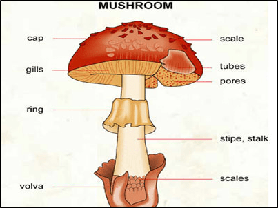
Fig : Fruiting bodies of various molds.

Studies by scientists from many different research fields discussing ecosystems around the globe have benefited from microbial ecology. The great interest lies in understanding the structure of microbial communities in the ecosystem at this period. It is essential to classify microbes present to attain this understanding; this can be done by using molecular methods even if the microbes have not been cultivated in the laboratory. Microorganisms’ enzymatic processes and microbial adaptations to the environment contribute to our understanding of microorganisms’ physiological ecology.

Roots of Microbial Ecology:

For millennia and long before bacteria were identified, people from various regions around the world used selective procedures to control the production of desired foods.

1. To make fermented milk, starter cultures were passed within a population, and traditional processes were used for fruit juice fermentation.

2. For food preservation, pickling processes involving natural fermentations are customary.

3. Increased rice production has resulted from unique practises in different regions of the world that we now understand are chosen for the growth of nitrogen-fixing cyanobacteria.

4. Some claim that microbiology began in 1675 with the reports by Anton van Leeuwenhoek (1632-1723) describing “very tiny animalcules” in the form of bacteria, yeast, and protozoa.

5. Saliva, dental plaque, and polluted water were the conditions that van Leeuwenhoek investigated.

6. As scientists in different countries studied the environment through direct observations or experimentation, knowledge of microorganisms gradually emerged.

7. Scientists’ contributions to disproving the “spontaneous generation theory” had a significant influence on microbiology, and the presentation by Louis Pasteur at the Sorbonne in Paris in 1864 was particularly important.

8. Pasteur emphasised the significance of microorganisms in fermentation by studying the function of microorganisms in diseases and their effect on our lives.

9. Many consider that Sergei Winogradsky and Martinus Beijerinck were the pioneers of microbial ecology, they were the first people to demonstrate the role of bacteria in nutrient cycles and to formulate concepts of soil microbial interactions.

10. Beijerinck invented the enrichment culture technique to isolate many bacterial cultures, including those now known as Azotobacter, Rhizobium, Desulfovibrio, and Lactobacillus.

11. The early studies of Beijerinck also contributed to the tobacco mosaic virus demonstration and offered insight into virology concepts.

12. Winogradsky was a Russian soil microbiologist who while working with nitrifying bacteria, developed the principle of chemolithotrophy. Winogradsky developed the notion of nitrogen fixation resulting from his experiment with Clostridium pasteurianum, and also demonstrated that bacteria could grow autotrophically with CO2 as the carbon source.

With the prominent interest in microbiology, it became evident that the relationship between microorganisms and also between microorganisms with their environment was highly complex. The study of microbial ecology today covers many different areas.

Current perspectives:

The study of microbial ecology covers the effect of the environment on microbial production and growth. Not only are microorganisms selected for physical and chemical changes in the climate, but biological adaptation helps bacteria and archaea to maximise the use of available nutrients to sustain development.

1. For early life forms, the prokaryotic cell was the ideal device because it had the facility for rapid genetic evolution.

2. Horizontal gene transfer between prokaryotes serves as the mechanism for the cellular evolution of early life forms to generate progeny with different genotypes and phenotypes, as we now understand.

3. While fossils provide evidence of the evolution of plants and animals, they also provide evidence of extinct early animal forms.

4. It is an irony in biology that dinosaurs and other prehistoric forms have engaged in the decomposition of the same prokaryotic organisms that evolved to create eukaryotic organisms. Not only does the prokaryotic mode of life survive today, but it prospers and continues to evolve.

5. It has been calculated that the top one inch of soil has more living microbial cells than several eukaryotic species living above ground.

6. It was estimated by William Whitman and colleagues that there are 5 × 1030 (five million trillion) prokaryotes on earth, and over half of the living protoplasm on earth is made up of these cells.

7. In the human body, the amount of bacteria that develop exceeds the number of human cells by a factor of 10. Although the function of each of these prokaryotic cells cannot be assessed, collectively, groups of prokaryotic cells can have a significant impact on eukaryotic life.

8. Human microbiome analysis shows that although the microbial flora of the skin is identical, each human being has a unique bacterial biome for that individual.

9. Not only are microorganisms involved in nutrient cycling, but they play an essential role in the organisation of the community and interactions with other types of life.

10. Without microorganisms, it would be difficult to imagine life on earth. It is useful to focus on the production of microbes on earth before discussing important divisions in microbial ecology.

TIMELINE:

The formation of the earth took place around 4.5 billion years ago, followed by the creation of the crust and oceans of the earth.

1. The Earth’s volcanic and hydrothermal activities have emitted different gases into the atmosphere. Dinitrogen (N2), carbon dioxide (CO2), methane (CH4), and ammonia (NH3) were the primary greenhouse gases in addition to water vapour, while hydrogen (H2), carbon monoxide (CO), and cyanide hydrogen (HCN) were present at trace amounts.

2. Scientists have critically examined the chemical discoveries of prebiotic earth that are important to life evolution. The anaerobic climate on earth gave decreasing power for the formation of the first organic compounds.

3. Early life forms were anaerobes that included chemolithotrophs, methanogens, and various microbes using thermophilic H2-showing dissimilar mineral reduction.

4. One of the earliest life forms is suggested to have been hyperthermophilic prokaryotes, and has gathered over 1500 strains of these species from hot terrestrial and submarine environments.

5. There is a significant abundance of these microorganisms in the environment, with 107 Thermoproteus cells found in a gram of boiling mud near active volcanoes, 108 Methanopyrus cells found in a gram of hot chimney rock, and 107 Archaeoglobus and Pyrococcus cells found in deep subterranean fluids below the North Sea per millilitre.

6. Although hyperthermophiles typically grow at 80o-113o C with a pH range of 0-9.0, one archaeal cell, Pyrolobus fumarii, resists in an autoclave with a temperature of 121o C for one hour.

7. Around 90 species of microorganisms are hyperthermophiles at present. The majority of hyperthermophiles are chemolithotrophic species that use molecular hydrogen (H2) as an energy-yielding electron source for reactions.

8. Although S0 is used as an electron acceptor by hyperthermophilic archaea, some hyperthermophiles may combine growth with the use of Fe3+, SO42-, NO3-, CO2 or O2 as electron acceptors.

9. For a few hyperthermophilic archaea, molecular oxygen (O2) is an appropriate electron acceptor and in these situations, only under microaerophilic conditions.

10. To sustain their anaerobic or aerobic growth, hyperthermophilic bacteria typically need organic material. Many anaerobes have active structures that use H2 as the donor of electrons.

The biological synthesis of methane is considered an ancient mechanism and the following reaction would have been attributed to catalysing prokaryotes:

4H2 + CO2 → CH4 + 2H2O

Methanogens might have developed methane from methanol, formate, or acetate if organic compounds such as acetate had accumulated in the environment. Only members of the Archaea domain are capable of the development of methane.

Based on the following reaction, chemoautotrophic microbes may have evolved to expand on energy from molecular hydrogen oxidation and carbon dioxide reduction:

2H2 + CO2 → H2O + [CH2OH]

Ultraviolet radiation may have produced H2 according to the following reaction, in addition to the output of H2 from geological formations:

2Fe2+ + 4H+ → 2H2 + 2Fe3+

The radiolysis of water due to alpha radiation will be another cause of H2. Heterotrophic prokaryotes metabolising organic carbon materials would have emerged sometime after the chemoautotrophs were formed with the accumulation of diverse organic compounds in the ecosystem.

Approximately 3 billion years ago, anaerobic photo-driven energy activities may have been present, using light to cause bacteriorhodopsin-like proteins to pump ions through cell membranes. The bacteriorhodopsin type of photo-driven energetics would have accompanied Chlorophyll-containing anoxygenic bacterial photosynthesis involving purple and green photosynthetic bacteria where H2S was the electron source. Although microbial evolution was initially in the aquatic world, about 2.75 billion years ago, microorganisms may have migrated to dry land. Cyanobacteria formed the aerobic atmosphere with oxygenic photosynthesis, and this was called the “great oxidation case.” Since the photocatalytic process produced O2 from water, the rate of O2 released was not limited by water availability.

Once molecular oxygen was released into the atmosphere, it reacted by microbial and abiotic processes with reduced iron and sulphur compounds (i.e. FeS and FeS2) to create oxidised inorganic compounds. The O2 level in the earth’s atmosphere steadily increased and oxygen respiration may have been used to help the development of the first single-cell eukaryotes by around 1.78-1.68 billion years ago.

The production of ozone (O3) from O2 due to an ultraviolet light reaction was another significant development in the aerobic atmosphere. Ozone absorbs ultraviolet light and forms a protective layer in the atmosphere to shield the earth from ultraviolet radiation’s harmful activities. Microorganisms may have been developing only in subsurface areas or habitats covered by rocks before the creation of the ozone layer.

BACTERIAL ENDOSPORES – FORMATION, STRUCTURE AND FUNCTIONS

BY: Reddy Sailaja M (MSIWM031)

Introduction

  • Microorganisms have the ability to adapt themselves to the changing conditions prevailing in the environment. Factors that influence microorganism’s survival could be physical, chemical or environmental.
  • Some microorganisms go in search of favorable conditions for survival, while some will become dormant till the favorable conditions arrive.
  •  One such mechanism adapted by certain gram-positive bacteria is the development of ‘endospores.  Gram positive bacteria, especially genera, Bacillus and Clostridium have the ability to form endospores in response to harsh conditions, nutrient deprivation in particular.
  •  When there is starvation due to nutrient deprivation, these bacteria produce most resistant and dormant ‘endospore ‘structures that preserve cell’s genetic composition to with stand the harsh assaults like high temperature, desiccation, UV radiation, chemical and enzymatic damage.
  •  Moreover, endospores are the most resistant form of “spores” or “cysts” produced by many bacteria and are resistant to most of the antibiotics. Altogether, endospores are resistant and dormant structures of life survival forms of bacteria and fight against harsh environments.


Formation of endospore

Figure 1: Development of Endospore

  • Bacillus subtilis is the model organism used to study and understand the development of endospore during the process is called sporulation.
  •  It takes many hours to complete endospore formation. Morphological changes that occur during this process are used as markers to classify stages of endospore development. Stage I is that when the bacterial cell is under favorable conditions.
  • Under unfavorable conditions, bacterial cell initiates endospores formation by asymmetric cell division and is called Stage II. Asymmetric cell division results in the formation of a larger mother cell and a smaller forespore (or pre-spore) with septum in between them.
  • Even though, these two cell types in stage II has varied developmental fates, intercellular communication system harmonize cell specific gene regulation by influencing specialized sigma factors in the cells.
  •  In stage III, peptidoglycan present in the septum gets dissolved and the mother cell engulfs forespore, which becomes a cell within a cell.
  • In the stages IV+V, cortex and the spore coat layers are formed around the forespore, leading to the production of endospore specific compounds.
  •  In the stages VI+VII, further dehydration and the maturation of the endospore happens. Finally, the mother cell dies by apoptosis (also called programmed cell death) and the endospore is release into the environment and remains dormant until favorable conditions prevail.

Endospore structure

Endospore structure comprises of multiple layers of coats that resist against harsh surrounds. The following table details various layers (from outer to inner), their compositions and functions.

Endospore layerCompositionFunction
ExosporiumCarbohydrates, proteins  and lipidsGives hydrophobic character to the endospore and is responsible for endospore pathogenicity
Spore coatCoat proteins cross-linked with disulfide bondsActs as primary permeability barrier and allows only smaller molecules like germinants
Outer membraneNot known
CortexPeptidoglycan without teichoic acids with low cross linkingStructural differences in the peptidoglycan of cortex and germ cell wall allow selective degradation of outer protection, germination of endospore and transformation of germ cell wall into vegetative cell.
Germ cell wallPeptidoglycan
Inner membraneSimilar to cell membrane composition. Germinant receptorsVaried fluidity and permeability and decreased mobility of the membrane lipids make the structure highly impermeable to the molecules including water, protecting core. Germinant receptors allow binding of germinants and begin germination and vegetative growth.
CoreBacterial DNA, RNA, ribosomes, essential enzymes, small acid-soluble spore proteins (SASPs), Dipicolinic acidDehydrated state protects enzymes and heat resistance. SASPs protect DNA from destructive chemicals and enzymes by forming shield. SASPs also function as carbon and energy source during germination into vegetative cell. Dipicolinic acid also protects endospore’s DNA against harsh environment.

Figure 2: Structure of endospore

Mechanism of sporulation

  • Sporulation of endospores is under the control of five kinases, namely KinA, KinB, KinC, KinD and KinE that act under phosphorelay signal transduction mechanism.
  • Each of these kinases gets activated based on specific environmental stimuli. Under a specific kind of environmental stimulus, one of the five sensor kinases undergoes autophosphorylation at conserved histidine residue by an ATP dependent reaction through a protein called Spo0F. Then the Spo0F transfers the phosphate to Spo0B, that act as a mediator and delivers signal to Spo0A.
  •  Spo0A further positively regulate genes necessary for sporulation and negatively regulate genes required for vegetative growth.

Figure 3: Mechanism of sporulation

Functions of endospores

  • Endospores mainly resist harsh conditions like high temperatures, disinfectants, radiation, etc.
  •  Endospores are reported to survive for millions of years. For example, viable endospores were isolated from gastrointestinal tract of a bee that was embedded in amber around 25-40 years ago.
  • Dipicolinic acid and SASPs are crucial in protecting core of the endospore that contains genetic material.

Infectious diseases caused by endospores

In spite of defensive mechanism, endospores also transmit some infectious diseases as follows:

i)Anthrax – caused by Bacillus anthracis endospores when inhaled, ingested will germinate under suitable conditions and spread the infection

ii)Botulism – Caused by Clostridium botulinum. Spreads through unprocessed food and infect

iii) Tetanus – caused by Clostridium tetani. Spread through anaerobic wounds and cause infection.

 Other infectious diseases like gas gangrene and pseudomembranous colitis are also popular.

SALMONELLA

BY: SREELAKSHMI (MSIWM012)

Salmonellas is an infectious disease caused by bacteria. It is also known as salmonellosis. Causes Enteric fever, Gastroenteritis, Septicaemia etc. Salmonella typhi rank Typhoid. Salmonella is two groups that include a group that includes the enteric flu that contains only typhoid & Paratyphoid bacilli or major human parasites and the other group includes a group that causes food poisoning but can infect people who cause stomach infections.

Causes: Often the cause of contaminated food or water. Food delivery can enter

Raw meat, poultry and seafood: Sewage enters immature meat and poultry causes contamination and disease. The transport of marine fish can be caused by water pollution in the agricultural environment.

• Raw Eggs: some infected chickens produce salmonella-containing eggs before the shell is formed. Raw eggs are used as versions of mayonnaise and hollandaise sauce.

Fruits and vegetables: New products like imported varieties, can be watered or washed during salmonella contamination. Contamination may also occur in the kitchen, where juice from raw meat and poultry comes in contact with uncooked food.

The Food and Drug Administration claims that some outbreaks of salmonella have been caused by spice contamination. Food can also be contaminated if it is prepared by people who do not wash their hands properly after using the toilet or changing diapers. Infection can occur if you touch anything contaminated including pets, birds and reptiles and put your fingers in your mouth.

Salmonella are negative gram sticks. With the exception of S. gallinarum pullorum they travel with peritrichate flagella. Salmonellae is an aerobic plant that grows in light media at a pH between 6-8 and a temperature of 15-41 ° C. They can be killed at 55oC for one hour or 60oC for 15 minutes. Boiling or dehydrating and dehydrating helps to eliminate bacilli. In dirty water it can live for weeks and freeze for months.

Salmonella infection is not a life-threatening disease. In some cases, especially when infants and young children, elderly adults, transplants, pregnant women, and people with weakened immune systems find this infection can be difficult.

It can be dangerous. It causes dehydration as you get diarrhoea and do not take a sufficient amount of water to compensate for water loss. Warning signs include reduced urine, dry mouth, sunken eyes, and decreased production of tears.

When salmonella infection enters the bloodstream, it can cause tissue damage throughout the body. When it affects the tissues around your brain and spinal cord it can lead to meningitis. If you have a heart attack or valve, it could be endocarditis.

 Infections in the bones or bone marrow can cause osteomyelitis. People who have been infected with salmonella are at greater risk of developing active arthritis. Also known as Reiter’s syndrome.

Active rheumatoid arthritis is the cause: Eye irritation, painful urination, painful joints Prevention can be done with care to avoid spreading the virus to others. Immunizations are really important when preparing food, caring for infants, the elderly and people with weakened immune systems. Washing hands thoroughly can help prevent the transmission of salmonella virus to your mouth from any food you prepare.

Salmonellae causes the following clinical disease in humans

 Enteric fever

It is also called typhoid fever. It is usually caused by Salmonella enteric serotype typhi and S. enteric serotypes paratyphi A, B, and C.

 They are phagocytosed by polymorphs and macrophages. They are able to withstand intercellular killings and multiply within cells. They enter the lymph nodes where they multiply .They enter the bloodstream through the thoracic tunnel causing bacteraemia.

 Bacillus is more abundant in the bladder as bile is a good culture. It is then continuously excreted in the intestine where it implants Peyer’s Patches and lymphoid follicles on the ileum, which is ulcerative and can lead to intestinal insufficiency & bleeding as a problem.

 The incubation period is usually 7-14 days but may occur in 3-56 days. Bacteraemia occurs early in the disease and blood pressure is very good in the first week of the flu. The diagnosis is collected about 5-10ml of blood and injected into a traditional bottle containing 50-100ml of 0.5 per cent of bile broth.

The sample was incubated overnight at 37oC. After that bile broth is a subculture in MacConkey agar, pale lactose fermenting colonies from this method were selected for chemical experiments. Salmonellae is usually motile, indole and urease non-fertile glucose and ferment. Another option is the display of typhoid bacilli in the blood or urine.

If Salmonellae is not found in the original small cultures, the smaller cultures should be repeated every other day until growth is achieved. If possible, if Salmonellae cannot be detected in the first subculture from the broth, subcultures should be repeated daily until growth is achieved.

MITOCHONDRIA

BY – SREELAKSHMI (MSIWM012)

MITOCHONDRIA

Mitochondria is majorly known as site of metabolism. It was discovered by collier in the flight muscles of insect. Term Fila was given by Fleming and term bio plast was given by Altmen .The term mitochondria was given by Benda.

 It is also called semi-autonomous organelle due its ability to perform certain activities. It is an important organelle in eukaryotes that produce adenosine triphosphate (ATP) which is the energy molecule for the cell. It is also called as the power house of cell. It is believed that mitochondria arises from free-living bacteria which were incorporated into cells.

Generally one mitochondria/cell is observed in unicellular organisms whereas 5 lakhs can be found in the flight muscle cells of insect which is the maximum observed till the date.

Structure of Mitochondria

  • Most common shape of mitochondria is disc or oval shape.
  • It have two membranes, an inner and outer membrane, which are made up of phospholipid layers.
  • Outer membrane has less proteins and they are permeable
  • Inner membrane is made up of several folds cristae, which increases the surface area. It also holds many proteins which supports the electron transport chain. It is semi-permeable. Many chemical reactions also take place in inner membrane. The increased surface area enhances the chemical reactions.
  • The inner area of mitochondria which is covered by inner membrane is called matrix. It is reach in enzymes foe cellular respiration and divalent ions like magnesium, ferric ions which are activators of enzyme present in matrix. It contains about 2/3 of total proteins. It is where the ATP production takes place. Space between inner and outer membrane is called perimitochondrial space.
  • Mitochondria have their own generic material and they produce their own RNA and protein. It also contains ribosomes.DNA present in mitochondria is reach in C and G nucleotides which results in the increase of denaturation temperature.
  • Mitochondria contains70s ribosomes .Generally 70s ribosomes are present in prokaryotes and this is the reason why Altman proposed a theory that eukaryotic mitochondria are either prokaryotic in origin or symbiotic.
  • But ribosomes in mammalian cells are 55s type. It has a larger subunit of 38s and smaller subunit of 28s.
  • It contains some nob like structures on cristae and this particles are called oxisomes.

FUNCTIONS OF MITOCHONDRIA

  • Most important function of mitochondria is to produce ATP.The simpler molecules are sent to mitochondria to be converted to ATP molecules. This process is called oxidative phosphorylation.
  • It performs major functions like like oxidation, dehydration, oxidative phosphorylation.
  • It also produces heat and ions of calcium or phosphate.
  • It also helps to maintain proper concentration of calcium ions within the cell compartments.
  •  Helps in building certain parts of blood and hormones
  • Mitochondria play an important role in apoptosis.

MITOCHONDRIAL DISEASE

Every year 1000 to 4000 children are born with mitochondrial disease in US. It is really difficult diagnosed.

In many cases, it is an inherited disease, it can also be due to environmental factors. Mutation in any one of the genes present in mitochondria can lead to mitochondrial disease. The mutation becomes so dangerous that it causes the proteins not to function properly.

This causes the mitochondria not to work properly which results in the decrease in the production of energy. Decrease in energy production can lead to any organ failure or even multiple organ failure.

There is no specific treatment for mitochondrial disease .It’s not having particular symptom or screening method which is the cause for misdiagnosis.

Symptoms can also include poor growth, developmental delays and muscle weakness. Mitochondrial diseases are generally transmitted by mother. Transmission of mitochondrial DNA mutation from mother can be reduced by mitochondrial replacement technique.

It is the replacement of mitochondria one or more cells. This technique uses healthy mitochondria of the donor’s egg. It is an in vito-fertilisatyion on which the mutated mitochondrial genes from mother’s cells is replaced by a third party.

The most common method in mitochondrial donation are maternal spindle fiber transfer using unfertilized egg and pronuclear transfer using fertilized egg. In maternal spindle fiber healthy nucleus of the mother is removed and from the donor egg is taken out without nucleus.

The healthy nucleus of the mother is now transferred to the donor egg and is fertilized. Pronuclear transfer involves the transfer of pronuclei from one zygote to another. It requires the fertilizes egg of donor also.

The healthy nucleus from the fertilized egg of parental couple is taken and the nucleus from donor egg is removed. Now the healthy nucleus is fused with the egg of donor. Mitochondrial disease include

  • Leigh Syndrome: It is a severe neuro disorder which is usually seen children. This leads to the eventual loss of mental and movement abilities. It results in patient’s death within 2-3 years due to respiratory failure. It can be due to the mutation or due to the deficiencies of pyruvate dehydrogenase enzyme. The symptoms also include weakness, muscle spasms. Lack of muscle ton, tremors which are symptoms of various other diseases also .Hence, it is really difficult to diagnose the disease.

VIRUS

BY: SREELAKSHMI (MSIWM012)

Virus are small structure which can pass its genetic material to living host where it can replicate. They can infect plants, animals and other microorganisms also. The study of viruses is called virology which is considered as a subspecialty of microbiology.  Martinus Beijerinck is the father of virology.

In 1898, Friedrich Loeffer and Paul Frosch was the first to find evidence of the nature of viruses, genetic entities of virus. They found the evidence from foot and mouth disease in livestock. The first identified human virus was yellow fever caused by mosquito Aedes aegyptii.Virus was discovered and reported in 1910 by a US Army physician.Veterniary virology gained importance after in1902 after cattle plague.

CLASSIFICATION OF VIRUS

Virus classification is the process of naming virus and places them into a taxonomic system. Virus are mainly classified by phenotypic characters like morphology, mode of replication, host organisms, nucleic acid type and the type of disease caused by the virus. A universal system for classifying viruses, and a unified taxonomy was established in1966 by International Committee on Taxonomy of Viruses (ICTV).vanRegenmortel (19) lists the following characters for discriminating between virus species:

  • Relatedness of genome sequence
  • Natural host range
  • Cell and tissue tropism
  • Pathogenicity and cytopathology
  • Mode of transmission
  • Physicochemical properties of viral proteins
  • Antigenic properties of viral proteins.

The system makes use of a series of ranked taxons,with the: order,family,subfamily,genus,species,order,family,genus,species.

 STRUCTURE BASED CLASSIFICATION OF VIRUS

It is classified based on shape and size. Based on the structure of nucleocapsid it’s divided into two namely helical morphology and icosahedral morphology. Helical morphology consist of a helical array of capsid proteins which is surrounded by a helical filament of nucleic acid. The number and arrangement of the capsomers are useful in identification and classification of icosahedral morphology Based on envelope it’s divided as enveloped and non-enveloped virus.

PROPERTIES OF VIRUS

  • It possess no membranes,cytoplasm,ribosomes or other cellular components
  • They cannot move or grow
  • They are really tiny, even smaller than cell and is only visible advanced electron microscopes.
  • It consist of two major parts which are a protein coat and a hereditary material which can be a RNA or DNA.

VIRAL REPLICATION

Viruses cannot replicate on its own and in order get multiplied it must infect a host. It replicates in host cell’s machinery to create more viruse.steps involved in replication includes:

  • Penetration: The virus gets engulfed by the cell or get attached to some other viruses which have surface proteins that bind to receptors on the host cell. It allows the host cell to either fuse with virus or absorb the virus. Once it reaches the cell, the genetic material is released to the cytoplasm.
  • Replication: The genetic material of the virus is copied several times.
  • Transcription: The genetic material act as a blue print for the cell to make messenger RNA which is used to make viral proteins.
  • Protein Assembly: It occurs in the cytoplasm (ribosome) where viral proteins are made.
  • Viral Assembly: The viral genetic material (after replication) will get surrounded by the newly made viral protein.
  • Release: Through budding viruses get emerged from the cell through cell membrane or by bursting out of the cell which will result in the death of host cell.

STRUCTURE OF VIRUS

Size of a virus is smaller than bacteria and size is variable. The larger viruses can vary their size about 300 A in diameters. It indicates that it may be as larger as a small bacteria. They occur mainly in three shapes which are helical (spherical or can be of complex symmetry. Virus lacks cytoplasm and hence lack cell organelles like mitochondria, Golgi bodies, ribosomes and enzyme systems are also present. Virus contains RNA or DNA whereas a normal cell contains both DNA and RNA. It contains a protein coating called capsid which acts as a protection for genetic materials by surrounding the genetic material. A virus particle is called virion.

Plant virus contains only RNA.Animal virus contains both RNA or DNA.wheras bacteriophages contain only DNA.

TYPES OF ANIMAL VIRUS

  • Double-Stranded DNA: Double-Stranded DNA virus generally have a polyhedral or complex structure. Examples are Papilloma Virus,Variola(smallpox)
  • Double-Stranded RNA: Double-Stranded RNA usually have polyhedral structure. Example is diarrhea virus.
  • Single –Stranded RNA: Single –Stranded RNA virus have two subunits which can serve as mRNA and other one which can serve as a template mRNA.Examples are Rhinovirus,HIV

ROLE OF VIRUSE IN CANCER

The tumor viruses change cells by integrating their genetic material with the host cell DNA.This is a permanent insertion in the genetic material which is never removed. The insertion mechanism can differ depending on whether the genetic material of the tumor causing DNA or RNA.In case if the genetic material is DNA then it can be directly inserted into the host DNA.In case of RNA as the genetic material, then RNA is first transcribed into DNA and then it is inserted into the host cell’s DNA.

MICROBIAL INTERACTIONS

BY: SAI MANOGNA (MSIWM014)

Biological interactions are the impact that species in the environment have on each other

  • The interactions between different microbes, plant and germ interactions to promote growth, animal interactions, interactions with human beings and water interactions occur in a whole range of microbes.
  • In any biological culture, microbial interactions are all distinctive, complex, critically important and are central in global biogeochemistry.
  • The relationships between these two populations are defined by whether the relations favour both populations and benefit one of them, or whether one or both populations are negatively affected

Microbial interactions are of two types :

  1. Positive Interactions : Mutualism, Syntrophism, Photoreception, Commensalism.
  2. Negative Interactions : Ammensalism (antagonism), Parasitism, Predation, Competition.

Positive Interactions :

  1. Mutualism : Mutualism is defined as interactions between organisms of two different species, in which each organism benefits in some way from interactions.

    i. These types of interactions are common and ubiquitous across all ecosystems, and scientists increasingly recognise the important role they play in ecology.

    ii. Mutualisms are also symbiotic partnerships. In such situations, the two animals live in close proximity to each other for part or all their whole lives; however, not all symbiotic relationships are reciprocal.

Examples of Mutualism:

Several known examples of mutualistic arrangements exist.

  1. Nitrogen fixing bacteria and Leguminous Plants : The relationship between nitrogen-fixing bacteria and leguminous plants
  • Intestinal flagellates and Termites : Intestinal flagellates and termites exhibit obligate mutualism, a strict interdependence in which protozoans digest the wood eaten by termites; Here neither partner can live under natural circumstances without the other.
  • Yucca Moths (Tegeticula) and Yucca Plants : Yucca moths depend on yucca plants and the other way around. The moth goes about as a pollinator while laying her eggs in yucca seed pods. The hatchlings incubate and feed on a few yet not all seeds. Here the two species have advantages such that the plant is pollinated and the moth has nourishment for its hatchlings.

II. Syntrophism : Syntrophy is the mutual dependency of various types of species on the fulfilment of their respective nutritional needs. The intermediate or end products metabolism of one organism are important materials for another. Syntrophism is exemplified in the mixed environment population.

Examples of Syntrophism:

  1. Dust mites on Human Skin : House dust mites fleece human skin. A healthy person produces about 1 gram of skin flakes daily. These mites may also produce chemicals that stimulate skin flakes. People may become allergic to these compounds.
  • Organisms on cow-dung : A cow eats lots of grass, which is converted into lipids by microorganisms in the large intestine of the cow.

III. Photoreception : It is a relationship in which the organisms are mutually beneficial and in association with each other. This relationship is similar to mutualism, but relations between the species in proto-cooperation are not necessary as in mutualism.

Examples of Photoreception :

  1. Interaction of Nitrogen fixing bacteria and Cellulomonas :

IV. Commensalism : In Biology,commensalism refers to the relationship between two species in which one species, without harming or helping the latter, obtains food or another benefits from the other.

    i. The species i.e, benefiting from the unaffected species (host), can obtain nutrients, shelter, protection or locomotion.

   ii. The relationship is predominantly between the larger host and the smaller commensal. In essence the host organism remains unchanged through contact, while the commensal species can exhibit great morphological adaptation.

Examples of Commensalism :

  1. Shark and Remora : remora attaches to the shark or other fish and rides on it. Remoras evolved on their heads as a flat oval shaped disc structure that adheres to their host bodies.

Negative Interactions :

  1. Ammensalism : Ammensalism is any relationship between two different species, where one organism is inhibited or eliminated, while the other remains consistent.

     i. One microbial population develops substances that are inhibitory to the other microbial populations which is antagonistic known as ammensalism are antagonism.

   ii. In nature, no organism can live its life in complete isolation. They must communicate in some way with other species and their environment.

Examples of Ammensalism : There are basically two types 1. Competition 2. antibiosis.

  1. Competition : A Larger more potent organism removes another organism from its source of food and shelter.
  2. Antibiosis : One organism secretes a chemical that destroys the other organism, but the one which secretes the chemical is unharmed.

       Example : penicillium on black walnut trees : The Mould that is capable of producing penicillin, which kills several types of bacteria that would also like to grow on this bread. The bacteria killing effects of penicillin that contributed to the use of penicillin as antibiotic. Penicillin doesn’t benefit from the death of other bacteria, making it an example of amensal antibiosis.

 II.  Parasitism : Parasitism is a relationship between two separate species, where one of them actually affects the other. An organism that harms the other is considered a parasite.

   i. The host-parasite relationship is characterised by a relatively prolonged period of interaction that may be physical or metabolic.

   ii. Some parasites live outside the host cell, known as ectoparasite, while others live within the host cell, known as endoparasite.

Examples of Parasitism :

  1. Ticks on Dogs and cats : Fleas or ticks that adhere to the skin of dogs and cats are parasites.
  • Lice :  This is another parasite that lives off the blood of host animals.
  • Aphids : Aphids are a type of insect parasite that feeds on the sap of host plants.

III. Predation : Predation refers to the relationship between organisms in which one organism kills and devours another.

   i. Predation gives energy to prolong life and to facilitate the reproduction of the organism that destroys the predator, to the detriment of the organism that is eaten, the prey.

  ii. Predation affects species in two ecological states.

  iii. At the level of an individual, the prey organism has a sharp decline in fitness as calculated by its lifetime productive success, since it can never produce again.

  iv. At the group level, predation decreases the number of individuals in the population of prey.

Examples of Predation :

  1. Carnivorous interactions :  The best known examples are Wolves hunting moose, Frogs eating flies, Owls hunting mice, Shrews hunting worms or insects.
  2. Group Predation : This occurs with ants and social spiders.

IV.  Competition : A Larger more potent organism removes another organism from its source of food and shelter.

  i. This interaction occurs when both species (populations) use the same resources, resulting in lower maximum density or growth rate for the microbial population.

Examples of Competition :

a. Competition between Paramecium caudatum and Paramecium aurelia : When these protozoa are brought together, both paramecium species feed on the same bacterial community.

BIOREMIDIATION

BY: RAHUL ANDHARIA (MSIWM001)

Introduction:

  • Bioremediation refers to use of naturally occurring microorganisms or introducing live microorganisms to break down environmental pollutants. This method is useful in reducing environmental pollution.
  • Enzymes, plants and microorganisms are used to prevent soil contaminants and to detoxify it from the environment.
  • The rate of natural microbial degradation of contaminants by supplementing indigenous microorganisms is enhanced by the process of bioremediation.

History:

  • Older versions of bioremediation dates back to 600Bc when bioremediation was first used by the Romans. They used bioremediation for the purpose of cleaning waste water.
  • Bioremediation was officially invented by George Robinson in the year 1960. First, large clean up of oil spill was initiated by him in the year 1968.

Principle involved in Bioremediation:

  • Microbes using contaminants like oil, solvents and pesticides as a source of food and energy relies on bioremediation for stimulation of their growth.
  • Contaminants consumed by microbes get converted to small amounts of water and carbon dioxide.
  • Combination of right temperature, nutrients and food is essential for effective bioremediation otherwise much longer time will be taken to cleanup contaminants.
  • Amendments can be added such as Molasses, vegetable oil, or simply air to make favorable conditions for bioremediation to take place.
  • Optimum conditions (most favorable conditions for growth) are created by these amendments for microbes to flourish and complete the bioremediation process.
  • Bioremediation is a slow process and can take anywhere from few months to several years. The amount of time required depends on factors like; size of the contaminated area, conditions like temperature and soil density, concentration of contaminants and whether the bioremediation process is in-situ or ex-situ.

Key Features of Bioremediation:

  • Contaminants in the soil matrix are destroyed in most of the bioremediation treatment technologies.
  • Generally, technologies are designed to reduce toxicity either by destruction or by transforming toxic organic compounds into lesser toxic forms.
  • Bacteria and fungi are most commonly used. In some cases, specific bacteria or fungi are used that can degrade the contaminant. (Biodegradable). Ozone or h202 (hydrogen peroxide) are added as electron acceptors to enhance growth and reproduction of indigenous organisms.

Categories of bioremediation:

  1. Microbial Remediation:
  2. Use of microorganisms to clean up contaminants is called as microbial bioremediation.
  3. Actinomycetes, fungi, bacteria, yeasts can be used in this process.
  4. Many elements present in microorganisms can be used as nutrients and microbes are easily available, omnipresent and highly diverse which makes them ideal candidates for bioremediation.
  5. Specific Microorganisms can be used for extreme environmental conditions and can be applied in both in-situ and ex-situ conditions.
  6. Mix culture approach is more suitable than pure culture approach in bioremediation as in pure culture synergistic interactions are seen while degrading crude oil present in soil.
  7. Examples: Cyanobaacteria, Pseudomonas, Nocardia, Acenetobacter and so forth. (These are the bacteria which are capable of degrading major pollutants).
  • Phytoremediation:
  • It refers to using different types of plants to transfer, remove, and stabilize/destroy contaminants present in soil and groundwater. Different phytoremediation methods are available which are as follows:
  • Rhizosphere biodegradation: Roots releases natural plant substances and supplies nutrients to microorganisms in soil. Biological degradation is enhanced by microorganisms.
  • Phyto-stabilization: chemical substances produced by plants immobilize contaminants rather than degrading them.
  • Phyto-accumulation: roots of plants accumulates contaminants along with nutrients. This process can also be called as phytoextraction. This method is best suited for wastes containing metals.
  • Hydroponic system for treating water streams: this method can also be called as Rhizofiltration. In this technique, plants used in cleaning process are raised in green houses with their roots dipped inside the water.
  • Phyto-Volatilization: organic contaminants are taken up by plants and are released into the air through leaves.
  • Phyto-degradation: plants destroys contaminants present in tissues through this technique.
  • Hydraulic control: in this method, groundwater movement is indirectly remediated by trees. Example: Poplar tree (30 gallons of water per day).

Types of Bioremediation methods:

  1. Natural bioremediation: occurs naturally. This is also called as intrinsic bioremediation.
  2. Biostimulation: fertilizers are added in the bioremediation process to increase bioavailability within the medium.
  3. In-situ technology: on site, treating of contaminated material is called in-situ technology. Large volume of soil can be treated at once. Example: Phyto-remediation.
  4. Ex-situ technology: contaminated material is treated at a place other than the site at which the process takes place, is called ex-situ technology. Treatment has more certainty in terms of uniformity. Example: slurry, solid phase bioremediation.
  5. Common examples of bioremediation technologies:

Phyto-remediation (using plants), bioventing (biodegrading contaminants by providing air or oxygen to microorganisms), bioleaching (extracting metals using microbes), bio-augmentation (Achaea cultures are added to speed up contaminant degradation and so forth.

Advantages of bioremediation:

  • Environmental friendly it uses green methods.
  • Cheaper than most of the cleanup methods.
  • It can be created for cleaning a specific site with the help of specific microbe.
  • Bioremediation is an underground process and hence microbes can be pumped underground to clean water and soil.

Limitations/Concerns of bioremediation:

  • Bioavailability of degradation products and toxicity of products cannot be known always.
  • If harvested plants contains heavy metals, it can pose a problem for disposal.
  • Plants may die, if concentration levels are too high.
  • Larger surface area is required for Phyto-remediation.  

Bioremediation is an emerging technology and has lots of prospects for specific type of cleaning but at the same time lacks few applications.

MEDIA FORMULATION and SCALE-UP

BY: RAHUL ANDHARIA (MSIWM001)

Growth medium is used for support of micro-organisms, cells and small plants. Microbial growth medium can be solid or liquid depending on the type of Micro-organisms to be grown and cultured. It is necessary to select an appropriate culture medium for in-vitro cultivation.

Components and media Formulation:

The requirement of media components depends on the type of cell lines used. Every component used has a specific role to perform. Basic components of media are glucose, amino acids, salts, vitamins, other nutrients, energy source, nitrogen source, water and carbon source.

Carbon and Energy Sources:

  • Carbon metabolism has a role to play in product synthesis. Formation of products directly depends on the rate at which carbon is metabolized.
  • Carbohydrates, oils and fats and hydrocarbons are some of the common carbon sources.
  1. Carbohydrates:
  2. Most commonly used source of carbon in fermentation processes.
  3. Maize, cereals and potatoes contains starch, which is an essential carbohydrate. Primarily used in fermentation of alcohol.
  4. Barely grains contains are rich in  amount of carbohydrates like sucrose, cellulose, starch and other sugars.
  5. One major source of sucrose is sugarcane and Molasses. Molasses(obtained after refining of sugarcane or sugar beets) are one of the major source of carbohydrates.

B.    Oils and Fats:

  • Oils provide more energy compared to sugars. Vegetable oil is a common source of carbon.
  • Oils and fats are generally used more as additives rather than as sole carbon source. It also has anti-foaming properties.
  • Examples include: vegetable oil, olive oil, linseed oil, soya been oil.

C.    Hydrocarbons:

  • Hydrocarbons used as carbon sources are C12- C18 alkanes.
  • They have more carbon and energy content per weight when compared to sugars. They are relatively cheap.
  • Can be used in antibiotics, organic acids, amino acids and protein fermentation.

Nitrogen Sources:

  • Most commonly used nitrogen sources includes, Ammonia, ammonium salts and urea.
  • For pH control, ammonia is used.
  • Soya meal, corn-steep liquor, peanut meal, cotton seed meal, amino acids and proteins.

Essential Metals and Minerals:

  • Na, K, Ca, P, S, Cl, Mg and Fe are known as macronutrients and are required in larger quantities.
  • Zn, Mn, Br, B, Cu, Co, No, V, and Sr are the micronutrients required in relatively less amounts.
  • Concentration of these elements used depends on the type of Micro-organisms to be cultured.

Inorganic Salts:

  • helps in maintaining osmo-regulatory balance.
  • Helps to Increase membrane potential by providing calcium, phosphate and sodium ions.

Growth Factors: common growth factors includes Vitamins, Minerals and fatty acids.

  1. Amino acids:
  2. Amino acids being building blocks of proteins, becomes an essential component of any type of culture media.
  3. Microbial cells cannot synthesise Essential amino acids, and hence it must be supplied for cell proliferation.
  4. Nitrogen for NAD and NADPH, and nucleotides is provided by L- Glutamine.
  5. As L-glutamine is an unstable amino acid, it gets converted to other form with time and hence it must be added to medium just before use.
  6. Non-essential amino acids can also be provided for those that are deprived of growth.

B.   Vitamins:

  • Cells cannot synthesise vitamins in sufficient quantities and must be supplied. Vitamins serves as important growth factor for cell proliferation.
  • B group vitamins like Thiamine, niacin Pantothenic acids are commonly added.
  • Serum is the major source of vitamin in media.

C.    Fatty acids: mostly important in serum free media, as fatty acids are commonly present in serum. Example- Linoleic acid.

Chelating Agents:

  • Insoluble metal precipitation can be prevented by using chelating Agents.
  • Chelating Agents forms complexes with metal ions present in media, and than this can be utilised by micro-organisms.
  • Example- EDTA( calcium and magnesium complex), citric acid, pyrophosphates.

Buffers: role of buffer is to regulate the pH of medium. Micro-organisms growth is affected by changes in pH and hence role of buffers is vital in any type of media Formulation.

  1. Natural Buffer systemBalance of CO2 along with co3/hco3 content of culture medium is termed as natural Buffer system. Air atmosphere has to be maintained in natural buffering system.(5-10% CO2).
  2. HEPES: can be used as buffering system. It increases the sensitivity of the media towards phototoxic effects.
  3. Phenol Red: it is used as an indicator in most of the commercially available media. pH of medium changes due to release of metabolites, as pH changes colour of the solution also changes. Phenol Red turns medium yellow at lower pH, while at higher pH it turns the medium purple.

Anti-foaming Agents:

  • Large amounts of foam is produced during microbial processes. Because of excess foaming cells gets removed from the media and leads to Autolysis.
  • Hence Anti-foaming agents are required to stop excess foaming and prevent cells from Autolysis.
  • Examples- Stearyl alcohol, cotton seed oil, linseed oil, silicones and sulphonates.

Selective agents:

  • Mostly  antimicrobials are used. This agents makes them selective for certain Micro-organisms.
  • They are added in a fixed concentration which is specific and prevents growth of unwanted Micro-organisms.
  • Examples- Selenite, bile salts, dye stuffs.

Gelling Agents:

  • Most commonly used gelling agent is Agar. It is mostly obtained from sea weeds like Gracilaria and gellidium.
  • Gelatine, polyacrylamide, carrageenan scan also be used as gelling Agents.

Serum:

  • Most important component of cell culture media. It is a complex mixture of Albumins, growth factors and growth inhibitors.
  • For cell cloning and fastidious growth of cell, fetal serum is used.
  • Due to its lower growth promoting ability, calf serum, is used for contact inhibition studies.
  • 2-10% of serum is present in normal media.
  • Serum provides multiple components like proteins(fibronectin), Albumins, amino acids, provides protease inhibitors(protects cells from proteolysis) and it can also act as buffer.

Scale Up of industrial Microbial process:

  • According to its name, Scale up simply means increasing something (process) in terms of size, production and it’s amount.
  • Scale up of industrial Microbial processes is essential to meet the customer needs and to mass produce a particular product or process for larger profit gains and for greater supply needs.
  • For scale up, any process developed in laboratory has to be converted into full manufacturing scale process. For example- 20000- 2000,000L fermenters are used for scale up process.
  • Scale-up industrial process is done in 2phases generally:

Pilot Scale- 100-10,000 L fermenters and downstream equipments. The main purpose of pilot Scale- is to convert lab based process to a smaller version of manufacturing process with medium production and scale up.

Demo Scale- 10,000-100,000 L fermenters, with downstream equipments. It minimises the larger investments risk by continuous supply chain, process validation and fulfilling market demand.

Roadmap for proper and successful scale-up process (For large scale plant)

  • For an extended period, fully integrated process, including recycle streams can be operated with full industrial materials and equipments.
  • Different design models of equipments such as fermenters and suppliers can be evaluated.
  • More number of people can be trained to operate large scale plant. Proper operation methods, if followed can lead to faster production.
  • Operating-know how and pilot plant data is used to create solutions and planning for preparing large scale plant.
  • Large quantities of product can be produced at end application, to build healthy customer relationship and increasing demand for higher commercial plant output.

Scale-up process is essential in terms of increasing production and fulfilling the demand based on increasing needs and supply chain of products.

MUSHROOM CULTIVATION

Content:

  • About
  • Morphology
  • Life cycle of mushroom
  • Mushroom cultivation requires

About:

  • Mushroom is a fungus which has a fleshy, spore bearing fruiting body, generally growing on the soil surface of on decaying woods.
  • The word ”mushroom” is used for the fungi which has a stem, a cap and gills on the lowerside of the cap.
  • The function of gills is to produce microscopic spore which further spread across the ground and give rise to many more mushrooms.
  • The spores produced by the gills are called basidiospores.
  • Mushroom lacks chlorophyll and is saprophytic in nature because they grow on dead organic matter.
  • Consist of mainly two parts stalk (stipe) and cap (pileus).
  • A mushroom develops from underground mycelium it is protected by a thin membrane which eventually raptured as the growing mushroom pushes upward, leaving fragment on the cap.
  • There is another membrane which is attached to the cap and the stalk, also raptured and develops into a remnant ring (annulus) on the stalk.
  • On the cap’s undersurface there is radiating rows of gills which bears club-shaped reproductive structures (basidia) give rise to minute spores basidiospores each spores germinates into a mushroom.

Morphology:

  • A mushroom develops from a primordium which is nodule or pinhead less than two millimetres in diameter, found nearly on the surface of the substrate.
  • The mass of thread like hyphae make up the fungus.
  • Mushroom consist of mycelia which absorbs nutrients from the soil, it doesn’t require sunlight for growth.
  • The primordium develops into a round structure of hyphae similar to an egg called “button”. Button has a roll of mycelium, the universal veil that surround the developing mushroom.
  • With the expansion of the mushroom the universal veil ruptures and remains as cup (volva) at the base of the stalk.
  • The cap like structure is called as pileus.
  • Mycelium is the underground part which contains numerous branched network of hyphae used to absorb nutrient from organic matter where it grows.

Life cycle of a mushroom:

Cultivators follow the path of the life cycle of mushroom as the life cycle is generally difficult to observe

Inoculation:

  • Spores from mushroom are spread on substrate and after getting favourable condition it germinates.

Spore germination:

  • Spores give rise to the hyphae; compatible hyphae then mate and create mycelium.

Mycelial expansion:

  • Work of mycelium is to break down organic matter and absorb nutrients from the surroundings.
  • During the growing stage of the mushroom the mycelium grows at an exponential rate, mycelium encounters many predators  which mycelium repels with a collection of enzyme and compounds hence, mycelium is also known as the immune system of the mushroom.

Hyphal knot:

  • Mycelium then condenses into hyphal knot which then give rise to primordia (baby mushroom).

Primordia formation:

  • Produces enzyme and optimizes the constituents of both mycelium and developing fruitbody.

Fruitbody selection:

  • During the development of mushroom a thousand of primordial formed, but the most promising is selected and developed into mature fruitbodies.

Mature fruitbody:

  • In this stage all the nutrient and energy is used to develop the fruitbody which then produces spores by sexual reproduction.

Spore release:

Spores produced by the mature fruitbody release into the environment for propagation those that land on the favourable substrate get germinated and begin the new life cycle.

Mushroom cultication requires:

  • Closed rooms with proper ventilation
  • Power/fuel supply for maintaining the temperature.
  • Well skilled labour.
  • Air cooler, humidifiers.
  • Contamination condition like sterilized paddy straw in hot air oven.

Mushroom cultivation process:

Spawn production:

  • The first process of mushroom cultivation is spawn production, the spores is bought from the market

Composting:

  • Compost is the key ingredient for growing mushroom; compost is made up of straw, gypsum, chicken manure and water added to the horse manure.
  • These ingredients play its unique role in the compost preparation, gypsum ensures proper acidity, straw improves structure and both the manure provides nutrient.
  • The compost is prepared in tunnels which prevent smell from it.
  • The fresh compost looks like dark brown (earth from forest).

Preparing the bag:

  • Packing of compost is done in the plastic bags of dimensions 12*24 inches.
  • Two inches of straw than sprinkle few spores on the top of the straw along the edges. Spores sprinkled in the middle will not grow so the spores are sprinkled along the edges.
  • Watering of inoculated compost filled trays:
  • Watering should be done twice a day or less depends on the moisture availability.
  • Water is spread on the newspaper to maintain humidity.
  • Room temperature should be at 24oC for 12- 15 days for good growth of the mycelium.

Harvesting of mushroom:

  • Harvesting is by twisting and uprooting the fruitbody,
  • The lower part of the stalk is removed where the compost remains attached.

Storage of mushroom:

  • The fruitbody is stored at 4OC for few days.

Nutritional value and medicinal value:

  • Most mushroom has high protein content
  • Fibre lowers the cholesterol and is necessary for the digestive system.
  • Vitamin D – absorption of calcium.
  • Having all essential amino acids.
  • Contain folic acid
  • Contain vitamins like B, C, D and K.

Some edible and poisonous mushrooms:

Edible mushrooms:

Agaricus brunnescens

Agaricus Campestris

Pleurotus edodes

Poisonous mushrooms:

Amanita phalloides

A. virosa (destroying angles),

 A. verna (fool’s cap),

 A. muscaria

CLASSIFICATION OF BACTERIA

Content:

  • Classification based on morphology of bacteria
  • Anatomical Based Classification
  • Classification Based On Staining
  • Classification Based Of Cultural Characteristics
  • Classification Based On Environmental Factors
  • Largest bacteria- Thiomargarita namibiensis
  • Longest bacteria- Epulopiscium fishelsoni
  • smallest bacteria- Mycoplasma genitalium

Classification Based On Morphology Of Bacteria:

  • Cocci: spherical shape, classified on the basis of arrangement
TypesExample
MonococciMonococcus
DiplococciStreptococcus pneumonia
StaphylococciStaphylococcus aureus
StreptococciStreptococcus pyogenes
TetradMicrococcus
Octardsarcinae
  • Bacilli: rod shaped, classified on the basis of arrangement
TypesExample
DiplobacilliMoraxella bovis
StreptobacilliStreptobacillus moniliformis
PalisadesCorynebacterium diphtheria
Chinese-letter formCorynebacterium glutamicum
CoccobacilliGardnerella vaginalis
  • Actinomycetes: they are bacteria but resemble fungi by exhibiting branching

Example: Actinomyces israeli

  • Spirochetes: these are spiral shaped long, slender, non- branched microorganism.

Example: Borrelia burgdorferi

  • Mycoplasma: lack rigid cell wall and are highly pleomorphic.

Example:Mycoplasma pneumonia

  • Rickettsiae and Chlamydiae: they are small and obligate parasite.

Example: Chlamydiae psittaci, Rickettsia rickettsii

Anatomical Based Classification:

  • Capsule:
TypeExample
CapsulateStreptococcus pneumonia
Non-capsulateViridans streptococci
  • Flagellate:
TypeExample
MonotrichousVibrio cholerae
LophotrichousPseudomonas fluorescens
Amphitrichousrhodospirillum rubrum
PeritrichousE. coli
  • Aflagellate: shigella spp.
  • Spore:
TypeExample
Spore-formingBacillus spp.
Non-sporingEscherichia coli

Classification Based On Staining:

  • Gram’s stain:
TypesExample
Gram-positive cocciStaphylococcus aureus
Gram-negative cocciNesseria gonorrhoeae
Gram-positive rodsClostridium
Gram-negative rodsEnterobacteriaceae
  • Acid-fast stain:
TypesExample
Acid-fast bacilliMycobacterium tuberculosis
Non acid-fast bacilliSalmonella typhi

Classification Based Of Cultural Characteristics:

  • Extra growth factors requirements:
TypeExample
FastidiousHemophilus influenza
Non-fastidiousEsherichia coli
  • Hemolysis on sheep blood agar:
TypesExample
Alpha-hemolysisStreptococcus pneumonia
Beta-hemolysisStreptococcus pyogenes
Gamma-hemolysisStaphylococcus saprophyticus
Alpha-prime hemolysisStreptococcus pyogenes
  • Utilization of carbohydrates:
TypesExample
OxidativeMicrococcus
FermentativeEscherichia coli
  • Growth rate:
TypesExample
Rapid growersVibrio cholera
Slow growersMycobacterium tuberculosis
  • Pigment production:
TypesExample
Pigment producerStaphylococcus aureus
Pigment non-producerEscherichia coli

Classification Based On Environmental Factors:

  • Temperature:
TypesExample
Psychrophiles(15-20o C)Pseudomonas fluorescens
Mesophiles (20-40o C)Salmonella enterica
Thermopiles (50-60o C)Bacillus stearothermophillus
Extremely thermophiles (as high as 250o C)Pyrococcus furiosus
  • Oxygen:
TypesExample
AerobeStreptococcus spp.
Obligate aerobesPseudomonas aeruginosa
MicroaerophilicCampylobacter jejuni
Facultative anaerobeE. coli
Obligate anaerobeClostridium
CapnophilicN. gonorrhoeae
AerotolerantStreptococcus
  • pH:
TypesExample
Acidophiles (pH less than 3)Lactobacillus acidophilus
Alkaliphiles (pH roughly 8.5-11)Vibrio
Neutralophiles (pH 6.5-8)Pseudomonas aeruginosa
  • Salt concentration:

Non-halophiles: cannot grow in high salt concentration

Example: E. coli

Halotolerant: Can tolerate low level upto 8% of salt concentration

Example:

Halophiles: can grow in high salt concentration

TypesExample
Slightly halophilic (require 0.5-3% concentration)Vibrio, pseudomonas
Moderately halophilic (requires 3-15% conc)Bacillus
Extremely halophilic (requires 15-30% conc)halococcus