DNA FINGERPRINTING

BY: RAHUL ANDHARIA (MSIWM001)

Technique to determine the nucleotide sequence at certain portions of DNA, which is unique to all is known as DNA fingerprinting. Human DNA is only 0.1% unique and 99%similar. This 0.1% makes every individual unique. DNA remains stable even after 1000 years. DNA Fingerprinting can also be called as DNA profiling. Sample of DNA can be collected and series of procedures are performed to determine the amount of polymorphism(difference in phenotypic expression)in noncoding repetitive sequence.

History:

By using Restriction digestion length polymorphism, in the year 1985, the concept of DNA fingerprinting was given by Alec Jeffery. He used Autoradiography as well as RFLP( Restriction fragment length polymorphism) methods for his work. DNA Fingerprinting work in India was revolutionized by Dr Lalji Singh, and hence he is known as father of DNA fingerprinting in India.

Principle of DNA fingerprinting:

  • 95% of the human genome is made up of noncoding, repetitive DNA, about only 5% is regulated by formation of proteins, we know it as genes.
  • By using Density Gradient centrifugation, this region’s can be separated as satellites, and can be called as satellite DNA.
  • Base repetition , is in tandem in the satellite DNA. Satellite DNA are of two types namely, microsatellites and minisatellites depending upon  number of repetitive tandem units, base composition and their length.
  • Polymorphism is shown by satellite DNA. Generally the term polymorphism is used when a variant at locus present with 0.01 frequency in a population.
  • Mutations gives rise to variations. Non coding region has mutations and these mutations gets build up with time, and forms basis of DNA polymorphism.
  • Length polymorphism, is the basis of the junk DNA region, showing changes in the Physical length of DNA molecule.
  • Number of tandem repeated varies with specific loci. This repeats are of two types based on their size, STR(short tandem repeats) and VNTRs(variable number of tandem repeats).
  • STRs have base repeats of 2-5bp while VNTRs has 9-80bp repeat.
  • Number of VNTRs will differ at particular region of DNA primarily due to deletions, insertions or mutations in base pairs because child receives 50% of DNA from mother and the other 50% from father.
  • Because of this reason, the VNTR component in each individual is different and this is the main principle in DNA fingerprinting concept.
  • Thus, the basics of DNA fingerprinting is that, DNA of individuals upon restriction digestion, results in fragment sites which will differ in their cleavage sites positions.

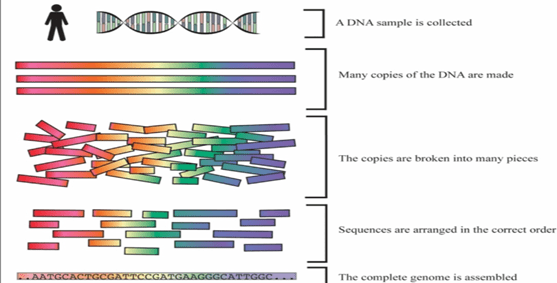
General Steps involved in DNA fingerprinting:

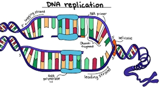
  1. First step is isolation of DNA. This isolated DNA is than treated with chemicals to break open the cell membrane to remove other materials, and extract pure form of DNA. Sample is generally taken from blood, cheek swabs.
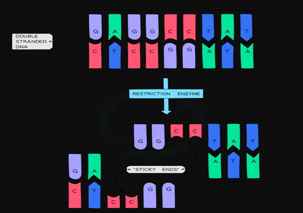
  2. The next step, is to digest the isolated DNA with restriction enzymes. Generally, repeated sequences(DNA portion with exact same sequence) are used so that they can be identified by same restriction enzymes. Common examples of DNA repeats used are VNTRs and STRs.
  3. As per the fragment size, digested fragments are than separated by using electrophoresis. (Separation based on size and charge by passing electric current).
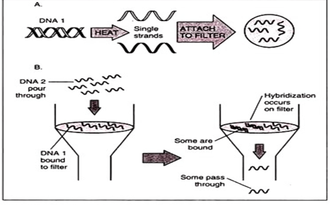
  4. Transfer (blotting) of separated fragment onto nitrocellulose of pvdf(polyvinyl difluoride) membranes. Gel is treated with weak acids, which break opens the DNA into individual nucleic acids which will easily get rub off onto paper.
  5. Labelled VNTR probes are used to hybridize the fragments.
  6. Autoradiography, is used to analyse the hybrid fragments.

Methods of DNA fingerprinting:

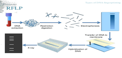
  • RFLP (Restriction fragment length polymorphism):
    • DNA isolation from sample material. Use large sample size for RFLP to obtain better results.
    • After sample isolation, subject the DNA for restriction digestion.
    • Separation of digested sample by Agarose gel electrophoresis.
    • Transfer of separated DNA to nitrocellulose membrane. The sample is than hybridised by labelled probe which is specific for VNTR region.(southern blotting)
    • After this, x-ray film is developed from the southern blot. On the x-ray film only hybridised regions(where the radioactive probe will bind) will be shown.
    • Analysis with other samples and studying band pattern and comparing different samples will give the result of DNA fingerprinting.

Advantages: Gives more accurate results compared to the PCR method. Generally because, the sample size used is large, sample DNA is fresh and also there is no chance of amplification contamination, which tends to happen in PCR

Limitations: Takes longer time to complete and proves to be a costly method for more frequent uses.

  • PCR Amplification of Short Tandem Repeats( STRs):
    • Particular variable region is amplified in thousands of copies.
    • Known repeat sequence of STR is amplified and separated with the help of electrophoresis.
    • Distance travelled by STR is noted.
    • Primers are specially designed for PCR amplification for the purpose of attaching highly conserved common non variable region of DNA which flanks the variable region of DNA.
    • STR sequence sizes are compared with various samples to give the result of DNA fingerprinting.

Advantages: Requires small amount of sample, shorter duration, faster results and less costly method.

Limitations: chancesofamplification contamination, not very accurate method.

Applications of DNA fingerprinting:

Generally, this technique is employed to identify individuals of same species by comparing their DNA.

  1. Forensic science:
  2. DNA is isolated from the crime scene and is generally useful in solving crimes. The isolated DNA is compared with the VNTR prototype.
  3. Common materials used for profiling of DNA are: blood, hair, semen, body tissue cells, etc.

       B.    Paternity and Maternity determination:

  • Dispute cases, inheritance cases and immigration cases are generally solved by parent-child VNTR analysis.
  • VNTR are generally acceded from his or her parents to the individual.

        C.    Personal identification:

  • DNA fingerprint pattern is unique in each individual.
  • Hence, this is used as a personal identification bench mark and as a genetic barcode of identification.

         D.   In diagnosis of inherited disorders:

  • Used in diagnosis of inherited disorders in new-born and prenatal.Some of this disorders include Huntington’s disease, haemophilia, cystic fibrosis, sickle cell anaemia.

          E.   Used to detect maternal cell contamination:

  • Maternal cell contamination is the common problem with prenatal diagnosis. Sometimes the amniotic fluid contains maternal tissues.
  • This type of contamination can increase the chance of false positive results and can hinder in carrier identifications.
  • So by using STR or VNTR markers and by performing gel electrophoresis, maternal cell contamination is identified during genetic pregnancy testing.

           F.   Breeding program:

  • Generally, breeders use phenotype to determine the genotype of plants or animals. It is difficult to determine whether plants are homozygous or heterozygous on the basis of appearance.
  • Thus, DNA fingerprinting method can be used for precise determination of the genotype.
  • This approach is used primarily in breeding hunting dogs and race horses.

DNA fingerprinting has many applications and is a useful technique in development of cures for inherited disorders, as the DNA prototype associated with disorder can be studied from the relatives. The accuracy for DNA fingerprinting relies more on RFLP method than in PCR methods.

GENOMICS AND PROTEOMICS

BY: RAHUL ANDHARIA (MSIWM001)

Genomics:

Complete set of genes or genetic material present in cell or an  organism is called a genome. Study of this genome is called as genomics. Genomics studies comprises of studying large number of genes through automated tools.

Genomics implies bioinformatics, sequencing methods, mapping methods, computational biology, recombinant DNA technology to study and analyse functions and structures of genomes.

History of Genomics:

Genome sequencing first started in the year 1970, when biochemist Frederick Sanger sequenced the genome for the first time. Virus and mitochondrion genomes were sequenced by Sanger in early 1970s.

 Types of genomic studies:

  1. Structural Genomics: It involves gene identification, construction of genetic and physical maps, annotating gene function and comparison of genome structures. It involves  determining structure of each and every protein encoded by genes.
  2. Functional Genomic studies:  understanding of how genes and intergenic regions of genome contribute to various biological processes. It also deals with the study of individual components of body and how it affects the phenotype. For example- functional genomics is useful in studying the dynamic expression of diseased genes.
  3. Comparative genomics: it is the comparison of genomes of different organisms. Basically it includes gene number, gene content and gene location comparison. The comparison between various genomes provides insight on conservation of different genomes and how the genome is being evolved in a particular organism.  This can also useful in genome evolutionary studies among various species.
  1. Mutation genomics: deals with studying the genome in terms of mutation that is it involves studying mutated genes and it’s effects and how gene mutations leads to diseases.

Methods in Genomics:

Genome mapping:

  • Mutations, relative locations in genes and traits on a chromosome can be identified by using genome mapping.
  • In this method, a particular gene is located or assigned at a particular position in the chromosome and than relative distance between the gene and the chromosome is mapped.
  • Linkage maps- It shows how genes and genetic markers are arranged in a chromosome.
  • Physical maps- It will show the physical distance between chromosomal landmarks and generally, physical maps represents a chromosome. The distance is measured usually in nucleotide bases.

Genome sequencing:

  • To sequence a genome, it is important to figure out the exact order of DNA nucleotides or bases. (A,T,G,C bases).
  • Sequencing the entire genome is a cumbersome task. The process involves breaking the DNA of genome into smaller pieces first,  than sequencing those each and every pieces and those pieces are than assembled as a single long consensus ( sequence with similar function and structure)
  • The DNA sequencing method in today’s modern era is very rapid and hence provides whole genome sequencing of various genomes of different species, which is instrumental in studying various diseases and evolutionary forms of life.

Genome sequence assembly:

  • Short sequence reads are generated from DNA clones.
  • These reads are around 500bases. Short sequence reads are joined together to form larger fragments after removing overlaps to assemble a whole genome sequence.
  • These new long sequences can be called as coting’s. They are around 5000-10,000 bases long.
  • Scaffold is formed from many number of overlapping contigs.( Oriented along a chromosome).
  • These scaffolds are than connected to make a final genome map.
  • Generally, computational tools are required to identify correct contigs and proper assembly of sequence reads.
  • Some common examples of assembly programs are Vecscreen, Phred, Phrap, TIGR assembler.

Genome annotation:

  • To analyse sequence for biological features before depositing it into the database, comments are provided for the sequences, which is called genome annotation.
  • Gene prediction and function assignment are the two types in annotating genomes which can be done using bioinformatic tools.

Human genome project: (HGP):

One of the most successful projects in scientific discovery, HGP was a major breakthrough, where scientists mapped the complete sets of genes, together called a genome. The HGP project was started in 1990 and completed in the year 2003. This was the first of its kind project that gave us the genetic blueprint for building a human being.

Applications of Genomics:

  1. Can be used in prenatal diagnosis.
  2. To analyse gene expression profiles of organisms.
  3. To identify diseased gene.
  4. Gene therapy –( defected gene is replaced by correct gene) used particularly to treat Genetic disorders.
  5. Genome editing ( to replace sections of DNA sequences, or to edit a particular sequence).

Proteomics:

It is the study of complete set of proteins expressed by the genome.(study of functions of proteomes) Set of complete proteins encoded by a particular genome can be called as a Proteome. This includes basically studying in depth about Protein-Protein Interactions, protein modifications and how a particular protein functions in different organisms.

History of proteomics:

Proteomics is still evolving as a field and is considered as a relatively new field of molecular biology. The history of proteomics dates back to 1995, when Mark Wilkins, for the first time coined the term proteome. There are around 400,000 proteins founded in human proteome than number of protein coding genes, which was one of the major findings of the Human genome project.

First protein studies were carried out in E.coli using 2 dimensional gel electrophoresis by O’Farrell in the year 1975.

Types of proteomic studies:

  1. Expression proteomics-  it involves studying gene expression between samples that differ by some variable. This can be useful in identifying particular proteins involved in disease.
  2. Structural proteomics- it involvesstudying structure and nature of protein complexes. It involves studying and analysing Protein-Protein Interaction networks.
  1. Functional proteomics: in this role of a particular protein or group of proteins is studied in different cellular pathways, signalling pathways and in PPI networks.

Categories for designing typical proteomics experiment: ( methods used for proteomics)

  • Isolation and protein separation- By using protein electrophoresis, complex mixtures of proteins can be resolved.
  • Acquiring protein structural information for identification and characterization of protein-

ES sequencing( Edman sequencing):

  • This sequencing was done to identify N-terminal sequences of protein.
    • It involves determining the sequence of N-terminal of a protein in order to predict it’s start site.
    • Proteins separated by SDS page can be more efficiently analysed by ED sequencing.
    • Major limitation with this technique is, we cannot modify the N-terminal of protein. For example, if a protein is blocked on the N-terminal, before the sequencing procedure, it will be very difficult to identify that protein.

Mixed peptide sequencing:

  • Cyanogen bromide is used to cleave protein to convert it to a peptide.
  • This is followed by ED sequencing.

Mass spectrometry:

  • Can identify gel separated proteins.
  • Sensitivity levels are very high in mass spectrometry, and that is the reason, this technique is preferred more over ED sequencing.
  • In the gel by using, trypsin, proteins are digested into peptides.
  • ESI(electrospray ionisation): in this approach of mass spectrometry, the peptides are fragmented and than ionised by using an electrospray in a tandem mass spectrophotometer. Sample flows from microcappillary tube to orifice, due to potential difference between the two, it results in generation of fine droplets. This has the ability to dissociate into carboxyl terminal, which gives fragments a and b.

Peptide mass mapping:

  • Eluted peptide mixture is used to obtain mass spectrum of the peptide eluted. This is followed by studying the peptide mass finger print resulting from the peptide spectrum.
  • In this method, as trypsin cleaves proteins at arginine and lysine amino acid, tripeptide mixtures are generally analysed in peptide mass mapping.

  Protein databases and utilization:

  • Submission of sequences and sequencing proteins or peptides leads to creation of protein assembly which are called as Protein data bases.
  • The database contains all the basic protein data.
  • The main aim of creating such database is to have accurate and quick results and to store information  at one place.

Applications of proteomics:

  1. Studying gene expression profiles of various proteins.
  2. To analyse Protein-Protein Interactions- how one protein interacts with other proteins and leads to changes or alterations, it’s expression and level of expression.
  3. To study post-translational modifications like glycosylation and phosphorylation.
  4. Molecular medicine- clinical proteomics information is used to design drugs. Proteins are considered as medical relevance for this studies and are used as targets to study their role in disease progression, and to develop better therapeutics.

CULTURE MEDIA FOR PLANT TISSUE CULTURE

BY: RAHUL ANDHARIA (MSIWM001)

Plant Tissue culture:

It is an in-vitro aseptic culture method in which plant tissues or cells are grown in an artificial medium under suitable environmental conditions generally to produce clones of plants. The medium used to grow this tissues or cells is known as culture medium. There are various culture medium used based on different requirements.

Principle of Plant Tissue culture and culture medium:

Many plants have the ability to regenerate into a new whole plant which is called as Totipotency. Protoplasts(plant cells without cell wall), pieces of leaves, single cell often is used for generation of new plant using appropriate plant culture medium. Specific environment is created to provide maximum growth and multiplication of the plant in the culture medium. The conditions include providing proper nutrient medium, maintaining temperature and pH and proper liquid and gaseous environment.

Culture medium facilities in-vitro growth and morphogenesis( differentiation of unfolding of undifferentiated cells) in plants. The success of tissue culture lies in the selection of plant culture medium. If an appropriate medium is selected, better results can be obtained. Generally, plants can synthesise their own food material, but the in-vitro plants are primarily heterotrophic, that is they cannot synthesise their own food, so it becomes essential that plant culture medium should consist of the same nutrients as required by the whole plant.

Composition of Culture medium:

Basically there are two factors inconsideration for the composition of plant culture medium:

  1. Species of the plant- composition of the medium very much depends on the species of the plant used in tissue culture. Each plant specie has different requirements and also requires different environmental conditions for their growth.
  2. Type of culture material- The composition also depends on the type of material used such as protoplasts, single cells or pieces of leaves. Each material requires different composition.

Thus, composition of culture medium is dependant on the specific requirements of the culture system and it’s formulation. The type of medium used can be solid or liquid medium. The choice of medium completely depends on the culture response and it’s growth.

pH of Medium:

Most optimum pH for tissue culture is in the range of pH 5-6. pH can be adjusted during sterilization, but usually falls after autoclaving. At a higher pH of 7 and lower pH of 4.5 plant cells will no longer grow in culture medium.

General method for medium preparation:

  • Prepare stock solutions using high purity chemicals and demineralised water.
  • Stock solutions can be stored in glass or plastic containers in freezer and can be used whenever needed.
  • As most of the growth regulators are not water soluble dissolve them first in NAOH or alcohol.

Rules for Selection of appropriate culture medium:

Each specie has different culture requirements and therefore selecting an appropriate medium is an integral part of plant tissue culture.  De Fosard Et al has described an experiment to select an appropriate culture medium. This is very beneficial especially if the culture system used is not tested.

The steps are as follows:

  1. Medium is divided into four different categories (a)  minerals, (b) auxins, (c) cytokines and (d) organic nutrients like sucrose, amino acids.
  2. Choose three different concentrations for each of the components that is high, medium and low.
  3. Try various combinations with the chosen four components. At the end it will lead you to 81 different treatments.
  4. Denote the four letter code the best among the 81 treatments. For example a medium containing medium salts, low auxin, low cytokinin and high organic nutrients can be represented as MLLH.
  5. At this stage, test the concentrations of auxins and cytokinins to  assess the suitable concentration of the growth regulators.

Types of Culture medium:

There are many number of culture media available. Some of them are listed below:

  • MS ( Murashige and Skoog) medium:

As the name suggests this medium was developed by the two scientists, Murashige and Skoog in the year 1962, while working on plant growth regulators. This is the most common culture medium used in labs. Basic formulation includes inorganic salts, nutrients and amino acids.

Purpose: This medium is used to induce callus culture, organogenesis, and micropropogation.

  • LS (linsmaier and Skoog) medium:

This medium was developed in the year 1965. Initially, the medium was used to optimise organic nutrient supplements in Tobacco culture. This medium contains Thiamine hypochlorite and inositol. Inositol is primarily involved in primary and secondary metabolism of plants and also is an cofactor in glycolysis and TCA cycle.

Purpose: used in induction of callus culture(growing mass of parenchyma cells is, callus) organogenesis, micropropogation and cell suspension.

  • Gamborg (BY) medium:

The medium was particularly used for cell suspension and callus generation of glycine max belonging to plant family, fabaceae by Gamborg in the year 1968. Formulation of medium generally includes inorganic salts, carbohydrates and vitamins. Generally, this medium has high concentration of nitrate and potassium and lower concentrations of ammonia. Soybean root callus formation is being induced by potassium nitrate, while ammonium sulphate helps in plant cell growth.

Purpose: used mainly in protoplasts culturing.

  • NN ( Nitsch and Nitsch )medium:

This medium was primarily first used by Nitsch, to develop anther culture in Nicotiana, which belongs to family solanaceae. This medium is rich in thiamine, folic acid and biotin which supports anther growth.

Purpose: Particularly used in in-vitro anther culture.

  • White’s medium:

The medium was developed for root culture of Tomato by P.R white in the year 1963. This was the first medium to be developed for root culturing. The medium has lower concentrations of salts and has higher concentration of Mgso4.

Purpose: widely used for shoot and callus culture. It is more appropriate for the culture of Musa and Daucus species.

Constituents of culture medium:

Elements play a vital role in plant growth and regulation. Elements differ in their physiological functions.

Inorganic nutrients:

It consists of micro and macro nutrients. Concentration of macronutrients is greater than  0.5mmol/1-,  while for micronutrients it’s less than 0.5mmol/1-.

  • Macronutrient elements:

It consists of six different types of elements mainly nitrogen, phosphorus, potassium, calcium and sulphur. Ideal concentration for calcium, phosphorus, sulphur and magnesium is 1-3mm mol, while for potassium and nitrogen it is 25mm mol.

  • Micronutrients:

They are required in smaller amounts by tissues and cells. Commonly  iron, manganese, copper, zinc, molybdenum and boron are the micronutrients. Iron requirement in the medium is very vital. Iron and copper in chelated form are used in the medium.

  • Carbon and energy sources: Tissues and plant cells in the culture are usually heterotrophic, so they require external source of energy and carbon for their growth. Sucrose is the most commonly used form of energy source. During autoclaving process, sucrose will hydrolyse into glucose and fructose. Plants uses more amount of glucose than fructose and glucose is considered as efficient as sucrose.
  • Organic supplements: They include vitamins, amino acids, organic extracts, activated charcoal, organic acids and antibiotics.
  • Vitamins plant cells produce vitamins but in less amounts. So it is necessary to add vitamins to the medium. Vitamins like thiamine, riboflavin, niacin, biotin, folic acid, pantothenic acid and ascorbic acid are added.
  • Amino acids: organic nitrogen in form of amino acids like L-arginine, L-glutamine, L-cysteine and L-arginine are more easily taken up by plants than inorganic nitrogen.
  • Organic extracts: Yeast Caseinhydrolysate, orange juice, tomato juice, coconut milk are used.
  • Activated charcoal: activated charcoal can stimulate growth of certain plant cells like carrot, tomatoes and orchids. Also it can remove certain toxic components from the medium which can otherwise hinder in cell growth.
  • Organic acids: succinate, citrate, fumarate and malate helps in plant growth.
  • Antibiotics: antibiotics bare generally added to prevent growth of microbes. Low amounts of kanamycin and streptomycin are added.
  • Growth Regulators: Phytohormones promote plant growth, differentiation and development.
  • Auxins: it induces cell elongation, cell division and formation of callus in cell cultures. At lower concentrations, auxins can promote root formation and high concentration it helps in callus formation.
  • Cytokinins: cytokinins are derivatives of adenine. They are involved in shoot differentiation, cell elongation and in somatic embryo formation. It also helps in promoting RNA synthesis and thereby stimulating enzyme and protein activity.
  • Gibberellins: Ga3 is the most commonly used gibberellin among 20 different types of gibberellins known. Ga3 helps in growth of cultured cells, enhances growth of the callus and makes dwarf plantlets to elongate. They are generally known to promote or inhibit tissue cultures depending on the species used.
  • Abscisic acid:   plays major role in induction of embryogenesis in the culture. Also helps in callus formation.
  • Solidifying agents: solidifying agents are used in preparation of solid medium or semi solid medium. Most commonly used agents are:
  • Agar: It is a polysaccharide extracted from seaweeds. Main advantages of using agar is that it cannot be digested by plant enzymes, remains stable at cultural temperature and doesn’t react with the media constituents. 0.5 – 1% concentration of agar can form gels.
  • Gelatine: it is used at higher concentration(10%) but has limited success, as it melts at lower temperature so has limited use.

Considering all the above factors, culture medium is the most important part of tissue culture. Without proper culture medium, tissue culture cannot be performed.

DNA: REPLICATION, TRANSCRIPTION & TRANSLATION

DNA: Replication, Transcription and Translation

Content:

About:

  • The process of producing identical replicas of DNA from original DNA molecule in known as DNA replication.
  • It is the most important part of inheritance and occurs in all living organisms.
  • DNA consist of two strands which are complementary to each other during the process of DNA replication these tow strand get separated and serve as template for the synthesis of new complementary strand this is known as semiconservative replication.
  • After cellular proofreading and error checking mechanism new DNA molecule is synthesized with one strand from parent DNA molecule and another is fresh.
  • Each cell of a human being contains 3 X 10⁹ base pairs of DNA distributed over 23 pairs of chromosomes.
  • In human genome 95% region do not code for any protein known as introns.
  • According to central dogma DNA makes RNA and RNA makes protein.
  • The process of producing RNA from DNA is called transcription and synthesis of protein from RNA is called translation

Steps Involved:

  • DNA replication
  • Transcription
  • Translation

DNA Replication:

  • When cell divides, the double stranded DNA splits into single strand and makes new copy of the genome which gets transferred into each cell.
  • Base pairing is according to the Watson-Crick paring of the DNA strand.
  • It is a complex process which includes array of enzymes.
  • Synthesis of DNA proceeds in the 5’-3’ direction and for attachment of DNA polymerase first DNA strand has to be unwind which is regulate by the helicase enzyme.
  • During the synthesis of the new strands by the DNA polymerase one strand is synthesized continuously and called leading strand on the other hand the opposite strand known as lagging strand synthesised in to the small fragments called okazaki fragments.
  • In bacteria there are three distinct DNA polymerase: pol I, pol II and Pol III. Pol III largely involve in chain elongation. DNA polymerase itself cannot initiate DNA synthesis it require a short primer with free 3’ hydroxyl group.
  • Once the primer attaches to the DNA strand Pol III then take over and synthesis of new strand begins.

Transcription:

  • The process by which DNA is copied to mRNA is called transcription.
  • The mRNA contains information for the protein synthesis.
  • Transcription occurs in two steps. In first step pre mRNA is synthesized with the help of RNA polymerase, the resultant RNA strand is reverse complement of the original DNA sequence. Then after pre messenger RNA is edited and forms the mRNA molecule by the process of RNA splicing.

RNA Splicing:

  • The pre-messenger synthesized contains introns and exons in it, introns are the sequence which does not code for any protein hence it is not required in protein synthesis.
  • Thus the pre- messenger RNA is chopped up and introns are removed from it which synthesise messenger RNA contain only exons.

Translation:

  • The mRNA synthesised in the transcription process is now transported outside the nucleus into cytoplasm to the ribosome.
  • Messenger RNA does not directly involve in the protein synthesis transfer RNA is required the process of protein synthesis is called translation.
  • The mRNA contains three base stretch called codon and each codon contains information for a specific amino-acid. While the ribosome passes through the mRNA each transfer RNA molecule anticodon interacts with codon of mRNA.
  • The transfer RNA contains amino acid at 3’ end which is added to the growing protein chain and the t-RNA expelled from the ribosome.

Transfer RNA:

  • Transfer RNA is tertiary structure which is represented in two dimensions as cloverleaf shape.
  • Each amino acid has its own unique t-RNA like t-RNA for phenylalanine is different from that of histidine.
  • Attachment of amino acid is at 3’- OH group of t-RNA.

Nucleic Acid Hybridization

BY: RAHUL ANDHARIA (MSIWM001)

Nucleic Acid Hybridization: It is a technique which involves interaction of single stranded Nucleic acids(DNA/RNA) to form complexes called Hybrids, which contain the similar complementary sequences as that of nucleic acids. This technology helps us to understand the sequence identity among nucleic acids and to determine and detect specific sequences.

History:

 In the year 1975, Grunstein and Hogness developed this method.

Principle:

In simpler terms it is a technique that simply detects specific DNA/RNA sequences using specific Probes. By using this technique we can determine sequence of  hundreds of clones from a particular colony which exhibits it. Bacterial Colonies are transferred from agar plates to nitrocellulose membrane where they are lysed by using alkaline solution.

After cell lysis, due to the negative charge of nitrocellulose membrane, the DNA and proteins gets adhere to the membrane. To remove the bounded protein, the membrane is dipped in Proteinase k solution. Upon exposure to UV rays, the DNA gets fixed to the nitrocellulose membrane. After baking, the membrane gets exposed to labelled RNA for hybridization. The hybrids can be monitored by Autoradiography. Colony giving positive result must be picked from the master plate.

Procedure overview of Nucleic acid hybridization:

  1. Probe, (DNA/RNA fragment which can be fluorescently radiolabelled ) is required which will anneal to the target nucleic acid.
  2. The next step is the attachment of target to a solid matrix, like for example a membrane.
  3. Denaturation (breaking of hydrogen bonds) of the target and the probe used.
  4. The denatured probe is than added to a target in a solution (Proteinase k sol)
  5. If sequence homology exists between the probe and the target, the probe will than hybridize or will anneal the target.
  6. The probe which is hybridised than can be visualised by methods like Autoradiography, Chemiluminescence or Colorimetry.

Hybridization Probes:

Probes are DNA or RNA fragments that can be radiolabelled. Probes help in identifying complementary segments in nucleic acid of Micro-organisms. The probe gets hybridised to single strands because of its complementarity with the target.

To make the probe hybridise with target sequence, it is labelled with molecular marker like radioactive 32-P ( radioactive isotope of phosphorus which incorporates into phosphodiester bonds of DNA).

Types of Probes:

  1. DNA Probe: Single stranded DNA is used to detect the complementary sequence amongother ss molecules. Therefore, it is a short sequence radiolabelled with radioactive isotope to detect complementary nucleotide sequence.
  2. RNA Probe: Single stranded RNA is used to detect the complimentary sequence among other ss molecules. RNA probes are also called as Riboprobes. RNA-RNA hybrids are known to be more stable than RNA-DNA hybrids generally because RNA hybrids shows more significant kinetic hydration.
  3. Oligonucleotide probes: refers to a short sequence of nucleotides which are synthesized to mimic the regions where mutations have occurred. They are synthesized in laboratory. Initially, a mononucleotide is attached to a solid support, and than one by one other mononucleotide are added to the 3’ end region. They are labelled with molecular markers at the 5’end.

Labelling of Hybridization Probes( DNA/RNA)

Involves 2 different types of methods:

  1. In vivo labelling: labelling of nucleotides in cultured cells.
  2. In vitro labelling: use of an enzyme to label the probe.

Strand synthesis labelling for DNA probes:

Most common method used to label DNA. In this method, enzyme DNA polymerase is used to attach labelled nucleotides to the DNA copies in the starting DNA. Any one of the nucleotide is labelled. There are various methods for DNA labelling:

  1. Nick Translation labelling: method in which DNA fragment is treated with DNase enzyme to induce single stranded nicks. Labelled nucleotides from nicked sites are incorporated by DNA polymerase I . Nick sites are replaced by DNA polymerase I that elongates 3’hydroxyl activity, thereby removing nucleotides by 5’-3’ exonuclease activity and replacing it with dntps.

  • PCR Method:  PCR method of amplification of DNA allows the resulting product to label either with modified nucleotide or oligonucleotide primers. With this, we can thus incorporate labelled nucleotides in the PCR reaction mixture which will than result in production of  labelled PCR products throughout its length. This method has higher yield even with a little template.

Strand synthesis labelling of RNA probes:

Most common method used to label RNA. In this method, enzyme RNA polymerase is used to attach labelled nucleotides to the RNA copies in the starting RNA. Any one of the nucleotide is labelled. There are various methods for RNA labelling:

  1. In vitro transcription from cloned DNA inserts: The RNA probe gets transcribed from a linear DNA template with the help of bacteriophage DNA dependant-RNA polymerase enzymes from bacteria like salmonella (sp6) and E.coli(T3 and T7). One major advantage is with this method the labelled probes are completely free of the vector sequences.
  • 3’end labelling RNA:  Short DNA template has to be designed to anneal 3’end of RNA, with a 5’overhang of 3’ TA-5′. The klenow portion( large fragment of protein) of DNA polymerase I can than extend 3’end of RNA by incorporating single 32P radio labelled dAtp.

Uses of Nucleic Acid hybridization:

  1. Testing frequency of relatedness between 2 DNA molecules:

Sample of DNA 1 is heated to melt it to single stranded DNA. The ssDNA is than  attached to a appropriate filter. Now chemical is being added to filter to block the sites, that can bind to DNA. After melting, DNA 2 sample is poured. Now some molecules from sample 1 will base pair with sample 2. The more closer the molecules are related, more hybrids it will generate. This method is highly useful, for example DNA of human gene, haemoglobin generated is melted and filtered, than it can be used to test the DNA from same gene of some different species.

  • Isolating Genes for Cloning:

Suppose we have say human haemoglobin gene and want to isolate corresponding monkeys gene, than human DNA is added to filter and the monkeys DNA is cut into segments with the help of restriction enzymes. The monkey DNA is than heated to make it to single strand and poured in filter. The DNA fragment carrying monkeys haemoglobin gene will now bind with human haemoglobin gene and gets stuck in the filter. Other genes will not hybridize. This is a very selective approach and provides hybridization of new genes from related or similar kind of genes.

GENETIC ENGINEERING

BY: RAHUL ANDHARIA (MSIWM001)

Genetic Engineering: It refers to altering the genetic material of an organism by insertion or removal of individual genes from another organism. In other terms it simply means manipulation of genetic make-up of an individual by artificial means. The organism that receives the genetic material is termed as transgenic organism.

History:

Father of Genetic engineering is Paul Berg. The process of Genetic engineering was first showcased by Paul Berg when he introduced the viral gene in bacteria with the help of lambda phage. Transgenesis, process of transferring of genes from one organism to other was first pioneered by Herbert Boyer and Stanley Cohen, in the year 1973.

Principle of Genetic Engineering:

There are several steps associated with Genetic Engineering:

  1. Isolation of gene of interest to be cloned from desired organism.
  2. Transfer of the isolated gene to create recombinant DNA molecule. This can be done by cutting the DNA molecules at specific sites by using Restriction Enzymes.
  3. Restriction enzymes cut DNA at specific locations. There are basically 4 types of restriction enzymes designated as I, II, III, and IV. All this four enzymes primarily differs in their structure, site of cleavage and some cofactors (substance which is required for enzyme activity).
  4. By the action of RE, DNA is left with sticky ends (short portion of unpaired bases). A bacterial plasmid is also cut with the same RE, so that the plasmid also has the similar sticky ends and can base pair. (Plasmids are extra-chromosomal DNA found inside the bacterial cell).
  5. The plasmid and the isolated gene are joined together by an enzyme called DNA ligase.
  6. The two pieces of DNA with same sticky ends (as cut by same RE) are being linked together by DNA ligase and forms a single, unbroken molecule.
  7. Than the genetically engineered plasmid is inserted into bacterial cell (Transformation). The gene insertion usually takes place by means of Vectors. (Molecules that carry the gene of interest.) choice of vectors depend on the type of gene to be inserted.
  8. When the bacteria reproduce, the plasmid will get copied and this recombinant plasmid spreads as bacteria multiply and expresses the gene and makes a human protein.

This is the basic procedure required to obtain a desirable gene and this whole process together refers to Genetic Engineering.

Various types of Genetic engineering techniques:

  1. Recombinant DNA technology:

Technique in which artificial DNA fragment is created by ligation of two different DNAs using various physical methods.

In this technique basically the gene of interest is inserted into a plasmid vector and can be used for gene transfer experiments. Two methods are commonly in use which includes:

  1. Plasmid method- In this method small pieces of circular DNA called plasmids are used for altering microorganisms like bacteria.
  2. Vector method- vectors are small carrier molecules that carry viruses. Generally, viruses have capsule, in which the DNA is present. When the viruses attach to the host cell they insert its DNA or RNA. The DNA in the host genome replicates itself by using host machinery and the gene inserted will become part of the host machinery.
  • Gene delivering:

This technique is employed to insert gene of interest directly into host genome. Some of the most popular gene delivering techniques which include:

  1. Electroporation- introduction of DNA into cells by using electric current. It is a rapid method to create more number of recombinants within a shorter duration of time.

The mixture containing cells and DNA are placed in a glass chamber. The glass chamber contains two electrodes. A single electric pulse of about 4000-8000 volts/cm is generated between the two electrodes for about 4-5 milliseconds. The electric current forms transient pores in the cell membrane through which the DNA seems to enter into the cells.

  • Liposome mediated gene transfer: liposome is a small vesicle made up of phospholipids used to deliver foreign DNA into cells. Lipids like phosphetidyl choline, Cholesterol are useful in making liposomes. Outer lipid bi-layer generally fuses with the cell membrane and releases the contents.
  • Gene editing:

The technique is used to edit the genome in which an undesired or diseased gene can be edited or replaced by a new gene in the host genome.

Some of the gene editing systems used are: CRISPR-CAS9, TALEN( Transcriptional activator like effector nuclease) and  ZFN( zinc finger nuclease).

Among this three CRISPR is the most widely used method currently. Bacteria generally possess this method in their genome to combat viruses. CRISPR normally  stands for clustered regulatory interspaced short Palindromic repeats. The nuclease, CAS9 cuts the DNA strands of foreign DNA and pathogens and thus destroys them.

Process overview of Genetic engineering:

  • Isolation of desired gene
  • Selection and plasmid construction
  • Gene transformation(uptake of DNA by cells)
  • Insertion of DNA into a suitable host
  • Insert confirmation- done by using various selectable markers.

Applications and future aspects of genetic engineering:

Genetic engineering has great prospects. It is highly practiced in Industrial and agricultural field. It has wide range of applications in the field of medicine, genetic research, and in therapeutic drug production.

  1. Plant Genetics: It is highly employed in plants to improve crops. Recombinant DNA technology in plants help to have better yield, to control pathogens, delayed fruit ripening, stress tolerant plants.

Classic example is Bt cotton, most famous Genetically modified crop.

  • Genetically engineered Insulin: human insulin gene is inserted into the gap in plasmid. The plasmid becomes genetically modified. The plasmid than upon insertion into new bacteria or yeast cell will produce insulin.

GENETIC CODE

GENETIC CODE:

CONTENTS:

  • Introduction
  • Protein synthesis (translation)
  • Variations in genetic code
  • Wobble
  • Universal code

INTRODUCTION:

BIOSYNTHESIS OF PROTEINS (TRANSLATION):

The final step in the expression of protein synthesizing genes is translation. The process of Protein synthesis is called translation because it is a decoding process of highly coiled substance. The information encoded in the language of nucleic acids must be written in the languages of amino acid forming proteins. During translation, the sequence of nucleotide is “read” in the appropriate sets of three nucleotide, each set being called a codon. Each codon codes for single amino acid.

  • The sequence of codons is “read” in only one way-the reading frame- to give rise to the amino acid sequence of a polypeptide. Deciphering the genetic code was one of the great achievements of the twentieth century.  

Close inspection of the code reveals several features that are related not only to the way cells use DNA to store information but also to why it is valuable for storing data.

VARIATIONS:

  • One feature is that the code words (codons) are three letters (bases) long; thus one small “word” conveys a significant amount of information. Each codon is re-organised by an anti-codon present on a tRNA molecule.
  • Another feature is that the code has “punctuation”. One codon, that is AUG, is always appears as the first codon in the protein synthesizing portion of mRNA molecules in very complicated and important process.
  • It is called the start codon because it serves as the start site for translation (a process of protein synthesis) by coding for the internal tRNA. Three other codons (UGA,UAG and UAA) are involved in termination of translation and are called stop or nonsense codons. These codons never encode for an amino acid and therefore they don’t have a tRNA bearing their own anti-codon.
  • Thus only 61 out of  64 codons in the code, are known as the sense codons which directs amino acid to incorporate into protein. Finally, the genetic code exhibits code degeneracy; that is, there are up to six different codons for a given amino acid.
  • Despite the existence of 61 codons, there are fewer than 61 different tRNAs. It follows that not all codons have a corresponding tRNA.
  • Cells can be successfully translated into mRNA using fewer tRNAs because there is not so strong pairing between the 5’ base in the anti-codon of the cell and the 3’ base of the codon which is almost tolerated.
  • Thus as long as the appearing first and second bases in the codon correctly base pair with an anti-codon, the tRNA which is bearing the correct amino acid will bind to all mRNA during translation process. This is evident on inspection of the code.

WOBBLE:

The codons for particular amino acids are most often different at the third position. This somewhat loose base pairing which is known as wobble, and it enhance cells the need to synthesize so many tRNAs. Wobble also decreases the effects of particular mutations.

UNIVERSAL CODE:

  • The description of the genetic code just provided is of the universal genetic code. However, there are exceptions to the code.
  • The first exceptions discovered were stop codons that encoded one of the 20 amino acids. For instance, some protists have a single stop codon (UGA); the other two stop codons are recognized by tRNAs bearing glutamine (Gln).
  • More dramatic deviations from the code have also been discovered. Proteins from members of all three domains of life have been discovered to contain the amino acid selenocysteine, the twenty-first amino acid.
  • Pyrrolysine, twenty-second amino acid, can be found in the proteins of several methanogens and at least one bacterium.
  • Genomic analysis indicates that pyrrolysine might also exist in many other bacteria and some eukaryotes. Selenocysteine is inserted at certain UGA codons, whereas pyrrolysine is inserted at UGA codons.

POLYMERASE CHAIN REACTION

polymerase chain reaction

Content:

  • About
  • Principle
  • Component
  • Steps in PCR
  • Types of PCR
  • Applications

About:

  • Polymerase chain reaction is a technique used to amplify the target DNA sequence from the source of DNA, it’s a rapid and a versatile in vitro methods
  • This method is introduced by Kary Mullis in 1985.
  • This method is able to amplify the specific sequence of the DNA from the heterogeneous collection of DNA molecules. For this selective amplification some prior information of the target DNA sequence is required.
  • Two primer synthesises by this sequence information get attached to the target DNA after denaturation (at the specific site). Primer initiates the DNA synthesis in the presence of heat stable DNA polymerase and DNA precursors (dnTPs).

Principle:

  • Technique based on enzymatic replication of DNA
  • Short segment of DNA is amplified by primers which are mediated by enzymes.
  • DNA polymerase synthesises complementary DNA from the template DNA. DNA polymerase adds nucleotide at 3’-OH group only so to attach DNA polymerase to the strand at specific site primers in required.

Components:

  1. DNA template: segment of the interested DNA from the sample
  2. DNA polymerase: a heat stable DNA polymerase which can withstand high temperature e.g. Taq polymerase.
  3. Oiligonucleotide primers: short stretch of DNA complementary to the 3’ ends of sense and antisense strands.
  4. Deoxyribonucleotide triphosphate: provide energy for polymerization and building blocks for the synthesis of DNA
  5. Buffer system: provide optimum conditions for DNA denaturation and renaturation, magnesium and potassium are the key component. Important for polymerase activity and stability.

Steps of PCR:

It is a cyclic process because newly synthesized DNA strand serve as the template for further DNA synthesis. It consist three steps:

Denaturation:

It is the initial step in which the reaction mixture is heated to the temperature about 90-980 C that ensures the separation of the DNA strands known as DNA denaturation. Duration of denaturation step usually is 2 min at 94oC.

Annealing:

In the steps the mixture is cooled to the temperature that permits the attachment of the primer to the complementary strand of DNA.

Primer attached at the 3’-ends of the two strands.

Duration for annealing is 1 min at 40-600 C.

DNA synthesis:

The temperature is so adjusted that DNA polymerase synthesizes the complementary strand using the primers for attachment at 3’OH end

Primers extended towards each other so that the DNA segment lying in between is copied.

Duration of this step is usually 2 min at 720 C.

Types of PCR:

  1. Nested PCR
  2. Quantitative Real-Time PCR
  3. RT-PCR
  4. Inverse PCR
  5. Anchored PCR
  6. RACE
  7. Touchdown PCR
  8. RAPD
  9. AFLP

Applications:

Forensic science

  • Genetic fingerprinting tool
  • Identification of criminal’s DNA collected from the crime site among the various people.
  • Tests for paternity

Research

  • Comparison of two organism’s genome for study
  • Gene mapping
  • Phylogenetic analysis of DNA from sources like fossils
  • Analysis for the expression of genes.

Medicine

  • Detection of the disease causing genes
  • Testing of mutation
  • Monitoring gene in gene therapy.

SEX LINKAGE

Sex Linkage

  • Content

Introduction

Characteristics

Inheritance of white eye in drosophila

Nondisjunction of X chromosome

Attached X chromosome in drosophila

Sex linkage in human and other organisms

Partial sex linkage

  • Introduction:
  • In Mendel’s crosses F1 and F2 from crosses show same results unaffected by whether it is a male or female both shows identical ratios.
  • But this rule is may not be applicable sometimes due to some exception produced by the sex linkage phenomenon.
  • Sex linkage is the phenomenon of gene being located on X or Y chromosome.
  • Inheritance of trait is determined by this gene on sex chromosomes.
  • First known case of sex linkage is haemophilia in human beings. In haemophilia, affected person bleed profusely even from minor cuts because their blood doesn’t clot on exposure to air.
  • There are many more exa mple of sex linked inheritance but it is clearly explained by Morgan in 1910( white  eye gene in drosophila).
  • Characteristics
  • Heterogametic sex has more no of individual showing recessive sex linked character than the homogametic sex.
  • Genes that control sex linked traits are not getting transferred from male parents to their male progeny directly.
  • Sex linked gene are transmitted from male parent to its entire daughter (daughter receives its half X chromosome from father) then daughter transmit this gene to half of its male progeny. As a result the gene transferred from male to female progeny and then to the half of male progeny. This transfer of male to female and back to the male is known as criss-cross inheritance.
  • Y chromosomes not carry sex linked genes. Heterogametic sex is present in hemizygous condition which lead to the expression of the recessive alleles of sex linked genes and in homogametic sex they have to be present in homozygous condition.
  • Inheritance of white eye in drosophila

Morgan in pedigree culture of normal dull red eye drosophila observed a single white eye male. He mate white eye male with red eye female and found F1 flies had all red eyes and in F2 3 red eyes and one white eye showing that white eye is due to a recessive gene.

  • But when he classified F2 flies on the basis of sex he found that all the female had red eyes and half of the male is red eyed and remaining half is white eyed. It seems as  if the eye colour related to the sex of the flies.
  • After that Morgan mated white eyed female with red eyed male he observed that half of them were red eyed and other half were white eyed, in F2 generation the ratio of red eyed and white eyed is 1:1, which same as the F1.
  • Later Moran reasoned that X chromosome carry white eye gene of drosophila,  and Y chromosome doesn’t carry any allele of this gene.

Nondisjunction of X chromosome

  • In 1916 Bridges, studying the inheritance of vermilion colour gene in drosophila. Observe that vermilion eye colour gene v (sex linked recessive gene) show the same pattern of inheritance as w
  • Cross between the vermilion females and red males, among the progeny of F1 some females were vermilion eyed and some males were red eyed. However, majority of females were red eyed and males were vermilion eyed.
  • Bridges explained these data and postulated that the X chromosome failed to separate during oogenesis of some oocytes of vermilion eyed females and both moved to the same pole and the opposite pole did not receive X chromosome such irregular distribution is known as nondisjunction.
  • Attached X chromosome in drosophila
  • Morgan in 1922, obtained all females with yellow body and all males with grey body when he mated yellow body female with grey body male drosophila
  • Yellow is recessive to the normal grey body located on X chromosome and determined by y.
  • Morgan reasoned that X chromosome nondisjunction is regularly takes place in oocytes and produced the following XvXand O unusual eggs
  • Such highly regular disjunction only takes place when the two X chromosomes shared the same centromere and behave as a single chromosome this fused form is known as attached X chromosome.
  • Sex linkage in human and other organisms
  • In addition to drosophila sex linked inheritance is also known in mice, cat , insects, cattle , in man
  • Drosophila had more than 150 sex linked genes  and over 200 genes in humans most of them cause genetic diseases. Example haemophilia ( failure in blood clotting ability), colour blindness ( inability to see one or more colour), optic atrophy (optic nerve degeneration), myopia(short-sightedness), etc.
  • For example like colour blindness it is a sex linked disorder occurs in 5-10% males and only 1% to the females. Human has there protein for red green and blue colour present on cone cells genes encoding for red and green light are located on x chromosome  and gene for blue light is placed on autosome.

Partial sex linkage

X and Y chromosome in human aur morphologically different. But during meiosis they pair in male cell

The paring  in two telomeric region is called as pseudoautosomal regions

  1. PAR1 major region of 2.6 Mb long, located at the tip of short arm of X and Y chromosomes, it is 70 times more than the normal recombination frequency. Example SHOX, WE7, Tramp
  2. PAR2 minor region of 320 kb located at the tip of the long arms of X and Y chromosomes, crossing over is not so frequent. Example IL9R and SYBL1.

X chromosomes contain PAR1 and PAR2 but this genes don’t show inheritance pattern of sex linkage because Y Chromosome but they resemble of autosomal genes.it  is known as partial sex linkage  gene X chromosome show  autosomal pattern

DNA REPLICATION

DNA replication

Content

  • Introduction
  • Types of replication
  • Model of DNA replication
  • Enzymes involved in DNA replication

Introduction

  • It is the process of producing two identical copy of DNA from one original DNA molecule.
  • DNA replication is one of the most essential or important properties exhibited by DNA.
  • Evolution of all morphologically complex form of life is based on the replication.

Types of Replication

  • There are possibly three modes of replication:
  • Dispersive
  • Conservative
  • semi-conservative

Semi Conservative

  • In this mode of replication, two  old parental strands serve as template for synthesis of new daughters strands and each new DNA contains one strand from the parent and one from newly formed progeny.
  • The evidence of semi conservative replication of DNA was first presented by Meselson and Stahl in 1958.

Dispersive

  • The old DNA molecule would break into several pieces, each fragment would replicate and the old and new segment would be combined to yield two progeny DNA molecules; each progeny molecule contains  both old and new strands along its length.

Conservative

  • According to the conservative replication, the old parental strands remains together and newly formed daughter strands are also together.

Model of DNA Replication

  • The brief discussion of DNA replication model is as follows:
  • DNA applications initiate at certain unique and fixed point called origin.
  • There is unwinding of complementary strands of DNA duplex with the help of two enzymes DNA gyrase and DNA helicase, this process is called Melting.
  • Single strand binding protein are attached to two single stranded region so that they not join to form duplex.
  • Due to melting in the origin region it produces two Forks(Y), in the DNA duplex and one fork is located at the end of the melted region. Generally, both the fox are involved in replication and become Replication Fork.
  • After formation of replication fork ,Primase enzymes initiate transcription of the strand in the 3’-5’ direction. This results in the generation of 10-60 long primer RNA.
  • The free 3’-OH of the  RNA primer provide initiation point for DNA polymerase for the sequential addition of deoxyribonucleotide.
  • The replication of the second strand (5’-3’) DNA is discontinuous, Hence 3’-5’strand of DNA is termed as the Leading strand and 5’-3’ is termed as Lagging strand.
  • Lagging strand generates small polynucleotides fragment called Okazaki fragments, during replication. This fragment is about 1000-2000 nucleotides long in E.coli.

Enzymes Involved in the DNA Replication.

  • There are some important enzymes which are involved in the replication of DNA :
  • DNA polymerase
  • Primase
  • Polynucleotide ligase
  • Endonuclease
  • Helicase
  • Single stand binding protein (SSB).

DNA polymerase

  • This enzyme synthesizes new strand on a template DNA strand.
  • It is also known as DNA replicase.
  •  Its activity was first explained by Kornberg in 1956.
  • In prokaryotes, there are five types of DNA polymerase:
DNA polymeraseGeneFunctions
1polAMajor repair enzyme
2polBMinor repair enzyme
3polCDNA replication
4dinBSOS repair
5umuD2’CSOS repair

Primase

  • It involves in the synthesis of RNA primer, which are required for initiation of DNA replication. It is RNA polymerase that are used only to synthesize primer during replication.

Ligases

  • This involves in the formation of phophodiester linkage and joining of two newly formed DNA strands.

Endonucleases

  • It produces an internal cut in a DNA molecule, while Restriction Endonucleases are those that cuts at only specific site or sequences.

Single strand binding protein

  • It prevents from forming duplex by binding to single strand DNA.