SYSTEMIC LUPUS ERYTHEMATOSUS (SLE)

BY: SAI MANOGNA (MSIWM014)

1. This is a chronic autoimmune inflammatory disease associated with a wide variety of physical findings and symptoms. 

2. It is characterised by a loss of self-antigens resistance, the development of immune complexes, and an interferon type I activated system. 

3. It is also characterised by nuclear and cytoplasmic antigen antibodies, inflammation of the multisystem, clinical manifestations of proteins, and a relapsing and remitting path. 

4. In females, more than 90% of SLE cases occur, mostly beginning at childbearing age. 

5. The first described genetic link to SLE was the major histocompatibility complex (MHC) on chromosome 6, which contains human lymphocyte antigens (HLA). 

6. In the immune system, protein products of the HLA genes are essential components of cell-to-cell contact. Indeed, HLA genes are more closely related to lupus-associated autoantibodies in some instances than to the illness itself.

Types Of SLE:

The most common and most extreme kind of lupus is SLE. The following are other forms of lupus: 

Cutaneous lupus (skin lupus): This type of lupus occurs in any part of the body, but typically appears when the skin is exposed to sunlight, affecting the skin in the form of a rash or lesion. 

Drug-induced lupus: SLE is similar to drug-induced lupus, but develops as a result of an overreaction to such drugs. Symptoms usually arise 3 to 6 months after the drug is initiated, and they disappear until the medication is discontinued. 

Neonatal lupus: This type of lupus develops when a mother with SLE passively acquires auto-antibodies from a child. Body, liver and blood disorders are healed by six months, but the most severe condition needs a pacemaker and mortality rate of around 20%.

Pathophysiology:

1. It is important to remember that there could be antibodies for several years before the onset of the first signs of SLE. 

2. There is a defect in apoptosis that causes increased cell death and disruption in immune tolerance is one long-standing proposed mechanism for the production of autoantibodies. 

3. During necrosis/apoptosis, the redistribution of cellular antigens contributes to the cell-surface display of plasma and nuclear antigens in the form of nucleosomes. 4. Intolerant lymphocytes subsequently start attacking intracellular antigens that are typically covered. 

5. The faulty clearance of the apoptotic cell debris allows for antigen and complex immune development to persist. 

6. It has long been believed that T cells play a central role in SLE pathogenesis, and T cells from lupus patients display defects in both signalling and effector function. 

7. These T cells secrete fewer interleukin (IL)-2 and, likely due to changes in the CD3 signalling subunits, one signalling defect appears to be associated with an increase in calcium influx. 

8. In T cells with SLE, the following tend to be adversely affected: effector activity such as CD8 cytotoxicity; T-regulatory, B-cell assistance; migration; and adhesion. 

In virtually all individuals with active SLE, serum antinuclear antibodies (ANAs) are detected. In order to diagnose SLE, antibodies to native double-stranded DNA (dsDNA) are relatively specific. It is unknown if there is polyclonal B-cell activation or a response to particular antigens, but much of the pathology include B cells, T cells, and dendritic cells. There is a decline in cytotoxic T cells and suppressor T cells (usually down-regulating immune responses). The polyclonal T-cell cytolytic activity generation is impaired. Helper (CD4 +) T cells have been enhanced. In animal lupus models, a lack of immune tolerance is shown. 

Aetiology: 

Gene-environment interactions and multiple genetic predispositions have been identified, although the specific cause of SLE is unknown. Perhaps the variable clinical manifestations in individuals with SLE are explained by this complicated situation. 

Less obvious are environmental and exposure-related causes of SLE. Possible risk factors in early life include the following: 

i. Low (< 2.5 kg) birthweight 

ii. Preterm birth (approximately one month early) 

iii. Childhood exposure to pesticides from agriculture 

The following are other potential factors: 

i. Smoking silica dust and cigarettes can increase the risk of the development of SLE 

ii. It appears that the use of oestrogen in postmenopausal women increases the risk of developing SLE. 

iii. Photosensitivity is an obvious precipitator of skin disease. Ultraviolet light stimulates keratinocytes, leading not only to the overexpression on their cell surfaces of nuclear ribonucleoproteins (snRNPs) but also to the secretion of cytokines that stimulate increased production of autoantibodies. 

A decreased risk of developing SLE is associated with breastfeeding. 

Although more recent data suggest that pregnancy outcomes are favourable and flares are rare among patients with inactive or stable mild-moderate SLE, pregnancy can be a time when lupus initially presents or flares. 

Vitamin D is involved in innate and acquired immunity, and autoimmunity and the development of rheumatic diseases, including SLE, have been implicated in vitamin D deficiency. 

Symptoms and signs of SLE : 

This chronic inflammatory disease that can affect nearly any organ system, although the skin, joints, kidneys, blood cells, and nervous system are primarily involved. Its course range and presentation from indolent to fulminant are highly variable. 

The clinical manifestations commonly found in childhood-onset SLE than in adults are: 

i. Ulcers / mucocutaneous participation 

ii. Renal involvement, proteinuria, the casting of urinary cells 

iii. Convulsions 

iv. Thrombocytopenia Patients 

v. Anemia 

vi. A fever.

The frequency ofRaynaud pleuritis and sicca are more prevalent in adults than in children and adolescents. In a woman of childbearing age, the classic presentation of a triad of fever, joint pain and rash should prompt investigation into the SLE diagnosis.

SLE diagnosis:

SLE diagnosis is based on a combination of laboratory clinical results and evidence. Diagnostic familiarity allows doctors to identify and subclassify SLE according to the target organ manifestations.

Testing: The following are laboratory studies used in the diagnosis of SLE: 

Assay on creatine kinase 

Creatinine serum

The ratio of Spot Protein / Spot Creatinine 

Urinalysis (microscopy)

Levels of ESR or CRP 

Levels for complement 

Tests for Liver Function 

Tests with Autoantibody 

Studies in imaging 

Patients with suspected SLE may be evaluated using the following imaging studies: 

Radiography at the Joint 

Chest radiography and CT scanning of the chest 

Echocardiogram 

MRI / MRA of the Brain 

MRI of the Cardiac 

Approaches 

Procedures that may be performed with suspected SLE in patients include the following: 

Arthrocentesis 

A puncture on the lumbar 

Biopsy of the renal

Treatment: 

A remedy for SLE does not exist. Therapy aims to relieve symptoms. Depending on the severity of symptoms and areas of the body SLE affects, treatment varies. 

Therapies can include: 

i. Anti-inflammatory joint pain and stiffness drugs, such as these online offerings, are available 

  1. Creams of hormones for rashes 
  2. To minimise the immune response, corticosteroids.
  3. Skin antimalarial medications and joint issues 
  4. For more extreme cases, disease-modifying drugs or targeted immune system agents

ii. Immunosuppressive medications (these medicines suppress the immune system). In severe lupus that affects the nervous system, the kidney or other organs, corticosteroids are used.

iii. Mycophenolate, azathioprine, and cyclophosphamide are the most widely used drugs. 

iv. Cyclophosphamide is restricted to a brief course of 3 to 6 months because of its toxicity. In certain instances, rituximab (Rituxan) is used as well. 

v. Blood thinners for clotting problems such as antiphospholipid syndrome, such as warfarin (Coumadin).

LEUKOCYTE MIGRATION AND INFLAMMATION

BY: SAI MANOGNA (MSIWM014)

Many leukocyte forms shift from one part of the body to the next. It refers primarily to lymphocytes that circulate in the blood and lymph continuously and travel through other types of leukocytes into the tissues at sites of infection or tissue damage. Not only does this recirculation increase the probability that lymphocytes specific to a specific antigen will encounter that antigen, but it is also crucial for inflammatory response development. The complex response to local injury or other damage is inflammation; it is characterised by redness, heat, swelling, and pain. Various immune system cells and various mediators are involved in inflammation. Without the regulated movement of leukocyte populations, assembling and controlling inflammatory responses would be difficult.

Recirculation of Lymphocytes:

1. Lymphocytes are capable of a remarkable recirculation stage, continually flowing to the different lymphoid organs via the blood and lymph.

2. Almost 45% of all lymphocytes are transferred directly from blood to spleen after a short transit period of approximately 30 minutes in bloodstream, where they live for approximately five hours.

3. Nearly identical numbers of lymphocytes (42%) exit from the blood into separate peripheral lymph nodes, where they live for about 12 hours.

4. A smaller number of lymphocytes of about 10% move into tertiary extra lymphoid tissues by crossing between endothelial cells that line the capillaries.

5. Typically, these tissues have few, lymphoid cells, but during an inflammatory response, they will import them.

6. Skin, pulmonary and genitourinary tract, and various mucosal epithelia of the gastrointestinal, interface with the external environment are known as immunologically active tertiary extra lymphoid tissues.

Mechanism:

1. The mechanism of continuous lymphocyte recirculation causes the antigen to be identified in the maximum number of antigenically committed lymphocytes.

2. An individual lymphocyte can make a complete circuit as much as 1-2 times per day from blood to tissues and back to lymph.

3. Since a particular antigen is recognised by only about one in 105 lymphocytes, a large number of T or B cells will tend to have affected the antigen in a given antigen-presenting cell within a short period in order to produce a specific immune response.

4. By thorough recirculation of lymphocytes, the small percentage of lymphocytes committed to a given antigen makes contact with that antigen when its presence is increased.

5. Factors that control, coordinate, and direct the circulation of lymphocytes and antigen-presenting cells also increase the probability of such contacts profoundly.

Cell Adhesion Molecules (CAMs):

The vascular endothelium controls the passage of blood-borne molecules and leukocytes into the tissues as an essential “gate-keeper.” The cells have to bind to and migrate between the endothelial lining cells of the blood vessels, in order for circulating leukocytes to penetrate inflamed tissue or peripheral lymphoid organs. This mechanism is called extravasation.

1. Endothelial cells release CAMs unique to the leukocyte.

2. Some of these membrane proteins are constitutively expressed; others are only expressed as a response to local cytokine concentrations generated during an inflammatory response.

3. Lymphocytes, monocytes, and granulocytes that recirculate carry receptors that bind to vascular endothelium CAMs, allowing these cells to extravasate into the tissues.

4. CAMs on leukocytes often help to increase the strength of functional interactions between immune system cells. They also play their role in adhesion of leukocyte to vascular endothelial cells.

5. It has been shown that different adhesion molecules contribute to the interaction between Th and B cells, Th and APCs, and CTLs and target cells.

6. A variety of CAMs of endothelial and leukocyte have been cloned and characterised, giving new information about the mechanism of extravasation.

7. Most of these CAMs belong to four protein families: the family of selectin, the family of mucin, the family of integrin, and the superfamily of immunoglobulin (Ig).

Mucin-like CAMsSelectinsIg superfamily CAMsIntegrins
GlyCam-1 CD34 PSGL-1 MAdCAM-1L-selectin P-selectin E-selectinICAM-1,2,3 VCAM-1 LFA-2 (CD2) LFA-3 (CD58) MAdCAM-1α4β1 (VLA-4, LPAM-2) α4β7 (LPAM-1) α6β1 (VLA-6) α Lβ 2 (LFA-1) α Mβ 2 (Mac-1) α Xβ 2 (CR4, p150/95)

SELECTINS:

1. The membrane glycoprotein selectin family has a distal lectin-like domain that allows these molecules to attach to particular groups of carbohydrates.

2. Selectins mainly interact with sialylated carbohydrate moieties, also associated with mucin-like molecules.

3. Three molecules named L, E, and P are part of the selectin family.

4. Most circulating leukocytes express L-selectin, while vascular endothelial cells express E-selectin and P-selectin.

5. The Selectin molecules are responsible for the initial vascular endothelium stickiness of leukocytes.

MUCINS:

1. Mucins are a group of strongly glycosylated serine- and threonine-rich proteins. 2. Their extended structure enables selectins to be presented by sialylated carbohydrate ligands.

3. For instance, sialylated carbohydrates on two mucin-like molecules (CD34 and GlyCAM-1) expressed on specific endothelial cells of lymph nodes are recognised by L-selectin on leukocytes.

4. The mucin-like molecule which is PSGL-1 found on neutrophils interacts with the inflamed endothelium expressed by E- and P-selectin.

INTEGRINS:

1. The integrins are heterodimeric proteins that are expressed by leukocytes consisting of ⍺ and β chain which promote both vascular endothelium adherence and other cell-to-cell interactions.

2. Integrins are classified according to which they comprise a subunit.

3. Different integrins are expressed by different leukocyte populations, allowing these cells to bind to various CAMs along the vascular endothelium which belong to the immunoglobulin superfamily.

3. Some integrins are activated before they bind to their ligands with high affinity.

4. Leukocyte-adhesion deficiency (LAD), and an autosomal recessive disorder shows the significance of integrin molecules in leukocyte extravasation.

5. It is characterised by repeated infections with bacteria and delayed wound healing.

ICAMS:

1. Several adhesion molecules contain a variable number of domains identical to immunoglobulins and are thus categorised within the superfamily of immunoglobulins.

2. ICAM-1, ICAM-2, ICAM-3 and VCAM, which are expressed in vascular endothelial cells and bind to different integrin molecules.

3. Both Ig-like domains and mucin-like domains have an essential cell-adhesion molecule called MAdCAM-1.

4. This molecule is expressed in the endothelium of the mucosa and directs the entry of lymphocytes into the mucosa.

5. It binds to integrins through its domain-like immunoglobulin and selectins through its domain-like mucin.

Extravasation of Neutrophils:

Different cytokines and other inflammatory mediators act upon the local blood vessels by which an inflammatory response develops, inducing increased expression of endothelial CAMs. It is then said to activate or inflame the vascular endothelium. The first cell type of binding to inflamed endothelium and extravasate into the tissues is usually neutrophils. Neutrophils must identify the inflamed endothelium and bind tightly enough so that the flowing blood does not sweep them away. The endothelial layer must then be penetrated by the attached neutrophils and migrate into the underlying tissue. By a similar mechanism, monocytes and eosinophils extravasate, but the best steps for the neutrophil have been identified, so here we concentrate on neutrophils.

It is possible to divide the neutrophil extravasation process into four sequential steps:

(1) rolling processes,

(2) activation by the stimulation of the chemoattractant,

(3) arrest and accession,

(4) migration of transendothelial.

Mechanism:

1. In the first step, via a low-affinity selectin-carbohydrate interaction, neutrophils bind loosely to the endothelium.

2, Cytokines and other mediators act upon the local endothelium during an inflammatory response, inducing expression of the selectin family’s adhesion molecules.

3. These E- and P-selectin molecules bind on the neutrophil membrane or with a sialylated lactose aminoglycan called sialyl Lewis to mucin-like cell-adhesion molecules.

4. This interaction briefly tethers the neutrophil to the endothelial cell, but soon the neutrophil is detached by the sheer force of the circulating blood.

5. Selectin molecules tether the neutrophil on another endothelial cell; this process is replicated so that the neutrophil tumbles end-over-end around the endothelium. This form of binding called rolling

6. different chemoattractants activate it as the neutrophil rolls; these are either permanent features of the surface of the endothelial cell or secreted locally by cells involved in the inflammatory response.

7. Members of a broad family of chemoattractive cytokines called chemokines are amongst the chemoattractants. Interleukin 8 (IL-8) and the macrophage inflammatory protein (MIP-1) are two chemokines involved in the activation process. Not all chemoattractants belong to the category of chemokines.

8. Other chemoattractants include platelet-activating factor (PAF), split-complement products such as C5a, C3a, and C5b67, and various N-formyl peptides produced during infection by breaking down bacterial proteins.

9. Binding to receptors on the neutrophil membrane of these chemoattractants triggers an activating signal mediated by receptor-associated G proteins.

10. This signal causes the integrin molecules in the neutrophil membrane to change conformationally, increasing their affinity with the endothelium Ig-superfamily adhesion molecules.

11. Subsequent contact between integrins and CAMs of the Ig superfamily stabilises the neutrophil’s adhesion to the endothelial cell, making it possible for the cell to bind tightly to the endothelial cell.

12. The neutrophil subsequently migrates into the tissues via the vessel wall.

The further steps in transendothelial migration and how it is guided are still largely unknown; they can be mediated by further activation or by a separate migration stimulus by chemoattractants and subsequent integrin-Ig-superfamily interactions.

MULTIPLE SCLEROSIS

BY: SAI MANOGNA (MSIWM014)

Multiple sclerosis (MS) is an inflammatory immune-mediated disorder that targets myelinated axons in the central nervous system, kills myelin and axon to various degrees and causes severe physical impairment in more than 30 % of patients within 20-25 years.

In most cases, with short-term episodes of neurological deficits that resolve entirely, the disorder follows a relapsing-remitting pattern. A minority of patients experiences the steadily progressive neurologic decline. 

The cause of MS is unclear, but it is likely to include a combination of genetic susceptibility and a suspected nongenetic trigger (e.g., viral infection, low levels of vitamin D) leading to a self-sustaining autoimmune condition resulting in repeated CNS immune attacks. A common misconception is that any CNS demyelination attack indicates a diagnosis of acute MS. If a patient has a first demyelination attack, the doctor does not rush to diagnose MS, since a variety of other diseases are included in the differential diagnosis. For instance, MS should be differentiated from other neuroinflammatory disorders. 

Classification: 

MS is divided into the following groups, primarily based on clinical criteria, including clinical recurrence frequency, disease progression period and MRI lesion development: 

Relapsing-remitting MS (RRMS): About 85% of cases 

Progressive MS Secondary (SPMS) 

Progressive Primary MS (PPMS) 

MS (PRMS) progressive-relapsing 

RRMS also contains the following two subgroups: 

Clinically isolated syndrome (CIS): A single episode of neurological symptoms.

Benign MS: MS with almost total recovery between relapses and no physical impairment accumulation over time. 

Pathophysiology: 

An inflammatory, demyelinating condition of the CNS leads to multiple sclerosis. The demyelinating lesions of MS, called plaques, occur as indurated areas in pathologic specimens, hence the name sclerosis. 

Examination of the spinal cord and demyelinating brain lesions in patients with MS indicates myelin degradation, oligodendrocyte destruction, and reactive astrogliosis, often with relative sparing of the axon cylinder. The axon is also actively destroyed in some MS patients, however. 

1. The location of lesions in the CNS typically determines the resulting form of clinical deficiency. 

2. Some remyelination occurs when neural inflammation resolves in MS, but some regeneration of function that takes place in a patient may be due to plasticity of the nervous system. 

3. Perivenular infiltration of lymphocytes and macrophages is also characterised by MS. Inflammatory cell infiltration occurs in the hippocampus, brainstem, spinal cord, and optic nerves. 

4. The breakdown of the blood-brain barrier is one of the first steps in the development of lesions. 

5. The increased expression of adhesion molecules on the surface of lymphocytes and macrophages appears to underlie the capacity of these inflammatory cells to cross the blood-brain barrier.

6. The elevated level of immunoglobulin G ( IgG) in the cerebrospinal fluid, which can be demonstrated on electrophoresis by an oligoclonal band pattern, indicates a significant humoral component to MS (i.e. B-cell activation). 

7. In fact, MS lesions have shown variable degrees of antibody-producing plasma cell infiltration. An outline of demyelination is given in the image below. 

Aetiology 

The cause of MS is unclear, but several factors are likely to work in concert to induce the disease or sustain it. It has been hypothesised that when an environmental agent or occurrence (e.g., viral or bacterial infection, chemical exposure, lack of exposure to the sun) operates in combination with a genetic predisposition to immune dysfunction, MS results. 

Genetic and biochemical aspects 

Among monozygotic twins, the concordance rate for MS is only 20-35%, indicating that genetic factors have only a modest effect. The involvement, along with environmental effects, of predisposing non-Mendelian factors (i.e., epigenetic alteration in 1 twin) plays an important role. The risk of developing the condition is seven times higher for first-degree family members (children or siblings) of people afflicted with MS than in the general population, but the risk of family lifetime excess is just 2.5 to 5 percent. 

Different variants of genes usually found in the general population, commonly referred to as polymorphisms, can result in different cellular expression gradations of those genes and, thus, of the proteins they encode. With MS susceptibility, an exaggerated response (e.g. elevated expression of a pro-inflammatory gene) to a given antigen may be produced by a polymorphism within the promoter region of a gene involved in immune reactivity, leading to uncontrolled proliferation of immune cells and autoimmunity. 

Environmental factors: In early childhood, any environmental factor must affect. If a person remains in a low incidence MS area until he or she is 15, he or she will be at low risk even if he or she is moved to a high incidence area afterwards. On the other hand, despite living in higher occurrence regions, some ethnic groups (e.g., Eskimos) have no high MS frequency. Therefore, the exact position of geography vs genetics is uncertain.

Levels of Vitamin D: 

Low levels of Vit D have been identified as one environmental factor leading to the growth of MS. By decreasing the development of pro-inflammatory cytokines and increasing the production of anti-inflammatory cytokines, vitamin D plays a role in regulating immune response; high circulating vitamin D levels also appear to be associated with a reduced risk of MS.

Signs and Symptoms 

Multiple sclerosis, with remissions and recurring exacerbations, is characterised by varying CNS deficits. Exacerbations average around 1 every 2 years, although they differ significantly in frequency. 

Although MS can progress and regress unpredictably, on the basis of that  there are typical progression patterns: 

Relapsing-remitting pattern: Exacerbations, when partial or full recovery occurs, or symptoms are stable, then they are alternate with remissions. Remissions can be months or years in length. Exacerbations can occur spontaneously, or an infection such as influenza may cause them. 

Primary progressive pattern: With no remissions, the disease progresses steadily, while temporary plateaus can occur during which the disease does not progress. There are no apparent exacerbations, unlike in the relapsing-remitting pattern. 

Secondary progressive pattern: It starts with recurrences alternating with remissions, accompanied by incremental disease progression (relapsing-remitting pattern). 

Progressive relapsing pattern: The condition develops steadily, but rapid, clear relapses disrupt development. This pattern is infrequent. 

The following are the most common initial symptoms of multiple sclerosis: 

In one or more extremities, in the trunk, or on one side of the face, paresthesia. 

A leg or hand weakness or clumsiness 

Visual disturbances

Slight stiffness or irregular weakness of the limb, slight gait disruptions, vertigo, and mild affective disruptions are other typical early symptoms of MS; all typically display scattered CNS involvement and may be subtle. 

Most MS patients have trouble regulating the bladder (e.g. frequency, urgency, hesitancy, incontinence, retention). Tiredness is widespread. Excess heat can temporarily intensify symptoms and signs.

Mild cognitive symptoms may be apathy, impaired judgement, or inattention. Affective disorders include emotional lability, euphoria, or depression. Depression can be reactive or partially due to MS cerebral lesions. There are seizures in a few patients.

Diagnosis: 

Based on clinical results and supporting data from ancillary studies, MS is diagnosed. The following checks include: 

Magnetic resonance imaging: the imaging tool of choice to validate MS and track the progression of the disease in the CNS 

Evoked potential: Used for subclinical lesions to be identified; findings are not unique to MS 

Lumbar puncture: can be helpful if MRI is not available or MRI results are not diagnostic; oligoclonal bands and intrathecal immunoglobulin G (IgG) development are evaluated for CSF. 

Treatment: 

Multiple sclerosis (MS) treatment has two aspects: immunomodulatory therapy (IMT) for the underlying immune deficiency and symptom-relieving or modifying treatments. IMT seeks to reduce the incidence of relapses and to delay development. Many disease-modifying agents have currently been approved for use only in relapsing types of MS. Mitoxantrone is also approved for the treatment of progressive and progressive relapsing MS secondary (long-term). 

While therapy with immunomodulatory medications for the clinically isolated syndrome (CIS) (a single episode of neurological symptoms) has not yet become standard practise worldwide, research such as the TOPIC trial indicates that early intervention may be acceptable. 

Acute Relapses Treatment:  

Methylprednisolone (Solu-Medrol) from an acute exacerbation of MS can accelerate recovery. There is no conclusive evidence that it affects the course of the overall disease. 

If steroids are contraindicated or ineffective, plasma exchange (plasmapheresis) may be used in the short term of severe attacks. Plasmapheresis is classified as “possibly successful” as a second-line treatment for recurrent MS exacerbations that do not respond to steroids in neurological diseases. 

Relapsing-remitting MS Immunomodulatory Therapy: 

Disease-modifying therapies have shown beneficial outcomes in patients with relapsing MS, including a reduced incidence and severity of clinical attacks. The development of disability and accumulation of lesions within the brain and spinal cord appears to be delayed by these agents. The disease-modifying agents for MS (DMAMS) currently approved for use by the US Food and Drug Administration (FDA) include the following:

Interferons (eg, beta-1a IFN, beta-1b IFN, beta-1a peginterferon) 

Receptor modulators for sphingosine 1-phosphate (S1P) (e.g., siponimod, fingolimod, ozanimod) 

Monoclonal (e.g., natalizumab, ocrelizumab, alemtuzumab) antibodies 

Miscellaneous immunomodulators (e.g. monomethyl fumarate, glatiramer, mitoxantrone, teriflunomide, cladribine, dimethyl fumarate). 

MYASTHENIA GRAVIS

BY: SAI MANOGNA (MSIWM014)

It is a rare autoimmune disorder acquired by an antibody-mediated neuromuscular transmission blockade that results in weakness in the skeletal muscle. 

1. The autoimmune attack occurs when autoantibodies form at the neuromuscular junction of skeletal muscles against the nicotinic acetylcholine post-synaptic receptors. 

2. Although the skeletal muscle nicotinic acetylcholine receptor (nAChR) is the main focus of autoimmune attack in most cases, other antigenic targets that are components of the neuromuscular junction (NMJ) have also been implicated. 

3. With regular use and regeneration of muscle strength after a period of rest, the decrease in the amount of AChRs results in a characteristic trend of gradually reduced muscle strength. Most often and most remarkably, the ocular and bulbar muscles are impaired, but most patients still experience some degree of fluctuating generalized weakness. 

4. Acute aggravation of fatigue, leading to neuromuscular respiratory failure is the most critical feature of MG in emergencies.

Pathophysiology:

In neuromuscular transmission, the following physiologic events occur: 

1. A potential for nerve activity propagates down the axon and depolarises the terminal of the presynaptic nerve. 

2. In response to depolarization, voltage-gage calcium channels open and there is calcium influx into the nerve terminal through these channels. 

3. With the presynaptic membrane, synaptic vesicles fuse, releasing ACh into the synaptic room. 

4. On the post-synaptic membrane, ACh molecules bind to AChR, allowing the receptors to undergo a conformational change, opening the ion channel. 

5. Increases membrane conductance to Na+ ions (Na+ goes in, K+ goes out) resulting in endplate area depolarization, causing endplate potential (EPP). 

6. An action potential is produced in the muscle fibre if the EPP is sufficient to depolarize the adjacent muscle membrane to the threshold. Conversely, it will not cause the muscle membrane to cross the threshold if it is of inadequate magnitude and thus fails to produce a potential for muscle fibre action. 

7. There is an occasional release of ACh molecules in the resting state around the primary synaptic cleft. 

8. On the post-synaptic membrane, the ACh molecules act as ligands and bind to the AChR. Two ACh molecules bind to and fuse with, the alpha subunits of the AChR. The bound AChR then undergoes a 3D conformational shift to open the funnel-shaped cation channel (whose inner wall is negatively charged) in the central ion channel portion of the AChR (M2, alpha-helical). 

9. This opening is small, about 1 ms, resulting in sodium ion influx while allowing potassium efflux along the opposite gradient of concentration at the same time. 

10. As a result, the muscle membrane will be shortly depolarized only in the junctional region, creating miniature endplate potential, known as post-synaptic non-propagative depolarization. 

11. The standard membrane resting muscle fibre potential is -80 mV (inside negative). The threshold in the muscle fibre for activating an action potential is -50mV to -65mV. 12. As a nerve action potential depolarizes the terminal axons, sodium ion conductance is increased, and voltage-gated calcium channels (VGCCs) are triggered at the same time, facilitating calcium ion inflow to the terminal portion of the axon. 

13. In the process of neuromuscular transmission, the entry of calcium ions is crucial (if Ca++ is withdrawn from extracellular space, the transmission of NMJ ceases). 

14. The introduction of Ca++ initiates a complex interaction with the presynaptic membrane of several proteins, including the SNARE protein complex at the nerve terminal, which enables the fusion of ACh-containing vesicles. 

15. Consequently, the discharge of ACh into the synaptic cleft occurs by exocytosis. The higher the concentration of calcium within the presynaptic terminal, the greater the amount of ACh released into the synaptic cleft. 

16. ACh molecules bind to AChRs, which results in more significant post-synaptic membrane depolarization, resulting in endplate potential (EPP). 

17. The EPP amplitude is typically high enough to activate the potential for action on the post-synaptic membrane. In the depths of secondary synaptic cleft, voltage-gated sodium channels promote the potential for action that is propagated through the muscle membrane of the NMJ.

18. As the transverse tubule system of the muscle is infringed by this action potential, another voltage-gated calcium channel (VGCC) is activated, triggering calcium ion influx and triggering mechanical contraction of the contractile muscle fibre apparatus. 

19. ACh’s activity on the post-synaptic membrane is short-lived and is terminated by hydrolysis by the enzyme acetylcholinesterase into acetic acid and choline within a few milliseconds of its release from the nerve terminal. The presynaptic membrane picks up the latter and is repackaged into new ACh molecules. 

The calcium ions are usually pumped within 100 ms from the terminal part of the axon, so they remain for a while and retain the axon terminal in a hyperexcitable state, enhancing the release of ACh should the axon be depolarized within this time frame by a second action potential.

Signs and Symptoms:

The most prevalent signs of myasthenia gravis are 

Ptosis, diplopia, muscular atrophy 

When the affected muscles are resting, fatigue resolves but returns when they are used again. In colder temperatures, fatigue due to myasthenia decreases. 

In 40 per cent of patients and finally, in 85 per cent, ocular muscles are initially affected and are the only muscles affected by 15 per cent. When generalized myasthenia occurs after ocular signs, it occurs within one year in 78 per cent of patients and within the first three years is 94 per cent. 

Handgrip (milkmaid’s grip) may alternate between low and regular. Muscles in the neck can become weak. It is normal to have proximal limb weakness. Some patients have signs of bulbar disease ( e.g. speech alteration, nasal regurgitation, choking, dysphagia). Deep tendon reflexes and sensations are common. In strength, manifestations fluctuate from minutes to hours to days. 

Myasthenic crisis, a severe generalized quadriparesis or life-threatening weakening of the respiratory muscle, occurs at least once in their lives in around 15 to 20 per cent of patients. It is also due to an infection that overcomes the immune system and reactivates it. Respiratory failure can occur quickly once respiratory insufficiency begins. 

Muscle fatigue that can occur when the dosage of anticholinesterase drugs (e.g., neostigmine, pyridostigmine) is too high is a cholinergic crisis. It can be challenging to distinguish a moderate crisis from a worsening of myasthenia. Typically, an extreme cholinergic crisis can be distinguished because it results in increased lacrimation and salivation, tachycardia, and diarrhoea, unlike myasthenia gravis. 

Diagnosis: 

  1. Checks are done by bedside 
  2. Levels of AChR antibody, electromyography, or both 

Symptoms and symptoms indicate the diagnosis of myasthenia gravis and are confirmed by tests. 

a. Checking Bedside:

The standard bedside anticholinesterase test using the short-acting drug edrophonium (< 5 minutes) is not used in the US and many other countries, and edrophonium is no longer available in the USA. 

Because weakness due to myasthenia decreases at cooler temperatures, the ice pack test can be used to test patients with ptosis. An icepack is applied to a patient’s closed eyes for 2 minutes for this examination, then removed. Complete or partial resolution of ptosis is a good outcome. The ice pack test typically does not work if ophthalmoparesis is present in patients. The rest of the procedure can be used to procedure patients with ophthalmoparesis. Patients are asked to lie passively in a dark room for 5 minutes with their eyes closed for this examination. After this rest, if ophthalmoparesis resolves, the outcome is positive. 

b. Testing against antibodies and electromyography: 

To validate the diagnosis, even if a bedside test is unambiguously positive, one or both of the following are required: 

i. Levels of Serum AChR antibody 

ii. EMG (Electromyography)

Treatment: 

Drugs with anticholinesterase to alleviate symptoms 

Corticosteroids, immunomodulating therapies (e.g., plasma exchange IV immune globulin [IVIG]), immunosuppressants, or thymectomy to minimize autoimmune reactions 

Supportive Treatment 

Anticholinesterase medications and immunomodulating therapies are not effective in patients with congenital myasthenia and should be avoided. Intubation and mechanical ventilation are required for respiratory failure patients. 

Symptomatic therapy:

Anticholinesterase medications are the foundation of symptomatic therapy but do not change the mechanism of the underlying disorder. Besides, they rarely alleviate all symptoms, and these medications can become refractory to myasthenia. 

Pyridostigmine is given orally and titrated on a symptom-based basis to a maximum of 120 mg/dose. Neostigmine can be substituted when parenteral therapy is required (for example, due to dysphagia). Anticholinesterase medications, which are treated with oral atropine or propantheline, can cause abdominal cramps and diarrhoea. 

Respiratory treatment is needed for patients who have reacted well to therapy and then deteriorated because they may have a cholinergic crisis, and anticholinesterase drugs must be stopped for several days. 

Immunomodulators: 

The immunosuppressants disrupt the autoimmune response and slow down the progression of the disease, but do not quickly relieve the symptoms. 

Corticosteroids are essential for many patients as maintenance therapy but have no immediate impact on the myasthenic crisis. After the start of high-dose corticosteroids, more than half of the patients worsen acutely. It can take several months to improve; the dosage should then be decreased to the minimum required to manage symptoms. 

Azathioprine can be as effective as corticosteroids, but for several months there may be no substantial benefit. The usual precautions include these medications. 

Methotrexate, cyclophosphamide, and mycophenolate mofetil are other medications that can be useful.

Monoclonal antibodies (e.g., rituximab, eculizumab) can be helpful but are expensive for patients with refractory disease. 

Thymectomy can be recommended for patients with generalized myasthenia if they are older than 80 years of age and should be performed in all thymoma patients. 

Plasma exchange or IVIG is useful before thymectomy for myasthenic crisis and for patients who are not drug-responsive.

tolerence and autoimmunity

TOLERANCE AND AUTOIMMUNITY

BY: SAI MANOGNA (MSIWM014)

Paul Ehrlich described the process of tolerance and autoimmunity. He further realized that the immune system could be incorrect and could concentrate its invasion on self-antigens instead of responding only to foreign antigens. A variety of chronic and acute illness, including multiple sclerosis, rheumatoid arthritis, lupus erythematosus, and certain forms of diabetes, may result from this disease, which he called ‘horror autotoxicus.’ Simply put, these diseases arise from the host’s humoral and cellular immune systems failing to differentiate self from non-self, resulting in autoantibodies and self-reactive T cells attacking self-cells and organs.

Tolerance: To protect a person from potentially self-reactive lymphocytes, various mechanisms exist; these are given the general term tolerance.

Central tolerance: Until the cells are allowed to mature, a primary mechanism called central tolerance deletes T or B cell clones if they have receptors that recognize self-antigens with more than a low threshold affinity. In the main lymphoid glands, bone marrow, and thymus, core tolerance takes place. There are additional precautions to restrict their operation since central tolerance is not complete, and some of the self-reactive lymphocytes find their way into the secondary lymphoid tissues.

Peripheral tolerance: Peripheral tolerance allows lymphocytes in secondary lymphoid tissues inactive or anergic, requires these backup safeguards. The lifetime of activated lymphocytes regulated by programs that cause cell death ( apoptosis) upon receipt of signals further limits the likelihood of damage from self-reactive lymphocytes.

Autoimmunity: T or B cells’ self-reactive clones are sometimes activated, producing humoral or cell-mediated responses to self-antigens, despite this layered regulation system. Such an inadequate immune system response to self-components is referred to as autoimmunity. Autoimmune reactions, often with fatal consequences, may cause severe cell and organ damage.

Establishment and maintenance of tolerance:

To avoid the reaction of its cells and antibodies with host components and the onset of autoimmune disease, many layers of defense exerted by immune system are known under general heading of tolerance, which is defined as a state of unresponsiveness to an antigen. The mechanisms that mediate this unresponsiveness will differ. Finding an antigen in the immune system leads to an immune response under normal circumstances, but presenting the antigen in some alternative form can lead to tolerance or non-responsiveness.

Instead of immunogens, antigens that induce tolerance are known to be, depending on how it is addressed to the immune system, the same chemical compound may be both an immunogen and a tolerogen. For instance, an antigen presented to T cells without adequate costimulation results in a type of tolerance known as anergy, while a potent immunogen may become the same antigen presented with costimulatory molecules present.

Factors that encourage tolerance rather than activation by a given antigen of the immune system include the following:

1. High antigen doses

2. Antigen persistence in the host

3. Intravenous presentation or oral presentation

4. Lack of Adjuvants

5. Low concentrations of costimulators

Mechanism of Central tolerance:

The dominant mechanism for preserving tolerance is deletion during early maturation of lymphocyte clones that can react with self-components. Consider the pathways in T-cell or B-cell receptors that produce diversity. The genetic rearrangements that give rise to a functional TCR or Ig occur through a mechanism through which any gene in the V-region can relate to any portion of the D or J gene. It implies that it is theoretically possible to produce V regions reacting with self-antigen. If this were permitted to occur frequently, mature functional T or B cells could be formed by certain TCR or Ig receptors that recognize self and follow autoimmune disease. Alternatively, this so-called central tolerance mechanism acts to suppress autoreactive B cells in the bone marrow and autoreactive T cells in the thymus by modifying or editing the receptors on those clones that respond with themselves, reducing their affinity for self-antigens below a critical threshold that would lead to disease. Although our understanding of the specific molecular mechanisms mediating central tolerance in T and B cells is not complete, it is known that B and T cells are subjected to a developmentally mediated event known as negative selection, leading to death in cells carrying potentially autoreactive receptors of TCR or Ig.

1. The elegant work of C. Goodnow and colleagues is one of the classic experiments showing that self-reactive lymphocytes are eliminated or inactivated after an encounter with self-antigen.

2. Mice expressing hen egg-white lysozyme (HEL) specific transgenic immunoglobulin were mated to transgenic mice expressing HEL B cells formed in the offspring encountered HEL in the bone marrow when anti-HEL * mice were mated to HEL animals.

3. They noticed that mature B cells expressing anti-HEL were absent from the F1 mice.

4. Subsequent studies have shown that autoreactive evolving B cells, for the most part, are deleted by apoptosis induction in the bone marrow.

5. These findings were expanded by David Nemazee and colleagues and demonstrated that some developing B cells would undergo a process called receptor editing.

6. The antigen-specific V region is edited in cells undergoing a different V-region gene segment swapped via V(D)J recombination for the autoreactive V gene segment (Editing occurs most often within the VL, rather than the VH region.)

7. Receptor editing and clonal deletion or apoptosis are believed to be one of the pathways leading to core deletion.

8. In growing B cells, tolerance. T cells growing in the thymus with too high an affinity for self-antigens are deleted, mainly through the induction of apoptosis and related mechanisms.

Mechanism of Peripheral tolerance:

Another important finding from the anti-HEL transgenic experiments was that it caused the clonal deletion of all immature B cells with anti-HEL Ig if HEL was expressed on the cells’ membrane. However, the B cells matured and exited the bone marrow and were located in the periphery when HEL was secreted and detected as a soluble protein. However, these cells were not receptive to the antigen HEL and lived in a condition known as anergy. Anergy can be characterized as a lack of antigenic stimulus responsiveness. As the HEL studies and numerous other examples demonstrate, central tolerance is not a fool proof approach.

It does not entirely eradicate all potential self-reactive lymphocytes since

(a) not all self-antigens are expressed in the central lymphoid organs where there is a negative selection, and

(b) there is a negative selection, until the clonal deletion is activated, the threshold requirement for self-antigens’ affinity enables some weakly self-reactive clones to survive the weeding-out process.

1. In certain instances, in the thymus or bone marrow, self-reactive T or B cells can escape deletion and appear in the periphery.

2. These cells are inactivated by a type of tolerance termed peripheral tolerance. Peripheral tolerance can be characterized as the inactivation of the periphery’s self-reactive T cells or B cells, making them unable to respond to themselves.

3. As with central tolerance, before the requisite experimental proof, peripheral tolerance was expected.

4. The HEL provided to mature anti-HEL B cells in peripheral tissues inactivates these B cells, and they never migrate to lymphoid follicles in the spleen or lymph node, using the expressing mouse models mentioned above.

5. Note that B cells in lymphoid follicles / germinal centers become antibody-secreting plasma cells after maturation and selection.

6. In these experiments, a significant aspect to appreciate is that T cells that recognize HEL in the HEL-expressing mouse are deleted before maturation and release because of central tolerance, the antigen can be recognized by the B cells, but T cells do not have any subsequent support.

7. These experiments showed that when mature B cells encounter soluble antigen, they become unresponsive, OFF, in the absence of T-cell assistance and never move to germinal centers and become anergic.

8. Early experiments carried out by M. K. Jenkins, D. Mueller, and RH. Schwartz demonstrated that CD4 + T-cell clones were unresponsive in vitro when stimulated solely via the TCR; to characterize this state of unresponsiveness, they used the term clonal anergy.

9. Subsequent evidence from several laboratories showed that the antigen-presenting cell interaction between CD28 on the T cell and B7 provided the requisite costimulatory signals needed to activate T cells.

10. A detailed review of costimulation, revealing the presence of inhibitory receptors such as CTLA-4, culminated in the understanding that CD28 / B7 signals provide necessary costimulatory signals for T-cell activation.

11. CTLA-4 binds to B7, like CD28, but CTLA-4 inhibits T-cell activation instead of supplying activating signals.

12. After T cells are activated, CTLA-4 expression is induced, maintaining control and regulation of T cells’ activation.

13. When the gene encoding this molecule was deleted, the role of CTLA-4 in tolerance was appreciated. Mice were missing CTLA-4 show massive lymphocyte proliferation and autoimmune disease, indicating that this molecule plays an essential role in maintaining peripheral tolerance.

Regulatory T cells:

1. Regulatory T (Treg cells) cells can also induce Peripheral Tolerance. Acting in and at secondary lymphoid tissues, Treg cells downregulate autoimmune processes due to inflammation.

2. Recall that Treg cells are a unique subset of high levels of the IL-2R alpha chain (CD25) expressed by CD4 + T cells; it has been shown that Treg cells originate from a subset of T cells that express receptors in the thymus with intermediate affinity for self-antigens.

3. Some of these cells up-regulate the Foxp3 and transcription factor and then grow into Treg cells that are capable of suppressing self-antigen reactions.

4. Early studies in non-obese diabetic (NOD) mice and BB rats (the first animal model of spontaneous autoimmune type I diabetes), two strains vulnerable to the development of autoimmune-based diabetes, demonstrated the ability of Treg cells to suppress an immune response.

5. When these animals are infused with normal CD4 + T cells by histocompatible donors, the development of diabetes in NOD mice and BB rats is delayed.

6. Further characterization of CD4 + T cells showed that a subset noteworthy for expressing elevated CD25 levels was responsible for the suppression of diabetes in these animal models.

7. The mechanisms by which Treg cells suppress immune responses are an intensive study area, but it is clear that suppression is regulated, at least in part, by productive cytokines, including IL-10 and TGFβ.

In preserving both central and peripheral tolerance, cell death plays a significant role. It is demonstrated by the growth of systemic autoimmune diseases in either the death receptor, Fas, or Fas ligand (FasL) mutations in mice that occur naturally. Increased Fas and FasL levels are expressed by activated T cells. The presence of Fas by FasL in both B and T cells induces a rapid apoptotic death known as activation-induced cell death (AICD). Mice carrying Fas (LPR / LPR) or FasL (gold / gold) inactivating mutations cannot participate in the AICD pathway and develop the autoimmune disease early in life.

AUTOIMMUNE DISORDERS

BY: SAI MANOGNA (MSIWM014)

The body protects against disease and infection with a robust immune system. However, if the immune system malfunctions, the healthy cells, tissues, and organs are wrongly targeted, leads to autoimmune disease. They affect any part of the body, weaken the body’s function, and even make it life-threatening. 

There are more than 80 autoimmune disorders. Some, such as multiple sclerosis, type 1 diabetes, lupus, and rheumatoid arthritis, are well known, while others are rare and hard to diagnose. Patients can suffer from rare autoimmune diseases years before they receive a proper diagnosis. There is no treatment for these diseases. To relieve symptoms, some need lifelong care.

Association of autoimmunity with a disease:

There is considerable diversity in the immune system, and since the repertoire of specificities expressed by the populations of B and T cells is produced randomly, it is bound to include those specific to self-components. Therefore, the body must develop mechanisms of self-tolerance to differentiate between determinants of self and non-self to prevent autoreactivity. However, there is a risk of a breakdown in the autoimmunity system. The processes of self-recognition are no exception, and a variety of illnesses have been reported. 

Types Of Autoimmune diseases:

Autoimmune diseases are of two types; 

They are: 

  1. Organ-specific autoimmune diseases
  2. Non-organ specific autoimmune diseases

Organ-specific: It typically affects a single organ, and the autoimmune response inside that organ is directed against antigens.

E.g., Hashimoto’s thyroiditis,

Type I diabetes mellitus, 

multiple sclerosis,

myasthenia gravis, 

vitiligo,

Goodpasture’s syndrome, 

Grave’s disease, 

autoimmune pernicious anemia, 

autoimmune Addison’s disease. 

Non-organ specific: They affect several organs and are typically associated with responses widely spread across the body toward self-molecules.

E.g., SLE, 

scleroderma, 

polymyositis,

Rheumatoid arthritis,

Primary Sjogren’s syndrome.

Autoimmune diseases – Spectrum:

Hashimoto’s thyroiditis-associated antibodies and primary myxoedema interfere only with the thyroid, so the resulting lesion is highly localized. On the other hand, the serum of patients with diseases such as systemic lupus erythematosus ( SLE) responds to a few, if not all, of the body’s tissues. In SLE, the cell nucleus is directed against one of the dominant antibodies. These two diseases define the poles of the autoimmune spectrum. The thyroid, adrenals, stomach, and pancreas are the specific target organs in organ-specific diseases. Non-organ-specific diseases, including rheumatological disorders, usually affect the skin, kidneys, joints, and muscles.

Hashimoto’s thyroiditis:

1. Autoimmunity occurs due to the abundant development of autoantibodies and autoreactive T cells. 

2. Hashimoto’s thyroiditis is one of the earliest cases in which the development of autoantibodies was associated with illness in a given organ. 

3. Thyroiditis has been particularly well-studied among autoimmune diseases. It is a thyroid condition that is most common in women of middle age and sometimes contributes to goiter formation and hypothyroidism. 

4. The gland is invaded by inflammatory lymphoid cells, often to an exceptional degree. 5. These are primarily mononuclear phagocytes, lymphocytes, and plasma cells, and it is normal to have secondary lymphoid follicles. 

6. The gland also exhibits regenerating thyroid follicles in Hashimoto’s disease. However, this is not a thyroid function in the associated disorder, primary myxoedema, in which comparable immunological characteristics are seen and where the gland is almost destroyed and shrinks. 

7. The serum of patients with Hashimoto’s disease naturally produces thyroglobulin antibodies. 

8. These antibodies, when present in high titers, are demonstrable by agglutination and precipitin reactions. 

9. There are also antibodies to the cytoplasmic or microsome antigen in most patients, which are also present on the follicular epithelial cells’ apical surface and are now considered to be thyroid peroxidase, the iodinating thyroglobulin enzyme.

HEPATITIS

BY: SAI MANOGNA (MSIWM014)

Introduction: 

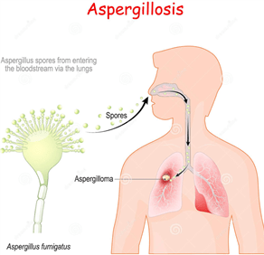
Hepatitis is the general term for liver inflammation, caused by several causes, both infectious such as bacterial, viral, parasitic and fungal, and non-infectious such as alcohol, autoimmune, drugs, and metabolic diseases. A disorder that happens when our body makes antibodies to liver tissue is autoimmune hepatitis. 

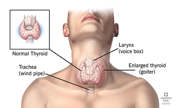
The liver is situated in the right upper region of the abdomen. It performs many critical functions in our body that influence metabolism, including: 

  • Production of bile.
  • Filtering the body’s contaminants.
  • Degradation of Carbohydrates, fats, and proteins.
  • Activation of enzymes, specific proteins that are important to the functions of the body.
  • Excretion of bilirubin (a red blood cell breakdown product), cholesterol, hormones, and drugs.
  • Blood protein synthesis, including albumin and clotting factors.
  • Glycogen, vitamins (A, D, E, and K) and minerals are processed. 

Types of viral hepatitis: 

Hepatitis A, B, C, D, and E contain viral infections of the liver known as hepatitis. A specific virus is responsible for each kind of virally transmitted hepatitis. Hepatitis A is often a short-term, acute infection, while hepatitis B, C, and D are most likely to become chronic and permanent. Hepatitis E is typically acute, but in pregnant women, it can be particularly hazardous. 

Hepatitis A Virus (HAV): 

Hepatitis A is due to a hepatitis A virus (HAV) infection. This form of hepatitis is most frequently transmitted from a person infected with hepatitis A by consuming food or water contaminated by feces. 

Hepatitis B Virus (HBV) 

HBV is transmitted through contact with infectious body fluids that contain the hepatitis B virus (HBV), such as blood, vaginal secretions, or semen.  

Hepatitis C Virus (HCV):

Hepatitis C comes from the virus (HCV) of hepatitis C. Hepatitis C is transmitted through direct contact with infectious body fluids, usually through sexual contact and injection drug use.

Hepatitis D Virus (HDV):

Hepatitis D also called delta hepatitis, is a severe hepatitis D virus (HDV)-induced liver disease. By direct contact with contaminated blood, HDV is contracted. Hepatitis D is a rare type of hepatitis that occurs only in combination with infection with hepatitis B. Without the involvement of hepatitis B; the hepatitis D virus cannot replicate. 

Hepatitis E Virus (HEV):

Hepatitis E is an illness caused by the hepatitis E virus (HEV) that is waterborne. Hepatitis E is found predominantly in poorly sanitized environments and usually results from the ingestion of fecal matter that contaminates water source. 

Hepatitis G Virus (HGV):

Recently, the hepatitis G virus (HGV, also called GBV-C) has been discovered and resembles HCV, but flaviviruses are more closely related. The virus and its effects are being studied, and it is uncertain about its role in causing disease in humans.

Incubation period:

The period between hepatitis infection and the onset of the illness is called the period of incubation. Depending on the individual hepatitis virus, the incubation time varies. The incubation period for HAV is approximately 15 to 45 days;

  hepatitis B virus is 45 to 160 days;

  hepatitis C virus, approximately two weeks to 6 months.

Pathophysiology:

Hepatitis A :

1. Several weeks of malaise, nausea, vomiting, anorexia, and elevated levels of aminotransferase mark the usual acute HAV infection cases. 

2. In more severe cases, jaundice occurs. In comparison to the classic image of elevated aminotransferase levels, some patients experience cholestatic hepatitis, characterized by the production of an elevated alkaline phosphatase (ALP) level. 

3. During a year, patients can experience multiple relapses. Infection with HAV does not spread and does not contribute to chronic hepatitis.

Hepatitis B:

The hepatitis B virus ( HBV) may be hepatocyte-directly cytopathic. Cytotoxicity mediated by the immune system plays a predominant role in causing liver damage, however. The immune attack is guided on the cell membranes of infected hepatocytes by human leukocyte antigen ( HLA) class I-restricted CD8 cytotoxic T lymphocytes that recognize the hepatitis B e antigen (HBeAg) and hepatitis B core antigen (HBcAg). 

Acute Infection :

1. Some patients have elevated serum levels of HBV DNA and a positive blood test for the presence of HBeAg but have normal levels of alanine aminotransferase ( ALT) and display minimal histologic evidence of liver damage, particularly those infected as newborns or as young children. These people are in the disease’s so-called “immune-tolerant phase.

2. years later, when the liver experiences aggressive inflammation and fibrosis, some but not all of these individuals can enter the “immune-active phase” of the disease in which the HBV DNA can remain elevated. 

3. In this time, an elevated ALT level is also noted. The immune-active process usually ends with HBeAg loss and the production of HBeAg (anti-HBe) antibodies. 

4. The “inactive carrier state”, which is formerly referred to as the “safe carrier state, can be reached by individuals who convert from an HBeAg-positive to an HBeAg-negative. 

5. These people are asymptomatic, have regular test results for liver chemistry, and have regular or minimally abnormal biopsy results for the liver.

Inactive carriers remain, via parenteral or sexual transmission, contagious to others. Ultimately, inactive carriers can produce anti-HBs and clear out the virus. 

Chronic Infection:

Either HBeAg-positive or HBeAg-negative can be considered for patients with chronic hepatitis. About 80% of chronic HBV cases are HBeAg-positive, and 20% are HBeAg-negative in North America and Northern Europe. 30-50 percent of cases are HBeAg-positive and 50-80 percent HBeAg-negative in Mediterranean countries and some areas of Asia. 

1. Signs of active viral replication are present in patients with HBeAg-positive chronic hepatitis with HBV DNA levels greater than 2 ×10⁴ IU / mL. Levels of HBV DNA can be as high as 1011 IU / mL. 

2. HBeAg-negative chronic hepatitis patients were probably infected at some stage with wild-type viruses. They also developed a mutation in either the precore or the viral genome’s main promoter region over time. 

3. HBV continues to replicate in such patients with a pre-core mutant state, but HBeAg is not formed. Patients with a core mutant state tend to have HBeAg production downregulated. 

4. The vast majority of HBeAg-negative chronic hepatitis B patients have a serum level of HBV DNA more significant than 2000 IU / mL. 

5. HBeAg-negative patients usually have lower levels of HBV DNA than HBeAg-positive patients do. The HBV DNA level is usually no higher than 2 ×10⁴ IU / mL.

Hepatitis C:

1. Patients with HCV-induced cirrhosis also have an increased risk of developing Hepatocellular carcinoma (HCC), particularly in the aspect of co-infection with HBV. 

2. HCC occurs in 1-5 percent of HCV-induced cirrhosis patients. In patients with HCV-induced cirrhosis, routine screening (e.g., ultrasonography and AFP testing every 6 months) is recommended to rule out HCC growth.  

Hepatitis D:

1. Acute hepatitis resulting from this can be mild or severe. Similarly, after acute exposure to both viruses, the risk of developing chronic HBV and HDV infection is the same as the incidence of developing chronic HBV infection after acute HBV exposure (approximately 5 percent in adults). 

2. Chronic HBV and HDV disease continues to lead to cirrhosis more quickly than chronic HBV infection alone. 

3. The introduction of HDV into a person already infected with HBV may have drastic implications. 

4. Superinfection may give the appearance of a sudden worsening or flare of hepatitis B to HBsAg-positive patients. Superinfection with HDV may result in FHF.

Hepatitis E:

Traditionally, it has not been assumed that HEV causes chronic liver disease. In organ transplant recipients, however, several studies have identified chronic hepatitis due to HEV. Dense lymphocytic portal infiltrates with interface hepatitis were revealed by liver histology, close to hepatitis C infection results. Some cases have moved into cirrhosis.

Etiology:

Hepatitis A Virus: 

The most commonly transmitted HAV through the fecal-oral pathway. Cases of HAV-associated transfusion or inoculation-induced disease are rare. 

HAV infection is prevalent, in the less-developed nations of Asia, Africa, and Central and South America, the Middle East has an exceptionally high prevalence. Most patients are infected when they are young kids in these areas. There is a chance of infection for uninfected adult travelers who enter these areas. 

Hepatitis B Virus: 

The presence of a surface antigen for hepatitis B (HBsAg) is characterized by HBV infection. Chronic HBV infection occurs in about 90-95 percent of neonates with acute HBV infection and 5 percent of adults with acute infection. The infection is removed in the remaining patients, and these patients develop lifelong immunity to recurring infections. 

Perinatal transmission: 

Perinatal transmission occurs in the vast majority of HBV cases around the world. During the intrapartum phase or, occasionally, in utero, infection tends to occur. Typically, neonates affected by perinatal infection are asymptomatic. The role of breastfeeding in the transmission is unclear, although breast milk may contain HBV virions. 

Sexual transmission: 

HBV is more readily transmitted than HCV or the human immunodeficiency virus ( HIV). Vaginal intercourse, genital-rectal intercourse, and oral-genital intercourse are associated with infection. Approximate 30 percent of HBV infected patients’ sexual partners also develop HBV infection. HBV cannot, however, be transmitted by kissing, touching, or household touch (e.g., towel sharing, eating utensils, or food). It is estimated that sexual activity accounts for as many as 50 percent of US cases of HBV. 

Sporadic Cases: 

The cause of HBV infection is unclear in roughly 27 percent of cases. Indeed, some of these incidents may be due to sexual reproduction or blood contact. 

Hepatitis C Virus: 

HCV is the most prevalent cause of non-A, non-B (NANB) parenteral hepatitis worldwide. In 0.5-2 percent of populations in nations around the world, hepatitis C is prevalent. In haemophilia patients and IDUs, the highest rates of disease prevalence are found. 

Transmission via a transfusion of blood :

The occurrence of transfusion-associated HCV infection has been significantly decreased by screening the US blood supply. Before 1990, the transfusion of infected blood products accounted for 37-58 percent of acute HCV infection (then known as NANB); currently, only about 4 percent of acute cases are due to transfusions. The chance of getting a hepatitis C-infected blood donation is 1 in 2 million. In dialysis units, acute hepatitis C remains a vital concern where the risk of HCV infection in patients is approximately 0.15 percent per year. 

Transmission by the use of intravenous and intranasal medications: 

IDU remains a useful HCV transmission mode. Around 60 percent of new HCV infection cases account for the use of intravenous ( IV) drugs and the sharing of paraphernalia used in the intranasal snorting of cocaine and heroin. HCV has been exposed to more than 90 percent of patients with a history of IDU. 

Sexual transmission: Roughly 20 percent of hepatitis C cases tend to be due to sexual contact. In comparison to hepatitis B, hepatitis C is contracted by about 5 percent of the intimate partners of those infected with HCV. 

HCV-infected individuals should be aware of the potential for sexual transmission. The presence of antibodies to HCV (anti-HCV) should be tested on sexual partners. For patients with multiple sex partners, safe-sex precautions are recommended. For patients with a steady sexual partner, existing recommendations do not encourage the use of barrier precautions. Patients can, however, avoid sharing razors with others and toothbrushes. Moreover, contact with the blood of patients should be avoided. 

Perinatal transmission: 

In 5.8 percent of infants born to mothers infected with HCV, perinatal transmission of HCV occurs. In children born to mothers co-infected with human immunodeficiency virus (HIV) and HCV, the risk of perinatal transmission of HCV is higher (about 18 percent). 

Hepatitis D Virus: 

HBV’s presence allows HDV to replicate, so HDV infection occurs only in patients who are HBsAg-positive. Patients may acquire HDV as a co-infection (at the same time that they contract HBV), or HDV may superinfect patients who are chronic carriers of HBV. In the United States, hepatitis D is not a reportable disease, so reliable data about HDV infections are scarce. It is estimated, however, that approximately 4-8 percent of acute hepatitis B cases include HDV co-infection. 

In IDU, the sharing of infected needles is considered the most common means of HDV transmission. HDV prevalence rates ranging from 17- 90% have been identified for IDUs that are also positive for HBsAg. It also explains sexual transmission and perinatal transmission. In Greece and Taiwan, the prevalence of HDV in sex workers is high. 

Hepatitis E Virus: 

The primary cause of enterally transmitted hepatitis from NANB is HEV., which is transmitted through the fecal-oral route, and in some parts of less developed countries, where most outbreaks occur, it tends to be endemic. HEV may also be vertically transmitted to HEV-infected mothers’ infants. High neonatal mortality is correlated with it. 

Anti-HEV antibodies in 29 percent of urban children and 24 percent of rural children in northern India were present in one study. Sporadic infections are found in individuals who migrate to these regions from Western countries.
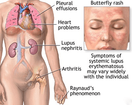
Symptoms:

There are little to no disease signs in many patients infected with HAV, HBV, and HCV. They do not have symptoms at first if they have contagious types of hepatitis that are chronic, such as hepatitis B and C. Symptoms cannot happen until the injury impairs liver function. 

Flu-like symptoms are the most common, including: 

Appetite deficit, Fever, Fatigue, Aching from the belly, Vomiting, Nausea, Feebleness 

Symptoms that are less common include: 

Dark-colored urine, fever, jaundice, and stools that are light-colored. 

Causes:

Alcohol as well as other toxins: 

Liver damage and inflammation can be caused by excessive alcohol consumption. It is referred to as alcoholic hepatitis occasionally. Liver cells are directly injured by alcohol, causing irreversible damage over time, leading to liver failure and cirrhosis, liver thickening, and scarring. Other toxic causes of hepatitis are drug overuse or poisoning and exposure to poisons. 

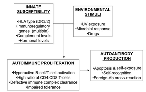
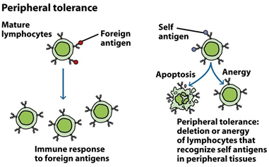
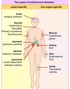
Autoimmune system responses:  

The immune system identifies the liver as a foreign entity in some situations and starts to attack it. It induces persistent inflammation that can vary from mild to severe, often impeding the liver’s function. In women, it is 3 times more common than in men.

Risk Factors:

People at the highest risk for the production of viral hepatitis. They are:

  • Staff in the health care professions
  • Patients with HIV 
  • Individuals with several sexual partners 
  • Intravenous drug users 
  •  Asians and islanders of the Pacific 
  • Workers in sewage and water treatment
  • People who receive blood clotting factors with hemophilia 

Once a common means of transmitting viral hepatitis, transfusion of blood is now a rare cause of hepatitis. Among lower socio-economic and poorly educated people, viral hepatitis is commonly thought to be 10 times more common. Approximately one-third of all hepatitis cases are from an undisclosed or unidentified source. To be infected with a hepatitis virus, a person does not have to be in a high-risk category. Food and water pollution with HAV raises risks in countries with low sanitation. Some daycare centers may become infected with HAV, so kids are at a higher risk of HAV infections at those centers.

Diagnosis:

History And Physical Examination 

The physician will first take history to assess any risk factors for contagious or non-infectious hepatitis to diagnose hepatitis. 

They gently press down on the abdomen during a physical examination to see if there is pain or tenderness and feel if the liver has been swollen. During the test, if skin or yellowish eyes were also examined. 

Tests for Liver Function: 

Tests for liver function use blood samples to assess how the liver functions effectively. The first indication might be inconsistent results from these tests, mainly if they do not display any physical exam signs for liver disease. High levels of liver enzymes can mean that liver is stressed, damaged, or not correctly functioning. 

Other Tests for Blood:  

Physicians would typically prescribe more blood tests to detect the cause of the problem if liver function tests are irregular. These tests can check for hepatitis-causing viruses. In conditions such as autoimmune hepatitis, they may also be used to search for normal antibodies. 

Ultrasound:

An abdominal ultrasound uses ultrasonic waves to closely inspect the liver and associated organs to create a picture of the abdomen’s organs. It will expose: 

Abdomen fluid: 

Harm to the liver or enlargement 

Tumors in the liver 

Gallbladder disorders 

The pancreas even shows up on ultrasound images occasionally. In identifying the cause of abnormal liver function, this can be a practical examination. 

Biopsy of the Liver:

An invasive procedure of taking a sample of tissue from the liver is a liver biopsy. It can be carried out with a needle through the skin and does not require surgery. Usually, before the biopsy sample is taken, an ultrasound is mandatory. 

This test helps to assess if the liver has been damaged by infection or inflammation. Some areas that appear abnormal in the liver may also be sampled using it.

Treatment:

Both Acute and chronic viral hepatitis are viewed differently. Acute viral hepatitis treatment requires sleeping, relieving symptoms, and ensuring sufficient fluid intake. Treatment of chronic viral hepatitis requires drugs to kill the virus and prevent further damage to the liver. 

Acute Hepatitis:

Initial treatment consists of relieving nausea, vomiting, and stomach pain in patients with acute viral hepatitis. Medications or substances that may have harmful effects on patients with abnormal liver function (for example, acetaminophen, alcohol) should be considered carefully. Only certain medicines that are deemed appropriate should be administered because the liver is not naturally able to remove drugs, and drugs may accumulate and reach toxic blood levels. Also, sedatives and “tranquilizers” are avoided because they can accentuate liver failure’s brain effects and induce coma and lethargy. As alcohol is harmful to the liver, the patient must abstain from consuming alcohol. To avoid dehydration caused by vomiting, it is sometimes essential to have intravenous fluids. For therapy and intravenous fluids, patients with extreme nausea and vomiting may need to be hospitalized. 

Acute HBV is not treated with medications that are antiviral agents. Though rarely diagnosed, acute HCV can be treated with some of the medicines used to treat chronic HCV. In 80 percent of patients who do not eliminate the virus early, HCV treatment is mainly recommended. The procedure results in virus clearance in most patients. 

Chronic Hepatitis:

Treatment of chronic HBV and HCV infections typically requires medicine or combinations of virus eradication drugs. In chronic hepatitis, alcohol aggravates liver damage and can cause more rapid progression to cirrhosis. Consequently, chronic hepatitis patients should avoid drinking alcohol. Cigarette smoking can also worsen liver disease and should be prevented.

Prevention :

Hepatitis prevention requires steps to prevent exposure to viruses, the use of immunoglobulin in case of exposure, and vaccines. Immunoglobulin administration is called passive defense since the patient receives antibodies from patients that have had viral hepatitis. As killed viruses or non-infectious components of viruses are given to stimulate the body to generate its antibodies, vaccination is called active defense. 

Avoidance of virus exposure:

Like any other disease, avoiding viral hepatitis is superior to depending on the treatment. Taking measures to avoid exposure to blood (exposure to dirty needles), semen (unprotected sex), and other bodily secretions and waste (stool, vomit) from another human can help prevent any of these viruses from spreading. 

By Immunoglobulins: 

Immune serum globulin (ISG) is a human serum which contains hepatitis A antibodies. In individuals that have been exposed to hepatitis A, ISG may be given to avoid infection. ISG operates immediately upon administration, and the security period is several months long. ISG is typically given to travelers to regions of the world where there is a high infection rate with hepatitis A and near or domestic interaction with hepatitis A patients. With few side effects, ISG is stable. 

The immune globulin of hepatitis B, or HBIG (BayHep B), is a human serum containing hepatitis B antibodies. HBIG is made of plasma (a blood product) believed to contain a high concentration of hepatitis B surface antigen antibodies. HBIG is almost always effective at avoiding infection if administered within ten days of exposure to the virus. However, even if administered a bit later, HBIG can reduce the severity of HBV infection. The defense against hepatitis B lasts for approximately three weeks after the administration of HBIG. HBIG is also given at birth to infants born to mothers confirmed to be infected with hepatitis B. HBIG is often given to individuals exposed to HBV by an infected person due to sexual contact or to healthcare workers inadvertently stuck by a needle known to be contaminated with blood.

Vaccinations:

Hepatitis A: 

In the US, the hepatitis A vaccine (Havrix, Vaqta) is available with two hepatitis A vaccines. They both contain the inactive (killed) virus of hepatitis A. For adults, it is preferable to take two doses of vaccination. After the first dose, protective antibodies develop within two weeks in 70% of vaccine recipients and within four weeks in nearly 100% of recipients. It is known that immunity from hepatitis A infection can last for several years after two hepatitis A vaccine doses. 

Hepatitis B:

A harmless hepatitis B antigen is given for active vaccination to activate the body’s immune system to develop protective antibodies against the hepatitis B surface antigen. Vaccines currently available in the U.S. are synthesized using recombinant DNA technology. These recombinant hepatitis B vaccines, the Energix-B and Recombivax-HB (hepatitis B) vaccines, are engineered to contain only a part of the surface antigen that is very potent in stimulating antibody production by the immune system. There is no viral component other than the vaccine’s surface antigen and can, therefore, not cause infections with HBV. Hepatitis B vaccines should be administered in three doses, 1 to 2 months after the first dose of the second dose, and 4 to 6 months after the third dose. Vaccinations are given in the deltoid muscles (shoulder) and not in the buttocks for the best performance. 

Both pregnant women should have a blood test for surface antigen antibodies to the hepatitis B virus. In addition to the hepatitis B vaccine at birth, women who test positive for the HBV risk transmit the virus during delivery. Thus, infants born to mothers with hepatitis B infection should receive HBIG. The rationale for offering both immunoglobulin and vaccine is that although the hepatitis B vaccine can provide long-lasting, active immunity, it takes weeks or months for immunity to develop. The short-lived, passive antibodies from the HBIG safeguard the baby before active immunity develops. 

Hepatitis C and D:

Currently, there is no hepatitis C vaccine. Due to the six different forms of hepatitis C, such a vaccine’s production is challenging. There is no hepatitis D vaccine available. However, since the hepatitis D virus requires live HBV to replicate in the body, the HBV vaccine will prevent a person not infected with HBV from contracting hepatitis D.

FUNGAL DISEASES

BY: SAI MANOGNA (MSIWM014)

Introduction:

Fungi: Fungi are eukaryotic microorganisms that do not photosynthesize and have a cellular wall. They parasitize or live as spores of organisms. Fungi invade keratinized tissue such as the horny cell layer, hair, and nails in superficial mycosis. 

1. A broad range of fungi that are commonly present in the environment cause fungal diseases. 

2. These invasive fungal infections occur rarely in healthy people, but in individuals with compromised immune systems, fungi may cause severe infections. 

3. In the soil and on plants and trees, as well as indoor surfaces and human skin, fungi live outdoors. 

4. Millions of different fungi species exist, but it is known that only a fraction of them make people sick. 

5. Anyone may get a fungal infection, such as a toenail infection or an athlete’s foot infection, but individuals with weakened immune systems are more likely to get severe fungal infections. 

6. An increasing danger to human health is fungal diseases. People living with HIV / AIDS, organ or stem cell transplants, cancer patients, and hospitalized patients are vulnerable to infection, while healthy people seldom suffer from severe fungal infections. 

7. Only four antifungal drugs exist, and fungal strains are emerging that are resistant to these drugs. At present, there are no licensed vaccines to prevent fungal infections.

Types of Fungal infections:

Many common fungal infections may infect the skin. Besides the skin, mucous membranes are another common place for fungal infections. Some examples of these are infections of vaginal yeast and oral thrush.

A. Candidiasis:

1. Candida is a yeast that can be found in the digestive tract, on the skin, on mucous membranes. 

2. Overgrowth of these yeasts can cause the development of symptoms that occur in the mouth or throat; for example, it is called “thrush” or candidiasis of the oropharynx. 

3. Usually, candidiasis in the vagina is referred to as a “yeast infection.” Overgrowth of these fungi can cause invasive candidiasis for individuals with certain risk factors, a severe infection that can damage the body’s blood, heart, brain, skin, bones, and other sections. 

4. The most common type of invasive candidiasis is candidemia, a bloodstream infection with Candida, and it often affects hospitalized patients. 

5. More than 20 Candida yeast species can cause human infection, but most infections are caused by Candida albicans, C. glabrata, C. parapsilosis. 

6. Tropicalis, Candida auris is a newly emerging fungi species that are difficult to detect and frequently avoids multiple antifungal drugs.

B. Aspergillosis:

1. A common mold found indoors and outdoors is Aspergillus. 

2. Without being sick, people breathe in various Aspergillus spores every day. 

3. People with compromised immune systems or lung disorders are, however, at risk of developing Aspergillus-induced health problems. 

4. Several kinds of aspergillosis range from mild to extreme illnesses. For instance, without causing infection, Aspergillus may cause inflammation of the lungs (allergic bronchopulmonary aspergillosis) or sinuses (allergic Aspergillus sinusitis). Invasive aspergillosis is a rare, severe lung or other body system infection and is a significant cause of mortality in immunocompromised people.

C. Cryptococcosis:

1. Cryptococcus fungi are present worldwide in the soil and are mostly related to bird droppings. 

2. Two key disease-causing species exist: Cryptococcus neoformans and C. gattii. 

3. These fungi cause infections in healthy individuals, but for people with weakened immune systems, such as those with HIV / AIDS, they can be severe. 

4. The infection happens typically when others inhale the fungus. The lungs and the nervous system are the most common sites of infection. The most prevalent cause of meningitis in adults is cryptococcal meningitis, a leading cause of death in people with HIV / AIDS.

D. Coccidioidomycosis (Valley Fever):

 1. It is an infection caused by the Coccidioides immitis and C posadasii fungi, also referred to as Valley Fever. These are soil-dwelling fungi found in arid, desert-like conditions. 

2. The fungi were discovered recently in south-central Washington. Infection usually happens by breathing Coccidioides spores into the lungs, and healthy individuals are at risk of Coccidioides infection, unlike most severe fungal diseases. 

3. In California and Arizona, the highest rate of infection has taken place. These states registered more than 11,000 Valley Fever cases in 2016 to estimate 15 to 30 percent of community-acquired pneumonia. 

4. Valley Fever can, however, be under-reported due to low testing rates. A self-limiting, moderate, flu-like disease can range from Valley Fever to severe disseminated infection that may require life-long therapy.

E. Athlete’s Foot:

1. Athlete’s foot infection is also called tinea pedis, which occurs on foot.

2. In wet, moist places such as shoes, socks, swimming pools, locker rooms, and public showers, the fungi grow best. 

3. In summer and hot, humid climates, they are always found. 

4. People who wear tight shoes, who do not change their sweaty socks, and use public baths and pools are more likely to do so. 

Causes: The fungi behind the foot of the athlete reside on the dead tissue of hair, toenails, and layers of the outer skin. There are four kinds of fungus which cause the infection. Trichophyton rubrum is the most common. 

Symptoms: The symptoms of an athlete’s foot vary from individual to individual such as, peeling, cracking of feet, blisters, black, softened, or broken-down skin, itching, and burning sensation.

F. Ringworm:

1. This ringworm is not a worm. It is a skin infection caused by moldlike fungi that live on skin, hair, and nails’ dead tissues. 

2. It can also cause infection on the scalp. 

3. That is what people call an athlete’s foot when they have it between the toes and also known as jock itch if it extends to the groin. 

Symptoms: A red, scaly patch or bump that itches is the telltale sign. The bump transforms into a ring- or circle-shaped patch over time. Maybe it will transform into many circles. Usually, the interior of the patch is transparent or scaly. The outside could be elevated and bumpy slightly. Ringworm appears to start as a lump or slight sore on the scalp. It may be flaky and scaly, and the contact may feel tender and painful on the scalp. It may also be noted that patches of hair are starting to fall out. 

Causes: The ringworm is incredibly infectious. In any of the following forms, can capture it by: 

From a different person: often, ringworm spreads by skin-to-skin touch. 

From the dogs: By rubbing sparky or grooming her, when it is done, the face should be washed. It is prevalent for cows, too. 

By touching things: On surfaces, clothing, towels, and in combs and brushes, the fungus that causes ringworm can linger. 

From the dirt: If people work or stand barefoot in soil tainted with the ringworm-causing fungus, they might get it.

G. Jock Itch:

1. Jock itch is caused by a form of fungus called Tinea. Often known as tinea cruris, the infection is Tinea is fond of wet, damp areas such as the groin, inner thighs, and buttocks. 

2. More commonly, infections occur in the summer or humid, wet climates. 

3. Jock itch is a red, itchy, frequently ring-shaped rash. 

Causes: jock itch is a mildly infectious, which spreads from person to person by direct or indirect contact through objects with the fungus on them. 

Symptoms: Jock itch signs include; On the groin or thigh, scratching, chafing, or burning, a circular, red rash with raised edges, redness in the thigh or groin, skin that flakes, peels, or cracks.

BACTERIAL DISEASE: TUBERCULOSIS (TB)

BY: SAI MANOGNA (MSIWM014)

Introduction : 

Mycobacterium tuberculosis, a tubercle bacillus, is the TB causative agent. It belongs to a closely related species, including M bovis, M africanum, and M microti, in the M tuberculosis complex. The most common site for TB development is in the lungs; 85 percent of patients with TB have lung complaints. Extrapulmonary TB can emerge as part of a primary, or late, generalized infection.

Types of Tuberculosis :

Tuberculosis is categorized into two types: active illness or latent infection. Lung disease is the most common type of active TB, but it can invade other organs, known as “miliary TB.”

TB – Active :

Active TB is a disease in which TB bacteria rapidly grow and invade the body’s various organs. Cough, phlegm, chest pain, fatigue, weight loss, fever, chills, and night sweating are common symptoms of active TB. Via airborne transmission of infectious particles coughed up into the air, a person with active pulmonary TB disease may spread TB to others. 

TB Miliary : 

Miliary TB is a rare type of active disease in the bloodstream when TB bacteria find their way. The bacteria rapidly spread across the body in tiny nodules in this form, affecting multiple organs at once. This form of TB can quickly become fatal. 

Latent Infection with TB : 

Many of those infected with TB may not experience overt illness. They have no symptoms, and that could be common for their chest x-ray. Reaction to the interferon-gamma release assay (IGRA) or tuberculin skin test (TST) may be the sole manifestation of this encounter. There is an ongoing possibility, however, that the latent infection can escalate to active illness. Other diseases, such as HIV or drugs that weaken the immune system, increase the risk.

Incubation Period :

The time of incubation can vary from two to 12 weeks or so. An individual may remain infectious for a long time (provided that viable TB bacteria are present in the sputum) and may remain infectious for several weeks before they have undergone sufficient therapy.

Pathophysiology : 

1. M tuberculosis infection occurs most often from exposure to infected aerosols from the lungs or mucous membranes. 

2. Droplets are 1-5 μm in diameter in these aerosols; a single cough will produce 3000 infectious droplets in a person with active pulmonary TB, with as few as 10 bacilli required to initiate infection. Droplet nuclei are deposited within the terminal airspace of the lung when inhaled. 

3. The organism will grow for 2-12 weeks until they reach several 1000-10,000, enough to evoke a cellular immune response, detected by a tuberculin skin test reaction. 

4. Mycobacteria are strongly antigenic and encourage a nonspecific immune response that is robust. 

5. Their antigenicity is due to the activation of Langerhans cells, lymphocytes, and polymorphonuclear leukocytes by multiple cell wall constituents, including phospholipids, glycoproteins, and wax D. 

Individuals infected with M tuberculosis may take 1 of several infection routes, most of which do not lead to actual TB. The host immune system can clear or suppress infection in an inactive type called latent tuberculosis infection (LTBI), with resistant hosts regulating mycobacterial production at distant locations before active disease output. Patients with LTBI cannot spread TB.

As earlier mentioned, the most popular site for TB production is in the lungs; 85 percent of TB patients have pulmonary complaints. As part of primary infection, or late, generalized infection, extrapulmonary TB may occur. The most prominent sites of extrapulmonary disease are: common location of tuberculous lymphadenitis is in the neck and the sternocleidomastoid muscle; it is typically unilateral and causes little to no pain; advanced tuberculous lymphadenitis cases may suppurate and shape a sinus drain. As in the kidneys, bones, meninges, skin, choroids, and the lungs’ apices, contaminated end organs usually have high regional oxygen stress. The fundamental cause of M tuberculosis infection tissue destruction is linked to the organism’s ability to incite extreme host immune reactions to the antigenic cell wall proteins. 

Lesions with TB :

An epithelioid granuloma with central caseation necrosis is the usual lesion of TB. The common site of the primary lesion in the lung’s subpleural regions is within alveolar macrophages. Locally, Bacilli proliferate and spread to a Hillary node via the lymphatics, forming the Ghon complex. 

The early tubers are spherical, 0.5- to 3-mm nodules with 3 or 4 cell areas, displaying the following characteristics: 

a. A necrosis of the central caseate 

b. Lymphocyte-admixed inner cell region of epithelioid macrophages and Langhans giant cells 

c. An outer cell region with plasma cells, lymphocytes, and immature macrophages 

d. A fibrosis rim (in lesions that heal) 

Initial Lesions: Until symptomatic illness occurs, initial lesions may heal, and the infection becomes latent. Smaller tubercles can be fully resolved. Fibrosis happens when hydrolytic enzymes destroy tubercles, and a fibrous capsule surrounds larger lesions. Typically, these nodules contain viable mycobacteria and can be reactivated. Some nodules calcify or ossify and are readily seen on radiographs of the chest. 

Proliferative Lesions: Where the bacillary load is small and host cellular immune responses dominate proliferative lesions form. These tubers are compact, mixed with activated macrophages, and surrounded by proliferating fibrosis lymphocytes, plasma cells, and an outer rim. Mycobacteria intracellular killing is successful, and the bacillary load remains low. 

Exudative Lesions: Exudative lesions predominate when large numbers of bacilli and there are low host defenses. Such loose aggregates of immature macrophages, neutrophils, necrosis of fibrin, and caseation are mycobacterial growth sites. These lesions progress without treatment, and the infection spreads. 

Etiology : 

M tuberculosis, a slow-growing obligate aerobic and a facultative intracellular parasite, are responsible for TB. In parallel groups called cords, the organism grows. After decoloration with acid-alcohol, based on the acid-fast stains used for pathological detection, it retains several stains. 

Fig: Acid-fast bacillus smear. 

In Mycobacterium tuberculosis, acid-fast bacillus smear shows aerobic, non-spore-forming, nonmotile, facultative, curved intracellular rods measuring 0.2-0.5 μm by 2-4 μm are mycobacteria, including M tuberculosis. Mycolic, acid-rich, long-chain glycolipids and phospho lipoglycans (mycocides) are contained in their cell walls that protect mycobacteria from cell lysosomal attack retains red basic fuchsin dye (acid-fast stain) after acid rinsing. 

Transmission :

The only known source of M tuberculosis has been humans. The organism is transmitted from a person in the infectious stage of TB, primarily airborne aerosol (although transdermal and GI transmission have been reported). 

1. Exposure to M tuberculosis in immunocompetent individuals results in latent/dormant infection 

2. Modifications in host immune systems resulting in a decreasing immune effectiveness may enable M tuberculosis species to reactivate, tuberculosis stemming from a combination of the direct effects of infectious organism replications, and subsequent host immune responses to tuberculosis antigens.

3. By restriction fragment-length polymorphism study, molecular typing of M tuberculosis isolates in the United States indicates that more than one-third of new patient TB occurrences result from person-to-person transmission. The remainder comes from latent infection reactivation. 

Symptoms and signs :

Symptoms associated with active pulmonary TB (older people with TB do not have the usual signs and symptoms) are as follows: 

i. Coughing ii. anorexia and fever 

iii. Sweats at Night 

iv. hemoptysis 

v. Chest pain (may also be caused by acute tuberculous pericarditis) 

vi. Tiredness 

The following may be signs of tuberculous meningitis: 

i. Headache, which for 2-3 weeks has been either sporadic or chronic 

ii. Fever that is low-grade or absent 

iii. Subtle changes in mental state that may progress towards coma over days to weeks 

The following may be signs of skeletal TB :

i. Pain in the back or stiffness

ii. Tuberculous arthritis, usually affecting just 1 joint (the hip or knee most frequently, followed by the foot, elbow, wrist, and shoulder) 

Genitourinary TB symptoms can include the following: 

Pain Flanking, DYSURIA, frequent urination, symptoms that resemble pelvic inflammatory illness in women, a painful mass of the scrotum, prostatitis, orchitis, or epididymitis in men 

Gastrointestinal TB signs apply to the contaminated site and can include the following: 

i. Non Healing mouth ulcers or anus ulcers 

ii. Malabsorption (with a small intestine infection) 

iii. Swallowing problems (with the esophageal disease) 

iv. TB-related physical examination results depend on the organs involved.

v. Mimicking peptic ulcer disease (with gastric or duodenal infection) with stomach pain 

vi. Pain, diarrhea, or hematochezia (containing colon infection) 

The following may be present in patients with pulmonary TB: 

i. Abnormal sounds of breath, especially over the upper lobes or areas involved, 

ii. Rales or signs of bronchial breath, suggesting consolidation of the lungs 

Depending on the tissues involved, symptoms of extrapulmonary TB vary and can include the following: Perplexity, chorioretinitis, coma, cutaneous Injuries, neurological deficit, lymphadenopathy pathology. 

Active TB is not excluded by the lack of any relevant physical findings. In high-risk patients, especially those who are immunocompromised or elderly, classic symptoms are often absent. 

Causes : 

M. Bacteria that cause tuberculosis to induce TB. When a person with pulmonary TB coughs, sneezes, spits, laughs or speaks, they will propagate in droplets through the air. The infection can be transmitted only by individuals with active TB. However, most people with the disease can no longer spread the bacteria after receiving sufficient care for at least two weeks. 

Factors of Risk : 

When deciding whether a TB infection is likely to be transmitted, the following factors help: 

i. Total of expelled species 

ii. Immune condition of the person exposed 

iii. Duration of time of exposure to polluted air 

iv. Organisms’ concentration 

A specific risk to non-infected individuals is posed by infected persons living in crowded or closed environments. Approximately 20% (positive tuberculin skin test) of household contacts develop an infection. Micro Epidemics have occurred on transcontinental flights and in closed settings such as submarines. Hospital workers, inner-city residents, nursing home residents, and inmates often include groups at high risk for contracting the infection. 

The factors increasing the risk of acquiring active tuberculosis in an individual are : 

i. Infection with HIV 

ii. Diabetes mellitus (3fold increase in risk) 

iii. Immunosuppressive counseling

iv. Abuse of intravenous ( IV) medications 

v. Renal End-stage Disorder 

vi. With alcoholism 

vii. Malignancies of hematologic origin

viii. Silicosis 

ix. Less than five years of age

x. Antagonists of tumor necrosis factor-alpha (TNF-alp) 

xi. Head and neck cancer 

xii. Surgery for intestinal bypass or gastrectomy 

xiii. Chronic Syndromes of Malabsorption 

xiv. Low body weight-In comparison, obesity has been associated with a lower risk of active pulmonary TB in elderly patients

xv. Smoking-To minimizes the risk of relapse; smokers who develop TB should be advised to quit smoking. 

TB in Children : 

The potential for developing fatal miliary TB or meningeal TB is a primary concern in children younger than five years old. In children with TB, osteoporosis, sclerosis, and bone involvement are more common than adults with the condition. As a result of their high vascularity, the epiphyseal bones may be involved. Children also do not infect other children because they rarely develop a cough, and sputum development is scarce. Cases of child-child and child-adult transmission of TB are, however, well known. 

Genetic considerations : 

Tuberculosis genetics are very complex, involving several genes. Some of these genes provide essential elements of the immune system, while others include more complex mechanisms by which Mycobacterium species communicate with the human body. The genes that follow have polymorphisms that are connected with either tuberculosis susceptibility or safety. Also, regions such as 8q12-q13, the gene has not yet been identified, are associated with increased risk. 

Diagnosis : 

Methods of screening for TB include: 

Mantoux tuberculin skin test for active or latent infection (primary method) with purified protein derivative (PPD) 

An interferon-gamma release assay (IGRA) in vitro blood test with antigens unique to Mycobacterium tuberculosis for latent infection 

Obtain the following laboratory examinations for suspected TB patients: 

Acid-fast bacilli (AFB) smear and sputum culture obtained from the patient: no positive smear result does not preclude active TB infection; the most specific test for TB culture is the AFB culture. 

Serology of HIV in all TB patients and uncertain HIV status: HIV-infected individuals are at increased risk of TB 

Other diagnostic tests, including the following, can justify consideration: 

Immunospot Specific Enzyme-Linked (ELISpot) 

Tests for Nucleic acid amplification 

Community of blood 

Drug susceptibility testing should be followed in supportive cultures; symptoms and radiographic results do not distinguish multidrug-resistant TB (MDR-TB) from completely susceptible TB. 

Such testing can include the following: 

a. Study of Direct DNA Sequencing 

b. Molecular Automated Testing 

c. Drug resistance (MODS) and thin-layer agar (TLA) assays for microscopic observation 

d. Additional quick tests (e.g., BACTEC-460, luciferase reporter assays, ligase chain reaction, FASTPlaque TB-RIF) 

e. To test for potential related pulmonary findings, obtain a chest radiograph. 

Treatment : 

TB is cured with early detection and effective antibiotics. 

The correct type of antibiotic and the period of therapy will depend on: 

i. The overall health and age of the individual 

ii. If they have active or latent TB 

iii. The position of the infection 

iv. If the TB strain is immune to drugs 

v. Latent TB treatment can vary. It may mean taking an antibiotic for 12 weeks once a week or for nine months every day. 

Treatment for active TB can require 6-9 months of taking several drugs. The treatment would be more difficult if a person has a drug-resistant strain of TB. Completing the full course of treatment is significant, even if the symptoms go away. Some bacteria can survive and become immune to antibiotics if a person stops taking their medication early. The person may continue to develop drug-resistant TB in this case. 

Prevention: Ways of preventing anyone from being infected with TB include: 

i. Having an early diagnosis and treatment 

ii. Staying away from other individuals until the risk of infection is no longer present 

iii. Wearing a mask, shielding the mouth and rooms with ventilation 

iv. Vaccinating against TB 

In individual nations, as part of a routine immunization program, children receive an anti-TB injection, the bacillus Calmette-Guérin ( BCG) vaccine. 

The live strain of Mycobacterium bovis developed by Calmette and Guérin for use as an attenuated vaccine to prevent tuberculosis ( TB) and other mycobacterial infections is Bacille Calmette-Guérin (BCG). The vaccine was first given to humans in 1921 and remained the only vaccine for general use against TB. 

The BCG vaccine is the world’s most commonly administered vaccine; it has been given to over three billion people, mainly in the form of compulsory newborn immunization (as dictated by World Health Organization guidelines)[1]. Several BCG vaccines are used worldwide, manufactured by various manufacturers, and administered under different schedules. The BCG vaccine is also effective against other diseases, including leprosy and Buruli ulcer, for safety. Moreover, it is used in the treatment of superficial bladder carcinoma as an immunostimulant.

BACTERIAL DISEASE : LEPROSY

BY: SAI MANOGNA (MSIWM014)

Introduction : 

A chronic infection caused by the acid-fast, rod-shaped Mycobacterium leprae bacillus is leprosy. Leprosy is also known as Hansen’s disease. Two related diseases that mainly affect superficial tissues, especially the skin and peripheral nerves, maybe leprosy. A mycobacterial infection initially triggers a broad array of cellular immune responses. The second component of the condition, peripheral neuropathy with possible long-term effects, is then elicited by these immunologic events.

As a highly infectious and debilitating disease, leprosy was once feared, but nowadays, it does not spread quickly, and treatment is very successful. However, nerve damage can result in hands and feet being crippled, paralysis, and blindness if left untreated.

Classification of Leprosy :

The number and sort of skin sores have determined leprosy. The type of leprosy depends on specific symptoms and treatment. The forms are: 

Tuberculoid:  A moderate type of leprosy, which is less severe. People with this type only have one or a few patches (paucibacillary leprosy) of flat, pale-colored skin. Owing to nerve damage underneath, the affected region of the skin can feel numb. It is less infectious to tuberculoid leprosy than other types. 

Lepromatous:  Come on. A more extreme form of the disorder. It brings widespread skin bumps and rashes, numbness, and muscle weakness (multibacillary leprosy). It can also impact the nose, kidneys, and male reproductive organs. It is more infectious than leprosy caused by tuberculosis. 

Borderline: The tuberculoid and lepromatous symptoms are present for people with borderline leprosy. 

You can hear physicians use this simplified classification as well: 

Paucibacillary – single lesion (SLPB): One lesion 

Paucibacillary (PB): lesions from two to five 

Multibacillary (MB): Six lesions or more

Incubation period :

The incubation period is called the interval between contact with the bacteria and the appearance of symptoms. Symptoms typically take about 3 to 5 years to appear after coming into contact with the leprosy-causing bacteria. Up to 20 years later, some individuals do not show symptoms. The long incubation period of leprosy makes it very hard for doctors to determine when and where a person with leprosy has been infected.

Pathophysiology :

Depending on the host’s reaction to the organism, leprosy can manifest in various ways. 

1. The tuberculoid type of the disease that usually affects the skin and peripheral nerves are present in individuals who have a robust cellular immune response to M leprae. 

2. They tend to be dry and hypoesthetic, and the number of skin lesions is small. 

3. Typically, nerve activity is asymmetric. Owing to the low number of bacteria in the skin lesions, i.e., < 5 skin lesions, with no organisms on the smear, this type of the disease is often referred to as paucibacillary leprosy. 

4. In these people, the findings of skin tests with antigens from killed species are positive.

5. The lepromatous type of the disease, characterized by extensive skin involvement, is for individuals with limited cellular immune response. 

6. Infiltrated nodules and plaques are often identified as skin lesions, and nerve involvement appears to be symmetric in distribution. 

7. The organism grows best at 27-30 ° C; therefore, skin lesions tend to develop in the body’s colder areas, with sparing of the groin, axilla, and scalp. 

8. Owing to no small number of bacteria present in the lesions, i.e.,> 6 lesions, with potential bacilli visualization on the smear, this type of disease is often referred to as multibacillary leprosy. 

9. Skin test results for antigens from killed species are non-reactive.

Patients can also have symptoms of both types (indeterminate or borderline leprosy) but typically evolve to one or the other over time. Interestingly, most people exposed to leprosy never contract the disease, possibly because more than 95 percent of individuals are naturally immune to this disease.

Epidemiology :

In 2018, 208,619 new leprosy cases were reported globally, according to WHO estimates based on data from 159 countries. The worldwide prevalence was 184,212 cases (rate, 0.2/10,000) reported at the end of 2018. In 2018, 79.6 percent of all new leprosy cases were in Brazil, Indonesia, and India. Furthermore, in 2018, 23 priority countries accounted for 96 percent of cases globally.

a. Mortality/Morbidity :

Leprosy is never lethal. The nerve damage and crippling sequelae are the main consequences of infection. 33-56 percent of newly diagnosed patients have already demonstrated symptoms of compromised nerve function[11], according to one report. According to reports, three million individuals who have undergone multidrug treatment for leprosy have suffered impairment due to nerve damage. While the skin and peripheral nerves are involved in both lepromatous leprosy and tuberculoid leprosy, tuberculoid leprosy has more severe manifestations. Nerve involvement contributes to sensory and motor loss of control, leading to repeated trauma and amputation. Most generally, the ulnar nerve is involved. 

Damage to the following nerves is related to characteristic leprosy impairment: 

i. Ulnar and Median-Hand Clawed 

ii. Tibial posterior-Plantar insensitivity and clawed toes 

iii. Peroneal Common-Foot Drop 

iv. Radial nerves of the cutaneous, nasal, and greater auricular

b. Race : 

Leprosy was once globally endemic, although no predilection for the race is recognized. The incidence of leprosy fell significantly in northern Europe and North America in the late 1800s, and the disease is now recorded mainly in tropical areas. 

c. Gender :

In males, leprosy is typically more common than in females, with a 2:1 male-to-female ratio. The prevalence of leprosy among females is equal to or greater than that of males in some areas of Africa. 

d. Age :

Leprosy can occur at any age, but the age-specific occurrence of leprosy in developed countries peaks in children younger than ten years, accounting for 20 percent of leprosy cases. In infants, leprosy is very rare; however, they are at relatively high risk of maternal leprosy, particularly in cases of lepromatous leprosy or mid borderline leprosy.

e. HIV Coinfection :

Preliminary data suggest that, unlike tuberculosis, HIV coinfection does not appear to affect leprosy. Besides, coinfection with HIV does not seem to affect the lepromatous to tuberculoid leprosy ratio.

Symptoms of Leprosy 

Leprosy mostly affects the skin and nerves, called the peripheral nerves, beyond the brain and spinal cord. 

Disfiguring skin sores, bumps, or lumps that do not go down for several weeks or months is leprosy’s principal symptom. The sores on the skin are pale-colored. Nerve damage leads to loss of sensation in legs and arms, weakness of muscles.

Causes :

Exactly how leprosy is transmitted is not clear. When an individual coughs or sneezes with leprosy, they can spread droplets containing M. The leprosy bacteria that someone else breathes in. For leprosy to be transmitted, near physical contact with an infected individual is required. Shaking hands, kissing, or sitting beside them during a meal on a bus or at a table, do not transmit the disease.

Pregnant leprosy mothers are unable to pass it on to their newborn babies. Neither is it spread through sexual touch.

Risk Factors :

Leprosy can permanently damage skin, nerves, arms, legs, feet, and eyes without care. 

Leprosy complications may include: 

Glaucoma or blindness 

Iritis 

Loss of hair 

For infertility 

Facial disfigurement (including permanent swelling, bumps, and lumps) 

Erectile dysfunction in males and infertility 

Failure of the kidney 

Weakness in the muscles contributing to claw-like hands or not being able to stretch feet 

The inside of the nose is permanently damaged, which can lead to nosebleeds and a chronic stuffy nose 

Permanent nerve damage, including those in the arms, legs, and feet, outside the brain and spinal cord 

Damage to the nerves can lead to a dangerous loss of feeling. Do not experience pain when one gets cuts, burns, or other injuries to hands, legs, or feet if they have leprosy-related nerve damage.

Diagnosis :

It is possible to distinguish Hansen’s disease by the presence of skin patches that may look lighter or darker than normal skin. The skin areas affected can often be reddish. The lack of sensation is prominent in these skin patches. For a needle, one cannot feel a light brush or a prick. 

The physician will take a sample of skin or nerve (through skin or nerve biopsy) and check for bacteria under the microscope and confirm your diagnosis, and may even conduct tests to rule out any skin diseases.

Treatment :

It is possible to treat leprosy. 16 million people with leprosy have been healed in the last 2 decades. The World Health Organization provides free care for all people with leprosy. 

Therapy relies on the type of leprosy they have. For treatment, antibiotics are used. Doctors recommend treatment on a long-term basis, usually for 6 months to a year. You may need to take antibiotics longer if you are suffering from severe leprosy. The nerve damage that comes with leprosy cannot be treated with antibiotics. 

A standard treatment for leprosy that combines antibiotics is multidrug therapy (MDT). That means you are going to take two or more drugs, mostly antibiotics: 

Paucibacillary leprosy: Two antibiotics, such as dapsone, are used every day, and rifampicin is used once a month. 

Multibacillary leprosy: In addition to daily dapsone and monthly rifampicin, you can take a daily dose of the antibiotic clofazimine. For 1-2 years, you will undergo multidrug treatment, and then you will be healed.

Antibiotics: The bacteria causing leprosy can be destroyed by antibiotics used during treatment. However, though medication may cure the condition and keep it from getting worse, nerve damage or physical disfigurement may have existed before a diagnosis is not reversed. Thus, before any irreversible nerve damage occurs, the condition must be detected as soon as possible.

Transmission : 

The exact way Hansen’s disease spreads between people is not understood. Scientists claim that it can happen if a person with Hansen’s disease coughs or sneezes, and a healthy person breathes in the droplets that contain the bacteria. Prolonged close contact is required with someone with untreated leprosy for several months. 

You do not get leprosy from casual contact with a person who has Hansen’s disease, such as: shaking or hugging palms, sitting for a meal together, sitting on the bus next to each other. 

During pregnancy, Hansen’s disease is also not transmitted from a mother to her unborn baby, and it does not spread by sexual contact. It is often challenging to locate the infection source due to the bacteria’s slow-growing nature and the long time to cause symptoms of the disease.

For general health purposes, if possible, avoid contact with armadillos. Speak to your physician if you have come into touch with an armadillo and are anxious about having Hansen’s disease. Over time, the doctor will follow up with you and conduct annual skin tests to see if the disease progresses. 

Prevention : 

In high-risk areas, awareness campaigns on leprosy are essential to enable patients and their families, who have been traditionally shunned from their communities, to come forward and receive care. 

Early diagnosis and multidrug therapy treatment is the most effective way to avoid leprosy disabilities and avoid further disease spread. However, the Bacille Calmette-Guérin ( BCG) vaccine is partially protective against leprosy.