COMPONENTS OF BLOOD

BY: Reddy Sailaja M (MSIWM030)

BLOOD

Blood is a specialized body fluid, comprises of plasma and the cells that circulate throughout the human body. It supplies oxygen, glucose, antibodies, vitamins, electrolytes, heat, hormones, and immune cells across the body for life survival. It removes carbon dioxide and other waste generated from the cells in the body.

BLOOD COMPONENTS

Blood is made up of three main components: plasma, blood cells (red and white) and platelets.

Plasma: Plasma occupies 55% of blood fluid in human beings. Plasma comprises of 92% water and the remaining 8% contains carbon dioxide, glucose, hormones, proteins etc.

 Blood cells: Blood cells comprise of 45% of total blood fluid. These are produced in bone marrow by the process called ‘hematopoiesis’ from a common precursor cell (hematopoietic stem cells). Then these blood cells mature into red blood cells (RBCs), white blood cells (WBCs) and platelets. The organs like lymph nodes, liver and spleen regulate the generation, destruction and regulation of blood cells.

Figure 1: Blood cells production by hematopoiesis

Red blood cells: RBCs are also called erythrocytes. They are double concave shaped structures without nucleus. Approximately, 4.5 – 6.2 million/microliter are present in the blood. Their main function is to carry oxygen from lungs to all parts of the body. RBCs contain a special protein called ‘Hemoglobin’ that aids in oxygen transport. RBCs have a life span of 120 days.

White blood cells: WBCs are otherwise known as leucocytes. WBCs are vital in fighting against invading pathogens and infections. Approximately 3700 – 10500/microliter are present in the blood. Apart from fighting infection, WBCs also help heal wounds by ingesting dead cells and debris, protects against foreign entity that enter blood stream and fights against cancerous cells.

The following are the types of WBCs that are produced in response to the kind of infection (bacterial vs fungal vs viral vs parasitic).  Life span varies from hours to days to years depending upon the type of WBCs. WBCs are majorly divided into two subtypes: Granulocytes and agranulocytes. Granulocytes contains protein containing granules in their cytoplasm. Eosinophils, basophils and neutrophils constitute granulocytes. Monocytes and lymphocytes constitutes agranulocytes. The following table explains the types of WBCs’, their nature and function.

Type of white blood cellPercentage of abundance in WBCS (%)Function
Basophils0.5 – 1Basophils produce in response to parasite infections, allergy and bone marrow damage. It secretes histamine – involve in allergic reactions and heparin – an anticoagulant that aids blood clotting at the site of injury and subsequent wound healing.
Eosinophils2 – 4Eosinophils defend against bacterial and parasite infections by releasing toxic substances and results in immflamatory reaction.
Neutrophils60 – 70First line of defense against invading pathogens. They attack the pathogens, engulf and digest them by phagocytosis process and maintain normal health.
Monocytes3 – 8Maintains tidiness of the blood and other tissues by clearing the dead pathogen particles and damaged cells and their debris.
Lymphocytes20 – 25B – cells produce antibodies against bacterial, viral and fungal infection. T – cells are two types, cytotoxic T-cells kills the antigens and helper T-cells aid antibody production from B-cells. Natural killer cells attack any foreign object that comes in contact with the body.

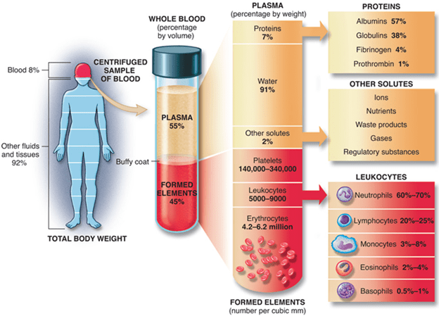
Figure 2: Blood and its components

Blood platelets: These are also called thrombocytes. Approximately 1,50,000 – 4,00,000 platelets/microliter are present in the blood. Platelets help clot the blood to stop bleeding during injury by protects the wound against further infection.

In brief, blood functions include:

  • Oxygen supply to cells and tissues
  • Supply essential nutrients – glucose, aminoacids, fatty acids,
  • Removal of waste material – carbon dioxide, urea, lactic acid
  • Fighting against infections
  • Regulating body temperature and pH balance
  • Transport hormones and transmits neuromessages

Blood disorders:

Blood disorders often cause life threatening situations as the infection spreads out throughout the body by blood circulation. General blood disorders are as follows:

RBCs disorder – Anemia: Low number of RBCs in blood cause anemic situation. This results in low oxygen supply in the body, fatigue and pale skin.

WBCs disorder – Cancer: Lymphoma, myeloma and leukemia are the major blood related cancers.

Platelets disorder – Internal blood clots: these clots block blood supply and can be dislodged and spread through various organs like lungs, heart, brain etc, which can be fatal to the body.

CYTOKINES

BY: Ria Fazulbhoy (MSIWM031)

Cytokines are an important group of proteins or glycoproteins which play a major role in cell-to-cell communication between cells like lymphoid cells, inflammatory cells and hematopoietic cells. They are secreted by white blood cells (WBCs) and other cells of the body. They respond to stimuli and assist in the regulation of development of immune effector cells and sometimes have a direct effect of their own as well. The cytokine binds to the target cell by the presence of specific membrane receptors present on the target cell. (very high affinity-cytokines work at picomolar concentrations)

Mode of action

  1. Autocrine

The cytokine is released from a cell and binds to the membrane receptor present on the same cell.

  • Paracrine

Cytokine is released from the producer cell and binds to the target cell which is in close proximity

  • Endocrine

Cytokine binds to the target cell which is in a distant part of the body.

Four major groups of cytokines are Hematopoietic family (interleukins-ILs), Interferons (IFs) family, Tumor necrosis factors (TNF) family and chemokine family.

A variety of cells secrete cytokines, but the major principal producer cells are Th cells and macrophages. Cytokines secreted from these cells activate an entire network of interacting cells.

   Macrophage and Th cells are major producers of cytokines in the body.

Some biological functions of cytokines include:

  • Cellular and humoral immunity development
  • Inflammatory response induction
  • Control of cellular proliferation and differentiation
  • Healing of wounds
  • Development of innate/acquired immunity
  • Hematopoiesis

NOTE: Cytokines have a non-antigen specific mode of action and have very short half-lives.

Functions of some cytokines

Cytokine secretion by Tн cell subsets: Tн1 and Tн2

Difference in the pattern of cytokine secretion amongst Tн cell subsets determines the immune biological response made to a particular antigenic challenge. These two subsets are Tн1 and Tн2, which secrete different cytokines and mediate in different ways. Both these subsets secrete IL-3 and GM-CSF.

Tн1 and Tн2 have the following functional differences:

1) Tн1 subset:

  • It is responsible for mainly cell-mediated immune responses like activation of Tc cells and delayed hypersensitivity reactions.
  • Helps in promotion of excessive inflammation and tissue injury
  • Helps in production of opsonization-promoting IgG antibodies.
  • Effective in viral infections and intracellular pathogens.
  • IFN-४, IL-12 and IL-18 are responsible for the development of Tн1 cell response.
  • E.g.: IFN-४ and TNF-ß mediates inflammation and delayed hypersensitivity.
  • E.g.: IL-2 and IFN-४ promote differentiation of cytotoxic cells Tc from CD8 precursors.

2) Tн2 subset:

  • Responsible for secretion of antibodies for immune response.
  • Stimulates eosinophil activation and differentiation
  • Helps B cells
  • Promotes production of large amount of IgM and IgG
  • Supports allergic reactions
  • IL-4 is essential for the development of Tн2 response.
  • E.g.: IL-4 and IL-5 induce production of IgE and helps eosinophil attack on helminth or roundworm infections.

Tн2 development is favoured over Tн1. The cytokines produced by the two subsets are cross regulated. The cytokines produced by a subset (Tн1 or Tн2) promote the growth of their subset and simultaneously inhibit the activity and development of the opposite substrate (cross regulation). Two transcription factors known as T- Bet and GATA-3 are important in determining the cross regulation of the two subsets

  • T-Bet drives cells to differentiate towards Tн1 cells.
  • GATA-3 drives cells to differentiate along Tн2 cells.

EXTRAVASATION OF LYMPHOCYTES

BY: K. Sai Manogna (MSIWM014)

At inflammatory sites and secondary lymphoid glands, different subsets of lymphocytes exhibit directed extravasation. Therefore, lymphocyte recirculation is closely monitored to ensure that sufficient populations of B and T cells are recruited into various tissues. Extravasation of lymphocytes involves interactions between a variety of cell-adhesion molecules, as with neutrophils. The overall process is similar to what occurs during the extravasation of neutrophils and involves the same four stages of touch and rolling, activation, arrest and adhesion and, eventually, transendothelial migration.

Sites of Lymphocyte Extravasation:

1. Some regions of vascular endothelium consist of specialised cells with a plump, cuboidal (‘high’) form in the postcapillary venules of different lymphoid organs; such regions are referred to as high-endothelial venules or HEVs.

2. In appearance, their cells contrast strongly with the flattened endothelial cells that line the rest of the capillaries. Each of the secondary lymphoid organs comprises HEVs, except the spleen.

3. There are about 1.4 × 10⁴ lymphocytes extravasate into a single lymph node every second through HEVs.

4. Cytokines developed in response to antigen capture influence the production and maintenance of HEVs in lymphoid organs.

5. In order to prevent the antigen from entering the node, the role of lymphocyte antigenic activation in the preservation of HEVs has been demonstrated by surgical blocking of afferent lymphocyte vasculature of the node.

6. The HEVs demonstrate impaired function within a short period and gradually return to a more flattened morphology.

7. High-endothelial venules express several cell-adhesion molecules. HEVs, like other vascular endothelial cells, express CAMs from the selectin (E- and P-selectin) family, the mucin-like (GlyCAM-1 and CD34) family, and the superfamily of immunoglobulins (ICAM-1, ICAM-2, ICAM-3, VCAM-1, and MAdCAM-1).

8. In a tissue-specific way, some of these adhesion molecules are distributed. These tissue-specific adhesion molecules have been named vascular addressins (VAs) because they help to guide the extravasation to specific lymphoid organs of various populations of recirculating lymphocytes.

Receptor Profiles and Signals Guided by Lymphocyte Homing:

1. Related to neutrophil extravasation, the general lymphocyte extravasation mechanism is similar.

2. The fact that different subsets of lymphocytes migrate differently into different tissues is a significant aspect that separates the two processes. This method is known as trafficking or homing.

3. The numerous lymphocyte subset trafficking patterns are regulated by unique combinations of adhesion molecules and chemokines; homing receptors are called receptors that guide the circulation of different lymphocyte populations to specific lymphoid and inflammatory tissues.

Researchers have established several lymphocytes and endothelial cell adhesion molecules that are involved in lymphocyte interactions with HEVs and endothelium at tertiary sites or sites of inflammation.

Recirculating Naive Lymphocytes into Secondary Lymphoid Tissue:

Until it has been triggered to become an effector cell, a naive lymphocyte is not able to mount an immune response.

1. In specialised microenvironments within secondary lymphoid tissue (e.g., peripheral lymph nodes, Peyer patches, tonsils, and spleen), activation of a naive cell occurs.

2. Dendritic cells catch antigen inside these microenvironments and present it to the naive lymphocyte, resulting in its activation.

3. Naive cells do not display a preference for a specific form of secondary lymphoid tissue but instead circulate indiscriminately across the body to secondary lymphoid tissue through recognising HEV adhesion molecules.

4. The initial attachment to HEVs of naive lymphocytes is usually mediated by the binding of the L-selectin homing receptor to HEV adhesion molecules such as GlyCAM-1 and CD34.

5. The naive cell trafficking pattern is designed to keep these cells continuously recirculating across secondary lymphoid tissue, the primary purpose of which is to trap antigen transmitted by blood or tissue.

6. They are activated and enlarged into lymphoblasts until naive lymphocytes encounter antigen trapped in secondary lymphoid tissue. Activation takes approximately 48 h, and the blast cells are retained in the paracortical area of secondary lymphoid tissue during this time.

7. The antigen-specific lymphocytes cannot be identified in the circulation during this process, called the shutdown phase.

8. During the shutdown point, rapid proliferation and differentiation of naive cells occur. Then the effector and memory cells that this process produces leave the lymphoid tissue and begin to recirculate.

Lymphocytes of Effector and Memory follow distinct patterns of trafficking:

1. Effector and memory lymphocyte trafficking patterns vary from those of naive lymphocytes.

2. By recognising inflamed vascular endothelium and chemoattractant molecules produced during the inflammatory response, effector cells appear to be home to regions of infection.

3. On the other hand, memory lymphocytes selectively house the type of tissue in which antigen was first encountered.

4. This presumably ensures that a specific memory cell returns to the tissue where the antigen it recognises is most likely to re-encounter a subsequent threat.

5. Memory cells and effector cells express increased levels of specific molecules of cell adhesion, such as LFA-1, which interact with ligands present in additional tertiary lymphoid tissue (such as skin and mucosal epithelial) and at inflammation sites, allowing these sites to be accessed by effector and memory cells.

6. Naive cells lack the corresponding molecules of cell-adhesion and do not house these sites.

7. A variety of adhesion molecules, including E- and P-selectin and the Ig-superfamily molecules VCAM-1 and ICAM-1, are expressed in inflamed endothelium and bind to receptors expressed at high levels in the memory and effector cells.

8. Subsets of the memory and effector populations display tissue-selective homing activity, unlike naive lymphocytes.

9. Such tissue specificity is imparted by multiple combinations of adhesion molecules rather than by a single adhesion receptor.

10. A mucosal homing subset of memory/effector cells, for example, has high levels of LPAM-1 (4 7) and LFA-1 (Lb2) integrins that bind to MAdCAM and various ICAMs on venules of intestinal lamina propria.

11. However, since they have low levels of L-selectin that would promote their entry into secondary lymphoid tissue, these cells prevent direction to secondary lymphoid tissues.

12. Preferential homing to the skin is shown by the second group of memory/effector cells. Low levels of L-selectin are also expressed in this subset, however high levels of cutaneous lymphocyte antigen (CLA) and LFA-1, which bind to E-selectin and ICAMs on skin dermal venules, are seen.

13. While effector cells and memory cells that express decreased L-selectin levels do not appear to reach peripheral lymph nodes via HEVs, they may enter peripheral lymph nodes via afferent lymph vessels.

Adhesion-Molecule Interactions Play Extravasation Vital Roles:

1. A multi-stage mechanism involving a cascade of adhesion-molecule interactions is the extravasation of lymphocytes into secondary lymphoid tissue or regions of inflammation, similar to those involved in bloodstream neutrophil emigration.

2. This shows the usual interactions in the extravasation of naive T cells into lymph nodes through HEVs.

Mechanism:

1. In the first stage, a selectin-carbohydrate interaction similar to that seen with neutrophil adhesion.

2. L-selectin, which acts as a homing receptor which directs the lymphocytes to specific tissues expressing a corresponding mucin-like vascular addressin such as CD34 or GlyCAM-1, initially binds naive lymphocytes to HEVs.

3. The rolling of lymphocytes is less pronounced than neutrophil rolling.

4. Although the initial interaction of selectin-carbohydrate is minimal, the slow rate of blood flow in postcapillary venules, especially in regions of HEVs, reduces the possibility that the tethered lymphocyte can dislodge the sheer force of the flowing blood.

5. In the second stage, chemokines that are either localised on the endothelial surface or secreted locally mediate an integrin-activating stimulus.

6. To maintain these soluble chemoattractant variables on the HEVs, the thick glycocalyx covering of the HEVs can work.

7. If, as some have indicated, HEVs secrete lymphocyte-specific chemoattractants, it will clarify why, while they express L-selectin, neutrophils do not extravasate into lymph nodes at the HEVs.

8. As happens in neutrophil extravasation, chemokine binding to G-protein – coupled receptors on the lymphocyte contributes to the activation of integrin molecules on the membrane.

9. The integrin molecules interact with the adhesion molecules of the Ig superfamily (e.g., ICAM-1) once activated, so that the lymphocyte adheres tightly to the endothelium.

10. In the final stage, molecular mechanisms involved in the transendothelial migration are poorly understood.