GENE THERAPY

BY: ABHISHEKA G (MSIWM013)

INTRODUCTION:

   Among all the advanced techniques in the field of biotechnology gene therapy is one of the important milestones. Gene therapy is a technique used in biotechnology in which new foreign genes are inserted into existing cells of living organisms to treat diseases. This technique is employed to replace the defective genes in the genome which causes diseases. The research on gene therapy was first started in 1972 by Friedmann and Roblin who published a paper entitled ‘gene therapy to cure genetic disorders. Later in the future, the research was developed by scientists, and on the 14th of September 1990, for the first time the gene therapy clinical trials were approved by the National Institute of Health (NIH), the US under the guidance of William French Anderson, which was performed on 4 years old girl who was suffering from severe combined immunodeficiency.

TYPES OF GENE THERAPY:

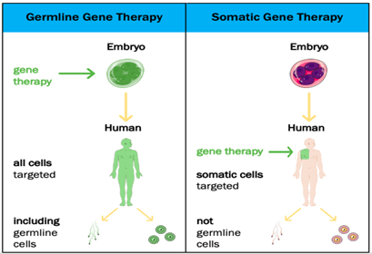
The technique gene therapy is classified into two types based on cells in which foreign gene is inserted, a) Germline gene therapy

                     b) Somatic cell gene therapy

Germline gene therapy: In this type, the foreign gene or therapeutic gene is inserted into the germ cells of the living organism. In this technique the inserted genes are passed from generation to generation, hence they are heritable.

 Example: The therapeutic cells are inserted into the germ cells like sperm and egg.

Somatic cell gene therapy: In this technique, the foreign gene or therapeutic gene is inserted into the somatic cells of the living organism. In this technique, the inserted genes do not pass from generation to generation.

Example: Insertion of genes into bone marrow, blood cells, skin cells, etc.

APPROACHES OF GENE THERAPY:

There are two ways of approaches are available in gene therapy:

a) In-vivo gene therapy

 b) Ex-vivo gene therapy

In-vivo gene therapy: In this technique, the target cell is fixed in the body of the living organism and the therapeutic gene or foreign gene is inserted into the target cell through a vector. The vector used to insert the therapeutic gene into the target cell is either a viral or non-viral vector. This technique is mainly used to treat genetic disorders.

Example: It is used to treat the genetic disorder cystic fibrosis.

Ex-vivo gene therapy: In this technique, the therapeutic genes are inserted into the cultured cells in the laboratory.

Procedure: The genetically defective cells are isolated from the living organism and they are cultured inside the laboratory. Then the therapeutic genes are inserted into the cultured cells in the laboratory. After insertion, the genetically corrected cells from the cultured cells are isolated and grown in the laboratory, then the modified cells are transplanted into the living organism.

Example: The first gene therapy was performed on 4 years old girl Ashanthi Disilva, who was suffering from severe combined immunodeficiency (SCID), which is caused due to defects in the gene which codes for adenosine deaminase. This is an example of ex-vivo gene therapy.

VECTORS IN GENE THERAPY:

Vector is the vehicle of gene delivery, an agent that carries to the therapeutic or desired gene into the target cell. There are two categories of vectors are employed in gene therapy.

  1. Viral vectors
  2. Non-viral vectors

Viral vectors:  The viruses are used as vectors to deliver the desired gene to the target cell of the living host.

 Example: Retrovirus: The genetic material of the virus RNA and it can carry a DNA of size less than 3.4kb and integrated it into the host genome of stable fashion, and it targets the cells in dividing the state.

Adenovirus: The genetic material of this virus is double-stranded DNA, they carry and integrate the desired DNA into target cells that are a non-dividing state.

Non-viral vectors: Some of the chemicals and physical instruments are used to carry desired DNA into the host cell are called Non-viral vectors.

Some of the physical methods to insert the desired DNA into the target cell are Electroporation, gene gun, etc.

Electroporation: In this technique, the electric shock of small pulses of high voltage is used to carry the DNA across the cell membrane of the target cell. After the small electric shock, the temporary pores are caused in the cell membrane to provide the passage for desired DNA into the host cell.

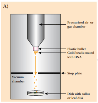
Gene gun: In this method, the gold particles or tungsten particles are coated with the desired DNA molecule, then the coated DNA is placed into a device called Gene gun which generates the high pressure necessary for the penetration of the desired DNA into the target or host cell and the gold and tungsten particles are left behind on stopping disc.

Sonoporation: In this method, the DNA is inserted into the target cell with help of ultrasonic frequencies.

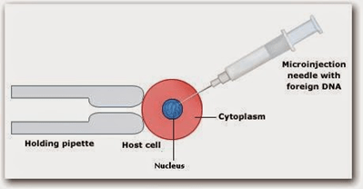
Microinjection: In this method, microscopic materials are inserted into the target cell with the help of a small microscopic device called Micromanipulator.

Some of the chemical methods used to carry desired into the target cell are lipofection and using detergent mixtures.

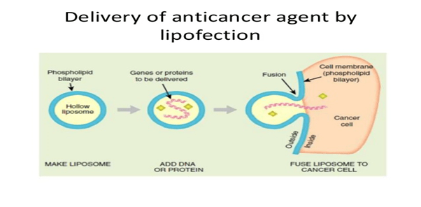
Lipofection: In this technique, the liposomes are used as a vector to insert the desired DNA into the target cell. The liposomes are artificial phospholipid vesicles used to transport various molecules into cells including DNA.

Detergent mixtures: Some of the detergent mixtures like calcium phosphate are mixed with cDNA molecules and they are inserted near to the target cells. Then these detergent mixtures disturb the cell membrane od the cell and increase the size of the pore through which the cDNA enters into the cell.

ADVANTAGES OF GENE THERAPY:

  1. Gene therapy plays an important role to replace the defective disease-causing genes in the genome.
  2. It also helps treat genetic disorders and some of the deadly diseases like cancer.

DISADVANTAGES:

  1. Diseases caused by defects in multiple genes are not treated by gene therapy.
  2. Some of the viral vectors cause toxicity and inflammatory reactions.
  3. Some of the ethical issues arise.


VIRUS

BY: SREELAKSHMI (MSIWM012)

Virus are small structure which can pass its genetic material to living host where it can replicate. They can infect plants, animals and other microorganisms also. The study of viruses is called virology which is considered as a subspecialty of microbiology.  Martinus Beijerinck is the father of virology.

In 1898, Friedrich Loeffer and Paul Frosch was the first to find evidence of the nature of viruses, genetic entities of virus. They found the evidence from foot and mouth disease in livestock. The first identified human virus was yellow fever caused by mosquito Aedes aegyptii.Virus was discovered and reported in 1910 by a US Army physician.Veterniary virology gained importance after in1902 after cattle plague.

CLASSIFICATION OF VIRUS

Virus classification is the process of naming virus and places them into a taxonomic system. Virus are mainly classified by phenotypic characters like morphology, mode of replication, host organisms, nucleic acid type and the type of disease caused by the virus. A universal system for classifying viruses, and a unified taxonomy was established in1966 by International Committee on Taxonomy of Viruses (ICTV).vanRegenmortel (19) lists the following characters for discriminating between virus species:

  • Relatedness of genome sequence
  • Natural host range
  • Cell and tissue tropism
  • Pathogenicity and cytopathology
  • Mode of transmission
  • Physicochemical properties of viral proteins
  • Antigenic properties of viral proteins.

The system makes use of a series of ranked taxons,with the: order,family,subfamily,genus,species,order,family,genus,species.

 STRUCTURE BASED CLASSIFICATION OF VIRUS

It is classified based on shape and size. Based on the structure of nucleocapsid it’s divided into two namely helical morphology and icosahedral morphology. Helical morphology consist of a helical array of capsid proteins which is surrounded by a helical filament of nucleic acid. The number and arrangement of the capsomers are useful in identification and classification of icosahedral morphology Based on envelope it’s divided as enveloped and non-enveloped virus.

PROPERTIES OF VIRUS

  • It possess no membranes,cytoplasm,ribosomes or other cellular components
  • They cannot move or grow
  • They are really tiny, even smaller than cell and is only visible advanced electron microscopes.
  • It consist of two major parts which are a protein coat and a hereditary material which can be a RNA or DNA.

VIRAL REPLICATION

Viruses cannot replicate on its own and in order get multiplied it must infect a host. It replicates in host cell’s machinery to create more viruse.steps involved in replication includes:

  • Penetration: The virus gets engulfed by the cell or get attached to some other viruses which have surface proteins that bind to receptors on the host cell. It allows the host cell to either fuse with virus or absorb the virus. Once it reaches the cell, the genetic material is released to the cytoplasm.
  • Replication: The genetic material of the virus is copied several times.
  • Transcription: The genetic material act as a blue print for the cell to make messenger RNA which is used to make viral proteins.
  • Protein Assembly: It occurs in the cytoplasm (ribosome) where viral proteins are made.
  • Viral Assembly: The viral genetic material (after replication) will get surrounded by the newly made viral protein.
  • Release: Through budding viruses get emerged from the cell through cell membrane or by bursting out of the cell which will result in the death of host cell.

STRUCTURE OF VIRUS

Size of a virus is smaller than bacteria and size is variable. The larger viruses can vary their size about 300 A in diameters. It indicates that it may be as larger as a small bacteria. They occur mainly in three shapes which are helical (spherical or can be of complex symmetry. Virus lacks cytoplasm and hence lack cell organelles like mitochondria, Golgi bodies, ribosomes and enzyme systems are also present. Virus contains RNA or DNA whereas a normal cell contains both DNA and RNA. It contains a protein coating called capsid which acts as a protection for genetic materials by surrounding the genetic material. A virus particle is called virion.

Plant virus contains only RNA.Animal virus contains both RNA or DNA.wheras bacteriophages contain only DNA.

TYPES OF ANIMAL VIRUS

  • Double-Stranded DNA: Double-Stranded DNA virus generally have a polyhedral or complex structure. Examples are Papilloma Virus,Variola(smallpox)
  • Double-Stranded RNA: Double-Stranded RNA usually have polyhedral structure. Example is diarrhea virus.
  • Single –Stranded RNA: Single –Stranded RNA virus have two subunits which can serve as mRNA and other one which can serve as a template mRNA.Examples are Rhinovirus,HIV

ROLE OF VIRUSE IN CANCER

The tumor viruses change cells by integrating their genetic material with the host cell DNA.This is a permanent insertion in the genetic material which is never removed. The insertion mechanism can differ depending on whether the genetic material of the tumor causing DNA or RNA.In case if the genetic material is DNA then it can be directly inserted into the host DNA.In case of RNA as the genetic material, then RNA is first transcribed into DNA and then it is inserted into the host cell’s DNA.

MICROBIAL INTERACTIONS

BY: SAI MANOGNA (MSIWM014)

Biological interactions are the impact that species in the environment have on each other

  • The interactions between different microbes, plant and germ interactions to promote growth, animal interactions, interactions with human beings and water interactions occur in a whole range of microbes.
  • In any biological culture, microbial interactions are all distinctive, complex, critically important and are central in global biogeochemistry.
  • The relationships between these two populations are defined by whether the relations favour both populations and benefit one of them, or whether one or both populations are negatively affected

Microbial interactions are of two types :

  1. Positive Interactions : Mutualism, Syntrophism, Photoreception, Commensalism.
  2. Negative Interactions : Ammensalism (antagonism), Parasitism, Predation, Competition.

Positive Interactions :

  1. Mutualism : Mutualism is defined as interactions between organisms of two different species, in which each organism benefits in some way from interactions.

    i. These types of interactions are common and ubiquitous across all ecosystems, and scientists increasingly recognise the important role they play in ecology.

    ii. Mutualisms are also symbiotic partnerships. In such situations, the two animals live in close proximity to each other for part or all their whole lives; however, not all symbiotic relationships are reciprocal.

Examples of Mutualism:

Several known examples of mutualistic arrangements exist.

  1. Nitrogen fixing bacteria and Leguminous Plants : The relationship between nitrogen-fixing bacteria and leguminous plants
  • Intestinal flagellates and Termites : Intestinal flagellates and termites exhibit obligate mutualism, a strict interdependence in which protozoans digest the wood eaten by termites; Here neither partner can live under natural circumstances without the other.
  • Yucca Moths (Tegeticula) and Yucca Plants : Yucca moths depend on yucca plants and the other way around. The moth goes about as a pollinator while laying her eggs in yucca seed pods. The hatchlings incubate and feed on a few yet not all seeds. Here the two species have advantages such that the plant is pollinated and the moth has nourishment for its hatchlings.

II. Syntrophism : Syntrophy is the mutual dependency of various types of species on the fulfilment of their respective nutritional needs. The intermediate or end products metabolism of one organism are important materials for another. Syntrophism is exemplified in the mixed environment population.

Examples of Syntrophism:

  1. Dust mites on Human Skin : House dust mites fleece human skin. A healthy person produces about 1 gram of skin flakes daily. These mites may also produce chemicals that stimulate skin flakes. People may become allergic to these compounds.
  • Organisms on cow-dung : A cow eats lots of grass, which is converted into lipids by microorganisms in the large intestine of the cow.

III. Photoreception : It is a relationship in which the organisms are mutually beneficial and in association with each other. This relationship is similar to mutualism, but relations between the species in proto-cooperation are not necessary as in mutualism.

Examples of Photoreception :

  1. Interaction of Nitrogen fixing bacteria and Cellulomonas :

IV. Commensalism : In Biology,commensalism refers to the relationship between two species in which one species, without harming or helping the latter, obtains food or another benefits from the other.

    i. The species i.e, benefiting from the unaffected species (host), can obtain nutrients, shelter, protection or locomotion.

   ii. The relationship is predominantly between the larger host and the smaller commensal. In essence the host organism remains unchanged through contact, while the commensal species can exhibit great morphological adaptation.

Examples of Commensalism :

  1. Shark and Remora : remora attaches to the shark or other fish and rides on it. Remoras evolved on their heads as a flat oval shaped disc structure that adheres to their host bodies.

Negative Interactions :

  1. Ammensalism : Ammensalism is any relationship between two different species, where one organism is inhibited or eliminated, while the other remains consistent.

     i. One microbial population develops substances that are inhibitory to the other microbial populations which is antagonistic known as ammensalism are antagonism.

   ii. In nature, no organism can live its life in complete isolation. They must communicate in some way with other species and their environment.

Examples of Ammensalism : There are basically two types 1. Competition 2. antibiosis.

  1. Competition : A Larger more potent organism removes another organism from its source of food and shelter.
  2. Antibiosis : One organism secretes a chemical that destroys the other organism, but the one which secretes the chemical is unharmed.

       Example : penicillium on black walnut trees : The Mould that is capable of producing penicillin, which kills several types of bacteria that would also like to grow on this bread. The bacteria killing effects of penicillin that contributed to the use of penicillin as antibiotic. Penicillin doesn’t benefit from the death of other bacteria, making it an example of amensal antibiosis.

 II.  Parasitism : Parasitism is a relationship between two separate species, where one of them actually affects the other. An organism that harms the other is considered a parasite.

   i. The host-parasite relationship is characterised by a relatively prolonged period of interaction that may be physical or metabolic.

   ii. Some parasites live outside the host cell, known as ectoparasite, while others live within the host cell, known as endoparasite.

Examples of Parasitism :

  1. Ticks on Dogs and cats : Fleas or ticks that adhere to the skin of dogs and cats are parasites.
  • Lice :  This is another parasite that lives off the blood of host animals.
  • Aphids : Aphids are a type of insect parasite that feeds on the sap of host plants.

III. Predation : Predation refers to the relationship between organisms in which one organism kills and devours another.

   i. Predation gives energy to prolong life and to facilitate the reproduction of the organism that destroys the predator, to the detriment of the organism that is eaten, the prey.

  ii. Predation affects species in two ecological states.

  iii. At the level of an individual, the prey organism has a sharp decline in fitness as calculated by its lifetime productive success, since it can never produce again.

  iv. At the group level, predation decreases the number of individuals in the population of prey.

Examples of Predation :

  1. Carnivorous interactions :  The best known examples are Wolves hunting moose, Frogs eating flies, Owls hunting mice, Shrews hunting worms or insects.
  2. Group Predation : This occurs with ants and social spiders.

IV.  Competition : A Larger more potent organism removes another organism from its source of food and shelter.

  i. This interaction occurs when both species (populations) use the same resources, resulting in lower maximum density or growth rate for the microbial population.

Examples of Competition :

a. Competition between Paramecium caudatum and Paramecium aurelia : When these protozoa are brought together, both paramecium species feed on the same bacterial community.

MOLECULAR BIOLOGY AND GENETICS

BY: SREELAKSHMI (MSIWM012)

Molecular biology is the study of the chemical and physical structure of biological macromolecules. Genetics is a branch of science dealing with the study of heredity and variation.

 Molecular Biology is an overlapping with other areas of biology and chemistry. It is the understanding of the interactions between DNA, RNA and protein. It is basically of two steps: transcription and translation. It is called Central Dogma. Synthesis of RNA from DNA is called transcription and synthesis of Protein and DNA translation.

TRANSCRIPTION

  • Transcription involves synthesis of new strand of nucleic acid complementary to a DNA template strand.
  • To transcribe a gene, RNA polymerase proceeds through a series of well-defined steps which are grouped into three phases- initiation, elongation and termination.
  • The bacterial core RNA polymerase can initiate transcription at any point on a DNA molecule.
  • RNA polymerase can initiate a new RNA chain on a DNA template and therefore do not require a primer.
  • The elongating polymerase is a processive machine that synthesize and proofreads RNA.
  • Ribonucleotides enters the active site and the added to are growing RNA chain.
  • Termination of transcription is activated by the presence of terminator sequence which results in the elongating polymerase to dissociate from the DNA and release the RNA chain.

TRANSCRIPTION

In eukaryotes, they have three different polymerases and several initiation factors.

TRANSLATION

Translation converts the genetic information present within the mRNA to a linear sequence of amino acids in proteins

The decoding of mRNAs into the language of proteins is composed of four components which are Trna,aminoacyl tRNA synthetasesand also ribosome.mRNa template provides the information that must be interpreted.Aminoacyl Trna synthetase couple amino acids to specific tRNAs that recognize the appropriate codon. The protein coding region of each mRNA have contiguous and non-overlapping string of codon called an open reading frame. Translation starts at 5’end and ends at 3’end.it starts with start codon and ends with a stop codon.AUG is usually a start codon, whereas UAG,UGA,UAA are stop codon.

GENETICS

Genetics is the study of hereditary and variation. The term was first introduced by W Bateson.

Gregor Mendel is known as the Father of Genetics.

Mendel was the first one who told that there are some factors which give you a particular phenotype (eye color, hair texture).The pioneering study on generics was by Mendel on pea plants. He looked at how the size, height, colur.He selected only pure breeds. From result of this experiment he came up with the hypothesis:

  • LAW OF SEGREGATION:

It is studied with the help of monohybrid cross. It states that the alleles of a given locus segregate into separate gametes. It is also called as law of purity.

MONOHYBRID CROSS

  • LAW OF INDEPENDENT ASSORTMENT:

It can be explained with the help of dihydrid cross. Mendel considered seed form and cotyledon color for the cross. This law states that the factors or alleles of each character assort or segregate independent of the factors of other character at the time of gamete formation and get randomly rearranged in the offspring.

DIHYBRID CROSS

POST MENDELIAN

Basics of genetics was given by Mendel, later studies provided information on various genetic interactions which also led to the studies on various genetic disorders.

COMPLEMENTARY GENES

If two genes present on different loci produce the same effect when present alone but interact to form a new trait when present together .They are called complimentary genes.

SUPPLEMENTARY GENES

They are pair of non-allelic genes one of which produces its effect independently in the dominant state.

LINKAGE

It is an exception of principle of independent assortment.

CHROMOSOME THEORY OF LINKAGE

It was given by Morgan and Castle. It states that

  • The genes which show linkage are situated in the same chromosome and remain bounded by chromosomes by material.So,they cannot be separated during the processes
  • The degree or strengthen of linkage depends upon the distance between the linked genes on the chromosomes, closely located genes show strong linkage.
  • Genes lie in linear order in the chromosome.

CROSSING OVER

It is one of the two exceptions of Mendel’s law of independent assortment. It produces new combinations or recombination of genes by interchanging of corresponding segments between non sisters chromatids of homologous chromosomes at prophase 1 of meiosis the non-sister chromatids in which exchange of segments has occurred are called cross overs.

MICROBIAL NUTRITION AND GROWTH

BY: SHREELAKSHMI S NIAR

Microorganisms require the supply of raw materials or elements to construct new cellular components, these raw materials are called nutrients. These nutrients are provided by the culture medium where the microbes grow. These medium provides the nutrients which is required for the energy, building of cell substances and biosynthesis of fermentation products. On the basis of requirement nutrition can be classified as

Macronutrients: Carbon, oxygen, hydrogen, nitrogen, sulphur, phosphorous, potassium, calcium, magnesium and iron are included in macronutrients. They are required by the microbes in large amount and constitutes over95% of cell dry weight.Potassium,calcium,magnesium,iron,iron exist in a cell which act as cofactor for enzyme whereas carbon, oxygen, hydrogen, nitrogen, sulphur, phosphorous are the major components of biomolecules like carbohydrtes,proteins,lipids and nucleic acid.

Micronutrients: they are also called as trace elements. They require only in a small amount for microbes. It includes manganese, zinc, cobalt, nickel, molybdenum and copper. They mainly form a part of enzyme and help in catalysis of reaction. Contamination present in water, glasswares are sufficient to provide these nutrients.

Culture media components include inorganic nutrients, nitrogen supplements, carbon sources and growth factors. Inorganic Nutrients include Potassium Phosphate, Magnesium sulphate,Ammonium sulphate  or phosphate,Calcium carbonate.,Cobalt,  Copper, Iron, Mn, Mo, Zn.

NUTRITIONAL TYPES OF MICROORGANISMS

ON THE BASIS OF SOURCES OF CARBON

  1. Autotrophs: They majorly use carbon dioxide and hence they can carry out photosynthesis.
  2. Heterotrophs: They use reduced, performed organic molecules from other sources.

ON THE BASIS OF ENERGY SOURCE

  1. Lithotrophs: They reduced inorganic molecules as a source for electron.
  2. Organotrophs: They extract electrons from organic molecules.

ON THE BASIS OF THEIR PRIMARY SOURCE OF CARBON AND ENERGY

  1. Photolithoautotrophy: They use light as energy source and carbon dioxide source.E.G. Algae, cyanobacteria.
  2. Photoorganoheterotrophy: They use organic carbon and light as there source.
  3. Chemolithoautotrophy: They use inorganic compounds like iron, sulphur, nitrogen and carbon dioxide as the source.
  4. Chemoorganoheterotrophy: They generally use Organic molecule and carbon as there source.

MICROBIAL GROWTH

Growth is the orderly increase in all the components of an organism such as size or population number. The microbes are grown in batch culture or closed system. Because of the limited increase in cell size and frequency of cell division, growth in microorganisms is measured by increase in cell number. Bacteria multiply by binary fission, the process in which parent cell splits into two daughter cells. Bacterial cells first elongate, then followed by the formation of transverse membrane and new cell wall. The new membrane and cell wall will grow inward from the outer layers. Cell divides into two daughter cells. The growth of microorganisms reproducing by binary fission in culture can is plotted as the logarithm of the number of viable cells on Y-axis and the incubation time on X-axis. The resulting curve is called as standard bacterial growth curve that have four phases of growth namely lag phase, log phase, stationary phase and death phase.

Characteristics of Each Phase

  1. The Lag Phase: when the microorganisms are inoculate into fresh medium they do not increase significantly in number and thus this phase is called lag phase. But, the microbes are metabolically active and thus they increase in size. Enzymes, essential cofactors are formed and accumulate until they are present in concentrations that permit growth. They also produce quantities of energy in the form of ATP.it shows that they are preparing for replication. The length of lag phase depends on the condition of microorganisms and nature of medium. Refrigerated culture or microbes inoculated from chemically different media takes more time to adapt
  2. The Log or Exponential phase: The organisms divide at their most rapid rate .The population of organism’s doubles in this phase. The cell division depends upon the composition of growth and conditions of incubation. It provides the optimal conditions growth. Exponential growth can be balanced or unbalanced. When all the cellular constituents are synthesized at the constant rates relative to each other, it result in balanced growth and when rate of synthesis vary relative to each other, it is unbalanced growth. The bacteria’s at this stages are suitable for biochemical and morphological identification, drug sensitivity test.
  3.  The Stationary Phase: The cell division decreases to the point that new are produced at the same rate as old cells die, the total number of viable cells remains constant. The culture is said to be in stationary phase is represented by a horizontal straight line in the curve. At this phase, population may simply cease to divide but is metabolically active. This phase may result due to depletion of essential nutrients, lack of biological space, accumulation of toxic waste products.
  4. The Death Phase: As the condition of medium become more detrimental, population reaches in death phase, in which cells lose their ability to divide and thus, they die. In the death or decline phase ,the number of viable cells decreases exponentially, i.e., a constant proportion of cells dies every hour. However, sometimes death is not logarithmic and can vary with both environmental conditions and microorganisms involved. The duration of this phase depends on the genetic characteristics of the organisms.

DESIGN OF FERMENTERS AND TYPES OF FERMENTERS

BY: SHREELAKSHMI S NAIR

FERMENTORS:

Fermenter is a system which provide controlled environmental conditions for the growth of microbes to obtain a desirable metabolite by preventing the entry of contaminants. Sterility is maintained by steam. Steam is maintained at 121 degree for a period of time and all the unwanted microbes are killed.

In designing a fermenter, following points must be considered

  1. The fermenter vessel must be able to work aseptically for a number of days.
  2. Evaporation loss should be less
  3. It must allow nutrient and reagent feed.
  4. Proper aeration and agitation should be provided.
  5. Temperature and pH control should be provided.
  6. Power consumption must be low.
  7. It should facilitate the growth wide range of microbes.

PARTS OF FERMENTOR:

  1. Impellers: Mixes the nutrient media so that each and every cell gets equal amount of nutrients, It also prevents the settling down of nutrients. This impeller is connected to the motor.
  2. Temperature Sensor and pH sensor: Microbes grow only at a particular temperature and pH.Hence a sensor is provided at the top side of fermenter to keep a track on it.
  3. Water jacket: Helps in cooling down of the fermenter when the temperature rises. Water jacket provides the circulation of water which controls water. The reactors are covered with water jacket.
  4. Sparger: It is hollow plate which consist of two plates in which the top one contains holes on it. Oxygen passes to the lower plate which moves to the upper plate from where it comes out in the form of bubble .This bubble is broken by impeller releasing oxygen.
  5. Baffles: It helps in preventing vortex formation. Vortex formation prevents efficient mixing of nutrients hence it’s important to have baffles.

TYPES OF FERMENTER

  1. Air lift Fermenter: Volume of the culture will be divided into two sections with the help of baffle. Only of the two section will be sparged with air or any other gas and this region where it is sparged is called riser. The other section is known as down-corner. Successive cycles of low and high pressure are created during growth medium circulation. Hence it is also called pressure cycle fermenter. It can be used for continuous process. It doesn’t require agitation and it’s cost effective.
  • Continuous Stirred Tank Bioreactor: It consists of a cylindrical vessel which is occupied with a motor driven central shaft which supports one or more impellers. Shaft is present at the bottom of the bioreactor. The air provided to the culture medium is through the spranger.Advantages of stirred tank reactors are that they provide efficient gas transfer for the growing cells.
  • Bubble Column Fermenter: It is simple to construct and operate. It consist of a cylindrical vessel with a ratio of 4:6 (height to diameter).The upper section of the fermenter is often widened to provide proper gas separation. The gas is spranger into liquid by the means of spranger and hence, adequate level of mixing is obtained. The liquid phase can be delivered by batch or continuous mode, which can either be countercurrent or concurrent.  They have very low maintenance cost and very little space for maintenance. It is widely used for waste water treatment, production of enzymes, proteins and antibiotics.
  • Fluidized Bed Fermenter: This type of reactor is used to carry out multiphase reactions. The fluid is passed through a solid material at high velocities to suspend the solids. It provides excellent mixing, increased mass transfer and enlarged surface area which increases the efficiency. Removal of heat and exchange if gases are easy. It requires more pumping power and also chances for erosion of internal components are more.
  • Hollow Fiber Membrane Fermenter: This fermenters are commonly used for suspension cell. Acyclic polymer or polysulphone fibers are used. Nutrients and oxygen from the intercapilary stream moves across the membrane into the extra capillary space. The product accumulates on the extra capillary site and thus can be harvested at considerably high concentration.it consist of a cylindrical vessel which consist of individual fibers which are held together in a shell and tube heat exchange arrangement.it allows simultaneous separation of cell from extra cellular product no wash out required because cells are trapped. High productivity per unit of volume is obtained due to high cell density.in this type of reactors diffusion may cause limitation in growth and accumulation of toxic products in fiber are observed.
  • Fixed Bed Fermenter: It is generally used for connecting an immobilized bio catalyst cell for enzyme with the substrate solution. The vessel is packed with a bed of immobilized enzyme particles. The substrate solutions is added from one side and the product is recovered from the other end. It is difficult to maintain pH and gaseous reactant.
  • Tray Fermenter: Generally its made up of wooden or plastic metal tray which are widely used for traditional solid state fermentation the substrate in trays area arranged one above the other so that steam sterilization of the substrate allow aeration by air, moistures applied to the substrate by double flow nozzle with water and air and forced aerator by a blower and can be carried out automatically.
  • Dialysis Unit Fermenter: It allows toxic waste metabolite or end product to diffuse away from the microbial culture and permits new substrate to diffuse through the membrane towards the culture.
  • Deep Jet Fermenter (High Pressure Consuming Bioreactor): The fermenter medium is circulated by pumps gas dissolution and the liquid movement. The gas mixed with high power jet of liquid is injected into the fermenter. Better gas dissolution is obtained in this design but it also leaves involvers more power consumption.

AMES TEST- TEST FOR MUTAGENICITY

BY: RAHUL ANDHARIA (MSIWM001)

Introduction:

  • Mutagenic potential of any compound can be assessed by using AMES test. This test can be also called as test for mutagenicity (refers to compounds or substances causing mutations).
  • Whether a given chemical can cause mutations in the DNA of test organism or not bacteria are utilized in this test.
  • To determine whether the chemical at hand is a mutagen or not, this test was developed in the year 1970 by Bruce N Ames.
  • Examples of mutagens: certain chemicals like Ethidium bromide, which is a carcinogenic agent, UV radiations and X-rays can be called as potential mutagens.
  • If the testing substance yields positive result, then it indicates that the substance is a potential carcinogen or mutagen.

Objective of the test:

  • This test is specifically employed to test mutagenic activity of chemicals.
  •  The test is employed in sample bacteria to study mutations which it confers and to identify the potential mutagen and its pattern of causing mutations.
  • This test is also utilized in testing plumbing products in contact with drinking water, using the reversion system of Salmonella typhimurium histidine (His).

Principle of Ames test:

  • Strains of bacteria carrying a particular mutation are used in Ames test. Strains of bacteria like E.coli, salmonella are used.
  • Histidine or tryptophan operon of salmonella or E.coili is induced with point mutations (change in single base) which make the bacteria deprived of producing the corresponding amino acid.
  • Organisms grow only when histidine or tryptophan is supplied, and this is because of the induced point mutations.
  • Media containing certain chemicals is used to culture His-salmonella which results in mutation in histidine encoding gene, making them able to regain the capacity to synthesize histidine. (His+).
  • This can be explained as, when base substitution or frame shifts (shift in the reading frame) occurs within the gene, it causes reversion to amino acid prototrophy due to reverse mutations.
  • The bacteria obtained from the reverted culture will then be able to grow in histidine- or tryptophan- deficient media.
  • By exposing amino acid requiring organisms to different chemical concentrations and selecting for reversion event, samples mutagenic potential can be thoroughly assessed.
  • For selection purpose media lacking the specific amino acids (histidine and tryptophan) are used to allow growth of only those cells that have undergone prototrophy reversion to histidine and tryptophan to grow and survive.
  • If the reversion is seen in the test sample used, it indicated that the substance used for testing is a mutagen. 

Procedure:

  1. Isolation of auxotrophic strain (strain which lacks ability to synthesize an essential compound or nutrient) from Salmonella for histidine. (His-).
  2. Test suspension is prepared with histidine negative (His-) Salmonella in a plain buffer along with the test chemical to be used. Small amount of histidine is also required to be added to the mixture. (histidine added in smaller amounts helps in the growth of bacteria. As histidine is applied in smaller amounts, once histidine (his) depletes only those bacteria mutated to gain the ability to synthesize histidine will form the colonies).
  3. Control isprepared with his- salmonella without adding test chemicals.
  4. Suspensions are incubated at temperatures of 37 degrees, which is the temperature set inside incubator for 20 minutes.
  5. After 20min, the suspension is taken and is spread on the agar plates. (Perform this under laminar hood.)
  6. Colonies can be observed after 48 hours. Count the number of colonies in each of the agar plates.

Interpretation of results:

  • Chemical mutagenicity is proportional to number of colonies observed.
  • Comparing to control, if there are larger number of colonies, then result can be interpreted as, the chemicals used for testing are potential mutagens.
  • If less number of colonies is observed, probably this could be due to point mutations which occurred spontaneously on histidine encoding gene.

Applications:

  • Screens chemicals that are potential carcinogens or mutagens. Example- AF-2 which is a food additive commonly known as Furylfuramide. This was used as food additives but later was withdrawn in the year 1974 from the market after been identified as mutagenic to bacteria in-vitro.
  • Ames test has the ability to detect mutants from a larger population of bacteria with higher sensitivity.
  • This test is commonly known as test for mutagenicity and not as Carcinogenicity but most of the mutagens, almost above 90% detected by Ames test are known to cause cancer.
  • The defective gene of bacteria can be mutated into functional gene, as Ames is a bacterial reverse mutation assay.
  • Several samples like dye, drugs, reagents, cosmetics are been tested using Ames test to detect mutagens present in them.

Merits:

  • Low cost affordable assay.
  • The assay is simple, rapid and less time consuming.
  • Can detect mutants with high sensitivity even from a very large population.

Limitations:

  • Some Cancer causing substances in laboratory animals does not give positive result for Ames test. Example- Dioxins, highly toxic chemical compounds.
  • This assay cannot be a perfect model for humans, as strains used are of Salmonella Typhimurium.

PLASMID AND TYPES OF PLASMIDS

BY: RAHUL ANDHARIA (MSIWM001)

Introduction:

  • The plasmids are defined as naturally occurring, stable extra-chromosomal genetic elements found in all three major groups of microbes. (Archaea, bacteria and eukaryotes).
  • Among eukaryotes, plasmids are found in fungal cells and mitochondria of some plants.
  • In addition to naturally occurring plasmids, a wide range of plasmids have been engineered for specific applications in Recombinant DNA technologies and in genetic engineering.
  • Plasmids may be composed of single stranded or double stranded DNA or RNA.
  • Plasmids may be linear or circular. The size of plasmids range from 1kb to over 200kb. Plasmids replicate independently of the chromosome of cell.
  • Plasmids provide bacteria with genetic advantages, such as Antibiotic resistance, which is conferred by genes present in the plasmids.
  • The other important known function of plasmids is they contain genes that enhance survival of organism, either by killing the organism or by producing toxins against it.
  • Certain plasmids provide selective advantage to the host under specific conditions.

History:

  • The term plasmid was coined for the first time in the year 1952 by Lederberg.
  • He worked on experiments with bacterial conjugation to map the genome and revealed two groups of linked genes; (a) Main bacterial chromosomes and (b) a second chromosome that is present only in some bacterial cells.
  • This class of extra chromosomal elements is termed as Plasmids.
  • Plasmids are believed to be the evolutionary ancestors of viruses.

Structure of Plasmids:

  • Plasmids are generally composed of circular double chains of DNA. The two ends of plasmids are held together by covalent bonds.
  • Origin of Replication (ori): it refers to the site at which replication begins. In plasmids, this ori is generally composed of A-T base pairs, which are much easier to separate during replication. As plasmids are smaller in size, they have one to few origins of replication sites. Regulatory elements are also present at the ori site. For example- Rep proteins.
  • Multiple cloning sites: this is also called as polylinker. A short DNA sequence consists of few sites for cleavage by restriction enzymes. At the cleavage site, strand can be cut by different polylinkers. One main advantage of multiple cloning sites in plasmids is that it does not hinder the rest of the plasmid during the process and also possess unique restriction enzymes, which can cut the plasmid at specific points to allow DNA insertion.
  • Antibiotic Resistance gene: This is one of the main components in plasmids which help in Drug resistance. By a process of conjugation, plasmids transfer from one bacteria to the other and during this process they are capable of conferring antibiotic resistance properties to the bacteria.
  • A Promoter region: this region helps in the process of transcription and in recruitment of transcriptional machinery.
  • Primer binding site: this is specifically used for PCR amplification or for DNA sequencing and generally refers to short sequence of DNA on a single strand.

Types of Plasmids:

  1. F Plasmid (fertility factor):
  2. It is the best studied conjugative plasmid. It plays a major role in E.coli conjugation.
  3. It is about 100kb in size.
  4. It has genes responsible for cell attachment and plasmid transfer.
  5. Tra operon (has tra genes responsible for nonsexual transfer of genetic material in bacteria) is important in F plasmid.
  6. It contains 28 genes and these genes direct the formation of sex pilus (helps in transfer of DNA in bacteria during bacteria conjugation).
  7. F factor also possesses insertion sequence (short sequences which can acts as transposable elements) that assists plasmid interaction into host chromosome.
  • Ti Plasmids:
  • It is present in soil bacterium, Agro bacterium Tumifaciens.
  • It induces tumor in plants and hence commonly called as Tumor inducing plasmid.
  • It is a large plasmid. Generally, its size varies from 180-250kb.
  • It contains T-DNA region of about 23-25kb. This region is generally transferred into plant cells.
  • Ti plasmids can be of 3 types based on kind of opines (carbon compounds found primarily in crown gall tumors) they encode for; Octopine, Nopaline, Agropine.
  • Opines are neither naturally found in plants and nor required by plants. Agro bacterium uses it as a source of carbon and nitrogen for its growth and multiplication.

Mechanism of infection:

  • Formation of wound is essential in plants for the infection by Agro bacterium Tumifaciens.
  • The lipopolysaccharides present on the bacterial cell walls and Polygalacturonic acid of damaged plant cell wall helps in the process of attachment of agro bacterium to plant cell.
  • Damaged plant cell wall also produces Acetosyringone, a low molecular weight phenolic compound. This compound induces transcription of Virulence genes (Vir genes) present on Ti plasmid.
  • Enzymes produced by virulence genes, makes a nick in one strand of plasmid at two points.
  • This produces single stranded DNA fragment which is then carried to plant cells.
  • T-DNA of Ti plasmid integrates with plant cell chromosome. As a result of this plant cells produces opines. This opines helps in growth and multiplication of Ti plasmid.
  • T-DNA also codes for phytohormones like auxins and cytokinin. This hormones leads to disorganized proliferation plant cells causing tumors called Crown Gall tumors.
  • R plasmid (resistance plasmids):
  • R plasmids are well studied group of plasmids. Their role is to confer antibiotic resistance and inhibits various other growth inhibitors.
  • R plasmids have genes that encode for enzymes that are able to destroy or modify antibiotics.
  • R plasmids evolve rapidly and can easily acquire additional resistant determining genes.
  • A single plasmid transfer can turn a drug sensitive bacterium into a multiple drug resistant strain.
  • Broad host range plasmids that carry multiple antibiotic resistant genes are of great medical concern because they can be transferred to a wide range of bacterial species.
  • Degradative plasmids:
  • Degradative plasmids are Plasmids that encode genes required for the metabolism of wide range environmental contaminants.
  • As they can be transferred between microorganisms, they can provide a means for the rapid horizontal spread of degradative genes among natural microbial populations.
  • Direct seeding of plasmids by Soil bioremediation by borne genes into native soil is a potential useful way to enhance the degradation of environmental pollutants.
  • 2-4-D plasmids were found in strains isolated by enrichment on 2-4-D as the sole source of carbon and energy and some of them were found to degrade herbicide with similar structure.
  • Strain of Pseudomonas Putida called NCIB was formed to possess plasmid PDTG1 with 83,042 base pairs. This plasmid also encodes enzymes for Naphthalene degradation.
  • COL plasmids (col-colicine bacteriocines):
  • Col plasmids are present in different genes of E.coli.
  • They contain genes that control the synthesis of proteins called Colicines (proteins which has the ability to kill other bacterial strains and are often used by host bacterium).
  • This colicines inhibit growth of related bacteria that lacks Col plasmid.
  • Different types of colicines exhibit different mode of action.
  • Col-B induces damage of cytoplasmic membrane of the target bacteria.
  • Example of Col plasmids- (Col E2 and Col E3) causes degradation of nucleic acids.
  • Col plasmids are may be self transmissible or non-self transmissible (this non self transmissible may be mobilized by F plasmids).
  • This means that when F+ cell contains Col E plasmid, this plasmid can integrate with F factor and gets transported to Fcell during conjugation.

DNA DAMAGE AND REPAIR MECHANISMS

BY: RAHUL ANDHARIA (MSIWM001)

Introduction:

  • DNA in a cell is target for various endogenous and exogenous agents that can damage the base or sugar phosphate backbone.
  • It is estimated that each day 104 or 106 lesions are produced in the DNA of a human cell. Hence, this lesions needs to be repaired in order to avoid mutations in the DNA.
  • Prokaryotic and Eukaryotic cells have specialized mechanisms to identify and correct various kinds of damage. The rate of this repair depends on the factors such as cell type, age and extracellular environment.
  • A cell that is no longer able to repair its DNA damage can enter one of these possible states: senescence (old age), Apoptosis (cell death), and Neoplasia (unregulated cell division).

Types of Damages:

  • There are agents like Endogenous and exogenous agents which can damage the DNA.
  • There are two major sources of endogenous DNA damage:
  • Reaction of components with DNA with extremely reactive metabolites, such as reactive oxygen species (ROS) and reactive nitrogen species (RNS) produced from biochemical pathway.
  • Errors in DNA replication or repair by DNA polymerase.
  • The main types of damage due to endogenous cellular processes are; single stranded and double stranded breaks, hydrolysis of glycosidic bonds leading to depurination or depyremidation.
  • Oxidation of bases due to ROS resulting in products such as 8-oxo-G also occurs due to endogenous cellular processes.

Exogenous agents that can cause damage includes:

  • Cosmic ionizing radiations.
  • UV solar radiations.
  • Fungal and plant toxins.
  • Man made mutagenic chemicals like intercalating agents.
  • Types of damages generally caused by exogenous agents include; single and double stranded breaks, pyrimidine dimmers, modified bases like ethenobase, oxidized bases.
  • Inter and intra strand crosslink’s are also caused by exogenous agents.

DNA repair systems and their mechanisms:

  1. Direct Reversal of Damage:
  2. Cells can eliminate three types of base damage by chemically reversing it. This type of repair does not require a template on the complementary strand (normal, non-damaged base).
  3. Photo reactivation by Photolyase:
  4. The absorption of UV light by DNA results in the formation of pyrimidine dimmers, mostly thymine dimmers.
  5. An enzyme called photolyase is activated by energy absorbed from UV light and directly reverses this damage to restore the pyrimidines to their original un-dimerized form.
  6. Photolyase has two types of chromospheres; FADH in all organisms, Folate- in E.coli and yeast. Photolyase is absent in placental mammals.

Mechanism:

  • The photo antenna in photolyase, N5, N10-Ethenyl-tetrahydro-folyl-polyglutamate absorbs a blue light photon at 300-500nm of wavelength.
  • It transfers the excitation energy to FADH in the active site of the enzyme.
  • The excited flavin (FADH) donates an electron to the pyrimidine dimmer. This creates an unstable dimmer radical.
  • The unstable radical undergoes electronic rearrangement to revert to monomeric pyrimidines.
  • The electrons are then transferred back to FAD+ to generate FADH.
  • Methyl Guanine Methyl Transferase:
  • O6 methyl guanine is a common lesion produced by alkylating agents. It is highly mutagenic because methyl-G tends to pair with with T rather than C during replication. These mispairing results in G: C to A: T transition.
  • Although alkylating mutagens preferentially modify the guanine base at N7 position, O6 is a major carcinogenic lesion in DNA.
  • The DNA adduct formed, is removed by the repair protein O6 methyl guanine methyl transferase through SN2 mechanism. Since it removes the alkyl group from the lesion, this protein is not a true enzyme.
  • The methyl acceptor in the protein is a Cysteine.
  • Base Excision Repair: (BER)
  • In this mechanism, damage is caused to a single nucleotide by oxidation, alkylation, hydrolysis or deamination.
  • There are several specific enzymes in BER mechanism, each able to identify a specific type of base lesion.
  • These are the DNA N-glycosylases, which can cleave the glycosidic bond between a damaged base and the deoxyribose moiety.
  • When glycosylase comes across a damaged base, it hydrolyses the glycosidic bond to generate AP (apurinic/apyrimidinic) sites.
  • Now, the AP endonucleases cleaves the phosphodiester bond at 5’ of AP site, leaving 3’ OH group and a 5’ deoxyribose phosphate residue, marking the abasic site.
  • This creates a site for binding of DNA polymerase I, which then uses the 3’OH terminus to replace a few nucleotides using the complementary strand as template.
  • If only one nucleotide is repaired, it is called as short patch repair. If several nucleotides (2-10) are replaced it is called long patch repair.
  • Most of the BER is via short patch repair.
  • As the DNA polymerase adds new nucleotides to the 3’end created by AP endonuclease, it displaces the nucleotides 3’ of the nick, leading to Flap.
  • This flap is cleaved by deoxyribose phosphatase in short patch repair and by flap endonuclease in long patch repair.
  • Finally, the DNA Ligase seals the nick between the patch filled in by DNA polymerase I and the residue exposed by removal of overhanging.
  • Nucleotide Excision Repair:(NER)
  • Nucleotide excision repair- repairs damage affecting longer stretch of DNA, comprising 2-30 bases. The enzymes in NER can recognize bulky, helix-distorting lesions, intra-strand cross links, oxidative damage as well as single strand breaks.
  • NER has two sub pathways; Global genomic NER and Transcription coupled NER.
  • NER is critical to survival of all free living organisms; especially those that lack the photolyase system for direct repair of UV induced pyrimidine dimmers.
  • In E.Coli the key enzyme complex for NER is the ABC exonucease. This complex comprises three subunits, UVrA, UVrB, and UVrC. The complex is called exonuclease as it catalyses two specific endonucleolytic cleavages, one on either side of lesion.
  • This lesion is detected by a complex of UVrA and UVrB proteins which scans the DNA and binds to the site of the lesion.
  • Once the lesion is recognized, the UVrA dissociates, leaving behind a tight UVrB-DNA complex.
  • Now, UVrC binds to UVrB and mediates two incisions.
  • UVrC mediates the incision at 8th phosphodiester bond on the 5’ side.
  • UVrB cuts at the 5th phosphodiester bond on the 3’end of the lesion.
  • Thus, a 12-13 nucleotide long fragment encompassing the lesion is cut out of the DNA.
  • In the next step, the UVrD helicase removes the 12-13 nucleotide fragment by unwinding it away from the dsDNA.
  • DNA Ligase seals the nick and complete the process.
  • Defects in genes for NER proteins results in genetic disorders like, Xeroderma Pigmentosum, Cokaynes syndrome.
  • Homologous Recombination Repair:
  • Double strand breaks of DNA double helix can be repaired by homologous recombination repair.
  • A DNA cleaving enzyme degrades the broken DNA molecule to generate 3’ end.
  • The single strand tails thus, generated will invade the unbroken homologous DNA duplex.
  • Invading strand base pairs with its complementary strand present in the other DNA molecule.
  • The invading strands with 3’oH group will serve as primers for new DNA synthesis.
  • Eventually, the second strand also invades and repairs the DNA from 3’end.
  • This will generate two junctions known as Holiday Junctions. These holiday junctions are produced because of Branch migration.
  • This recombination intermediate will be further resolved by cleavage.
  •  Mismatch Repair:
  • This repair mechanism recognizes and repairs errors in insertions, deletions, and in mis-incorporation of bases. It plays a vital role in Homeostasis and genomic stability.
  • The mismatch is recognized and the DNA is kinked towards major groove. MUTs detects the mismatch with the help of conserved motif, Phe-X-Glu.
  • MUTs and MUTL interaction occurs and forms a bridge for other protein complexes.
  • Ternary complex is formed by MUTs, MUTL and mismatched DNA.
  • MUTS alpha activates EXO1 and removes mismatch base.

DNA repair mechanisms are essential to ensure species survival by enabling faithful inheritance of parental DNA. DNA repair mechanisms if failed to work can lead to cancer and mutations, which may lead to genetic disorders.

cDNA AND GENOMIC DNA LIBRARY

BY: RAHUL ANDHARIA (MSIWM001)

Introduction:

  • In higher eukaryotes, gene expression is tissue-specific. Moderate to high expression of a single gene or only certain cell types show a group of genes. This can be explained through an example, (genes encoding globin proteins are expressed only in erythrocyte precursor cells, called reticulocytes).
  •  A target gene can be cloned by isolating the mRNA from a specific tissue using this information. Bacteriophage vectors are used to clone specific DNA sequences synthesised from mRNA copies of particular cell type. A fully transcribed mRNA results in cDNA (complementary DNA) which contains only the expressed genes of an organism and replicas (Clones) of such DNA copies of mRNAs are called  cDNA clones.
  • A combination of cloned cDNA fragments constituting some portion of the transcriptome of an organism is termed as cDNA library, which can be inserted into a number of host cells. Before translation into protein, mRNA is spliced in eukaryotic cells. The DNA synthesized from the spliced mRNA does not have introns or non-coding regions of the gene, so because of this, the protein under expression can be sequenced from the DNA.

Following steps are involved in the construction of cDNA library:

1.  mRNA isolation

2.  First and second strand of cDNA synthesis

3. Incorporation of cDNA into a suitable vector

4.Cloning of cDNAs- the cDNA thus obtained is cloned.

Isolation of mRNA:

Total mRNA from a cell type or tissue of interest is isolated. The obtained mRNA is increased in copies by following methods:

  • Purification of mRNA by chromatography- in this method oligo-dT column is used which retains mRNA molecules, resulting in their enrichment.
  • Density gradient centrifugation method- this method is used to spin down mRNA.
  • A string of 50 – 250 adenylate residues (poly A Tail) is present in the 3′ ends of eukaryotic mRNA, which makes the separation easy from the much more prevalent rRNAs, and tRNAs using a column containing oligo-dTs tagged onto its matrix.
  • mRNAs bind to the column due to the complementary base-pairing between poly (A) tail and oligo-dT when an cell extract is passed through oligo-dT column. In an unbound fraction ribosomal RNAs and transfer, RNAs flow through. A low-salt buffer is used to elute the bound mRNAs.

First and second strand of cDNA synthesis:

  • As, mRNA is single-stranded it cannot be cloned as such and is not a substrate for DNA ligase. It is first converted into DNA before insertion into a suitable vector, which can be achieved using reverse transcriptase (RNA-dependent DNA polymerase or RTase), obtained from avian myeloblastosis virus (AMV).
  • Annealing of a short oligo (dT) primer to the Poly (A) tail on the mRNA takes place.
  •  Enzyme, (Reverse transcriptase) extends the 3´-end of the primer using mRNA molecule as a template producing a cDNA: mRNA hybrid.
  • By RNase H or Alkaline hydrolysis mRNA from the cDNA: mRNA hybrid can be removed to give a ss-cDNA molecule.

Cloning of cDNAs:

Vectors generally used to clone cDNAs are phage insertion vectors. The advantages of using Bacteriophage vectors over plasmid vectors are as follows;

  • When a large number of recombinants are, required bacteriphage vectors are more suitable for cloning low-abundant mRNAs as recombinant phages are produced by in vitro packaging.
  •  As compared to the bacterial colonies carrying plasmids, bacteriophage vectors can easily store and handle large numbers of phage clones. Particularly in the isolation of the desired cDNA sequence involving the screening of a relatively small number of clones, plasmid vectors are used extensively.

Applications of cDNA libraries/cloning:

  • To discover novel genes.
  • Gene functions in vitro studies by cloning full-length cDNA.
  • To determine alternate splicing in various cell types/tissues.
  • Various non-coding regions from the library can be removed with the help of cDNA libraries.
  • For detection of the clone or the polypeptide product, gene expression is required and is the primary objective of cloning.
  • To study the expression of mRNA.

 Disadvantages of cDNA libraries:

  • Parts of genes found in mature mRNA are present in cDNA libraries.For example, (those involved in the regulation of gene expression), will not occur in a cDNA library which are sequences before and after the gene.
  • For isolating the genes expressed at low levels, cDNA library cannot be used, as there will be very little mRNA for it in any cell type.

Applications and uses of cDNA library:


Due to the removal of non-coding regions, storage of reduced amount of information can be possible.

  • In prokaryotic organisms, cDNA can be directly expressed
  • In reverse genetics, cDNA libraries are useful, where the additional genomic information is of less use.
  • Gene coding for particular mRNA can be isolated using cDNA library.

cDNA library and Genomic DNA library differences:

  • Non-coding and regulatory elements found in genomic DNA are absent in cDNA library.
  • Detailed information about the organism can be obtained using genomic libraries, but are more resource-intensive to generate and maintain.

Construction and applications of genomic library

Introduction:

  • An organism specific collection of DNA covering the entire genome of an organism is considered as a genomic library. DNA sequences such as expressed genes, non-expressed genes, exons and introns, promoter and terminator regions and intervening DNA sequences are present in a genomic library.
  • Some common steps in construction of a genomic DNA library includes; purification and fragmentation of genomic DNA, isolation of gDNA followed by cloning of the fragmented DNA using suitable vectors.  Protease and organic (phenol-chloroform) extraction is used to digest eukaryotic cell nuclei. Thus, the genomic DNA obtained is too large to incorporate into a vector and needs to be broken into desirable size fragments. Physical methods and enzymatic methods are used for fragmenting DNA. Representative copies of all DNA fragments present within the genome can be collectively obtained in a genomic library.

 Mechanisms for cleaving DNA

(a)  Physical method

In this method, genomic DNA is sheared mechanically using a narrow-gauge syringe needle or sonication to break up the DNA into suitable size fragments that can be cloned. About 20 kb fragment is desirable for cloning into Lambda based vectors. Variable sized DNA fragments may result due to random DNA fragmentation. Large quantities of DNA are required in this method.

b)  Enzymatic method

  • In this method, restriction enzymes are used for the fragmentation of purified DNA.
  • The action of restriction enzymes will generate shorter DNA fragments than the desired size and hence this method is limited by probability distribution.
  • Multiple recognition sites for a particular restriction enzyme are present in a gene to be cloned; the complete digestion will generate fragments that are generally too small to clone and hence the gene may not be represented within a library.
  • Partial digestion of the DNA molecule is usually carried out using known quantity of restriction enzyme to obtain fragments of ideal size, to overcome the problem of multiple sites.
  • Type of ends (blunt or sticky) are the two factors that governs the selection of restriction enzymes to be used and are generated by the enzyme action and susceptibility of the enzyme to  modification of bases like methylation (chemical modification) can inhibit the enzyme activity.
  • Agarose gel electrophoresis or sucrose gradient technique is used to generate fragments of desired size and then ligated using suitable vectors.

(Figure –The complete (a) and partial (b) digestion of a DNA fragment using restriction). 

Cloning of genomic DNA:

There are different types of vectors available for cloning large DNA fragments. Î» phage, yeast artificial chromosome, bacterial artificial chromosome etc, are suitable vectors for larger DNA and Î»replacement vectors like Î»DASH and EMBL3 are preferred for construction of genomic DNA library. Selected DNA sequence into the vector is ligated by using T4 DNA ligase.

Advantages of genomic libraries:

  • It is used to identify a clone encoding a particular gene of interest.
  • Prokaryotic organisms having relatively small genomes can be mapped using genomic libraries.
  • Genome sequence of a particular gene, including its regulatory sequences and pattern of introns and exons can be studied from Genomic libraries from eukaryotic organisms.