HYPERSENSITIVITY

BY: RAHUL ANDHARIA (MSIWM001)

Hypersensitivity:

It refers to excessive, undesirable damage posing, discomfort producing and sometimes fatal reactions produced by the body’s own natural  immune system. In simpler terms it simply refers to allergic reactions caused by immune system.

Introduction:

  • During hypersensitivity, the host is in pre-sensitized immune state. This simply means that the host was primarily exposed once to the antigen, and comes in contact again for the second time with the same antigen.
  • Based on the mechanisms involved and time taken for allergic reaction, hypersensitivity reactions are classified into four types: Type I, Type II, Type III, and Type IV hypersensitivity reactions.

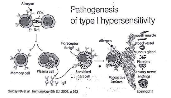
Type I Hypersensitivity:

It is also known as immediate or anaphylactic or atopic hypersensitivity. Type I hypersensitivity is generally caused due to reexposure to the specific type of antigen or allergen(substance which causes allergy). It can lead to systemic or local reactions and involves eyes(conjunctivitis), skin(eczema), nasopharynx(rhinitis) and gastrointestinal tracts(gastroenteritis). Symptoms generally varies from mild to even fatal and can lead to death or anaphylactic shock. The time of reaction ranges from 10-15 minutes after the action of allergen, but sometimes there is delayed onset(10-12 hours).

Mechanism:

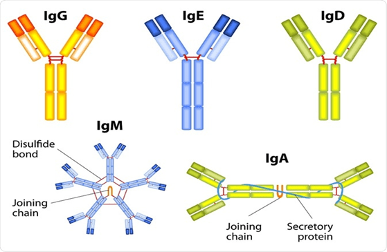
  1. The reaction is mediated by IgE antibody. Primary cellular component involved is mast cell or basophil cells.
  2. Upon very first exposure to allergen, APC(antigen presenting cells) processes the antigen and present it to Th2 cells.
  3. Upon released of IL-4 and IL-12 by Th2 cells, B cells gets activated.
  4. After that, the B cells differentiate into plasma cells. Plasma cells synthesise and secretes IgE antibody (type of antibody usually involved in allergic reactions).
  5. IgE binds to FC(fragment crystallization) of mast cells and sensitize it.
  6. Upon subsequent exposures to the same antigen, now the mast cells bind to IgE antibody and releases the inflammatory molecule. This will result in allergic symptoms.

Diagnosis:

  • Involves skin tests( pricks and intradermal samples)
  • Specific and total IgE antibody measurements against the suspected allergen by using ELISA(Enzyme linked immunosorbant assay).
  • Increased level of IgE in the ELISA test, will confirm allergy or atopic condition.

Treatment:

  • Anti-histamines for symptomatic treatment. Anti-histamines can block the histamine receptors.
  • Cromo Lyn sodium, a chemical can inhibit mast cell degranulation. It does it by inhibiting ca+2 influx.
  • IgG antibodies can be used as it can bind with IgEs FC portion and prevents mast cell sensitization.

Examples of type I hypersensitivity: Conjunctivitis, Eczema, Eosinophilia, Angioedema.

Type II Hypersensitivity:

This type II hypersensitivity is also known as antibody dependant or cytotoxic hypersensitivity and is known to  affect many organs and tissues. The antigens are normally endogenous. However, some exogenous chemicals or molecules such as haptens(substance which combined with proteins can produce antibodies) can also induce type II hypersensitivity. Reaction time is usually from few minutes to hours.

Mechanism:

  1. IgG and IgM antibodies bind to the antigen and form complexes. This complexes than results in activation of compliment pathway(pathway through which foreign antigens are destroyed).
  2. At the site of membrane attack complexes, mediators of acute inflammation are generated, which will lead to cell death and lysis.
  3. Phagocytes, expressing FC receptors can induce phagocytosis.
  4.  The FC receptors, recognises surface bound antibody and compliment proteins .
  5. ADCC(antigen dependant cell mediated cytotoxicity) is another form of hypersensitivity where tags of IgG and IgM antibodies bind. Macrophages and NK(natural killer cells) than recognises this tags and kills them.

Diagnosis:

  • It includes detection of circulated antibodies against the tissues involved.
  • Detection of presence of antibodies in the liquid biopsy through immunofluorescence.

Treatment:

  • Anti-inflammatory agents
  • Immunosuppressing agents

Examples of type II hypersensitivity:  Erythroblastosis Fetalis, Hashimotos thyroiditis, transfusion reactions, Rheumatic fever.

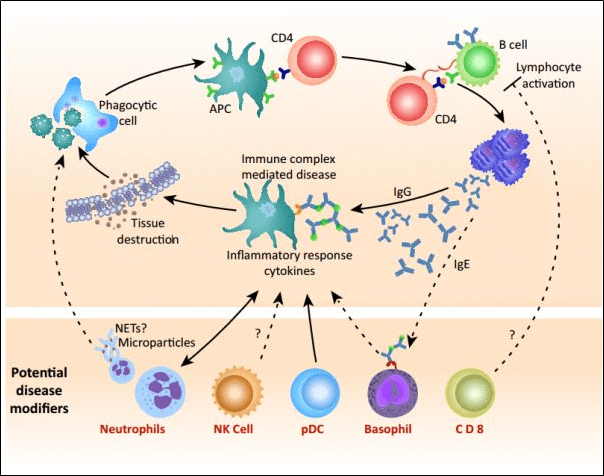
Type III Hypersensitivity:

Also called as immune complex hypersensitivity. The reaction is general or may involve organs such as skin, blood vessels, kidneys and joints. This reaction is caused primarily by micro-organisms. The reaction time is 3-10 hours after being exposed to the allergen.

Mechanism:

  1. Mediated by soluble immune complexes. Mostly the complexes are of IgG class but sometimes they can be of IgM class.
  2. Exogenous(viral or chronic infections) or endogenous (non specific autoimmunity) antigens.
  3. Primary components are compliment proteins( C3a, 4a and 5a).
  4. Major damage is caused by platelets and neutrophils.
  5. Macrophages and neutrophils are present in the lesions.
  6. Antibody deposition triggers an immune response according to classical compliment pathway.  There is formation of 2complexes(complex is formed when IgG and IgM are bounded to antigen)The larger complex gets eliminated but the first complex remains and the antigen antibody complex will spread and deposit.

Diagnosis:

  • Examinationof tissue biopsies for immunoglobulins and compliment by immunofluorescence microscopy.
  • Raji cell test and polyethylene glycol tests can be performed to measure immune complexes.

Treatment:

  •  Anti-inflammatory agents.

Examples of type III Hypersensitivity: Arthur’sreaction, Rheumatoid arthritis, symptoms of malaria.

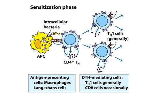
Type IV Hypersensitivity:

Also can be called as delayed type or cell mediated hypersensitivity. Classic example of this type of hypersensitivity is tuberculin reaction(montoux) which peas after 48 hours of antigen injection. Erythema and induration(thickening of skin) are the characteristics of the lesion.

Mechanism:

  1. Antigen in the complex is recognised by cytotoxic Cd8 T cells and Cd4 helper T cells by MHC I or MHC II complex.
  2. IL-1 secreted by macrophages further helps in proliferation of Cd4 T cells.
  3. Th1 mediated response is elucidated upon reexposure to antigen.
  4. Cd4 T cells secretes IL-2 which further induces the release of other type 1 cytokines and thus induce immune response.
  5. Cd8 cells when activated, destroys target cells upon contact. Macrophages produces hydrolytic enzymes and when comes in contact with intracellular pathogens, macrophages transforms into multinucleated giant cells.

Diagnosis:

  • Montoux test and patch test in-vivo is generally performed.
  • Some of the in-vivo tests for delayed hypersensitivity can be mitogenic response, nephro-cytotoxicity, and production of IL-2.

Treatment:

  • Corticosteroids
  • Immunosuppressive agents

Examples of type IV Hypersensitivity: symptoms of tuberculosis, coeliac disease, symptoms of leprosy.

Type V Hypersensitivity:

There is an additional type of hypersensitivity that is, type V(5). This usually occurs when the IgG antibodies have a effect towards their target. In this, the antibody binds to the cell surface receptors rather than binding to cell surface components and as a result it prevents the binding of the intended ligand to the receptor and thus impairs cell signalling.

Example of type V: Graves disease.

  • Diagram showing all four hypersensitivity reactions.

CULTURE MEDIA FOR PLANT TISSUE CULTURE

BY: RAHUL ANDHARIA (MSIWM001)

Plant Tissue culture:

It is an in-vitro aseptic culture method in which plant tissues or cells are grown in an artificial medium under suitable environmental conditions generally to produce clones of plants. The medium used to grow this tissues or cells is known as culture medium. There are various culture medium used based on different requirements.

Principle of Plant Tissue culture and culture medium:

Many plants have the ability to regenerate into a new whole plant which is called as Totipotency. Protoplasts(plant cells without cell wall), pieces of leaves, single cell often is used for generation of new plant using appropriate plant culture medium. Specific environment is created to provide maximum growth and multiplication of the plant in the culture medium. The conditions include providing proper nutrient medium, maintaining temperature and pH and proper liquid and gaseous environment.

Culture medium facilities in-vitro growth and morphogenesis( differentiation of unfolding of undifferentiated cells) in plants. The success of tissue culture lies in the selection of plant culture medium. If an appropriate medium is selected, better results can be obtained. Generally, plants can synthesise their own food material, but the in-vitro plants are primarily heterotrophic, that is they cannot synthesise their own food, so it becomes essential that plant culture medium should consist of the same nutrients as required by the whole plant.

Composition of Culture medium:

Basically there are two factors inconsideration for the composition of plant culture medium:

  1. Species of the plant- composition of the medium very much depends on the species of the plant used in tissue culture. Each plant specie has different requirements and also requires different environmental conditions for their growth.
  2. Type of culture material- The composition also depends on the type of material used such as protoplasts, single cells or pieces of leaves. Each material requires different composition.

Thus, composition of culture medium is dependant on the specific requirements of the culture system and it’s formulation. The type of medium used can be solid or liquid medium. The choice of medium completely depends on the culture response and it’s growth.

pH of Medium:

Most optimum pH for tissue culture is in the range of pH 5-6. pH can be adjusted during sterilization, but usually falls after autoclaving. At a higher pH of 7 and lower pH of 4.5 plant cells will no longer grow in culture medium.

General method for medium preparation:

  • Prepare stock solutions using high purity chemicals and demineralised water.
  • Stock solutions can be stored in glass or plastic containers in freezer and can be used whenever needed.
  • As most of the growth regulators are not water soluble dissolve them first in NAOH or alcohol.

Rules for Selection of appropriate culture medium:

Each specie has different culture requirements and therefore selecting an appropriate medium is an integral part of plant tissue culture.  De Fosard Et al has described an experiment to select an appropriate culture medium. This is very beneficial especially if the culture system used is not tested.

The steps are as follows:

  1. Medium is divided into four different categories (a)  minerals, (b) auxins, (c) cytokines and (d) organic nutrients like sucrose, amino acids.
  2. Choose three different concentrations for each of the components that is high, medium and low.
  3. Try various combinations with the chosen four components. At the end it will lead you to 81 different treatments.
  4. Denote the four letter code the best among the 81 treatments. For example a medium containing medium salts, low auxin, low cytokinin and high organic nutrients can be represented as MLLH.
  5. At this stage, test the concentrations of auxins and cytokinins to  assess the suitable concentration of the growth regulators.

Types of Culture medium:

There are many number of culture media available. Some of them are listed below:

  • MS ( Murashige and Skoog) medium:

As the name suggests this medium was developed by the two scientists, Murashige and Skoog in the year 1962, while working on plant growth regulators. This is the most common culture medium used in labs. Basic formulation includes inorganic salts, nutrients and amino acids.

Purpose: This medium is used to induce callus culture, organogenesis, and micropropogation.

  • LS (linsmaier and Skoog) medium:

This medium was developed in the year 1965. Initially, the medium was used to optimise organic nutrient supplements in Tobacco culture. This medium contains Thiamine hypochlorite and inositol. Inositol is primarily involved in primary and secondary metabolism of plants and also is an cofactor in glycolysis and TCA cycle.

Purpose: used in induction of callus culture(growing mass of parenchyma cells is, callus) organogenesis, micropropogation and cell suspension.

  • Gamborg (BY) medium:

The medium was particularly used for cell suspension and callus generation of glycine max belonging to plant family, fabaceae by Gamborg in the year 1968. Formulation of medium generally includes inorganic salts, carbohydrates and vitamins. Generally, this medium has high concentration of nitrate and potassium and lower concentrations of ammonia. Soybean root callus formation is being induced by potassium nitrate, while ammonium sulphate helps in plant cell growth.

Purpose: used mainly in protoplasts culturing.

  • NN ( Nitsch and Nitsch )medium:

This medium was primarily first used by Nitsch, to develop anther culture in Nicotiana, which belongs to family solanaceae. This medium is rich in thiamine, folic acid and biotin which supports anther growth.

Purpose: Particularly used in in-vitro anther culture.

  • White’s medium:

The medium was developed for root culture of Tomato by P.R white in the year 1963. This was the first medium to be developed for root culturing. The medium has lower concentrations of salts and has higher concentration of Mgso4.

Purpose: widely used for shoot and callus culture. It is more appropriate for the culture of Musa and Daucus species.

Constituents of culture medium:

Elements play a vital role in plant growth and regulation. Elements differ in their physiological functions.

Inorganic nutrients:

It consists of micro and macro nutrients. Concentration of macronutrients is greater than  0.5mmol/1-,  while for micronutrients it’s less than 0.5mmol/1-.

  • Macronutrient elements:

It consists of six different types of elements mainly nitrogen, phosphorus, potassium, calcium and sulphur. Ideal concentration for calcium, phosphorus, sulphur and magnesium is 1-3mm mol, while for potassium and nitrogen it is 25mm mol.

  • Micronutrients:

They are required in smaller amounts by tissues and cells. Commonly  iron, manganese, copper, zinc, molybdenum and boron are the micronutrients. Iron requirement in the medium is very vital. Iron and copper in chelated form are used in the medium.

  • Carbon and energy sources: Tissues and plant cells in the culture are usually heterotrophic, so they require external source of energy and carbon for their growth. Sucrose is the most commonly used form of energy source. During autoclaving process, sucrose will hydrolyse into glucose and fructose. Plants uses more amount of glucose than fructose and glucose is considered as efficient as sucrose.
  • Organic supplements: They include vitamins, amino acids, organic extracts, activated charcoal, organic acids and antibiotics.
  • Vitamins plant cells produce vitamins but in less amounts. So it is necessary to add vitamins to the medium. Vitamins like thiamine, riboflavin, niacin, biotin, folic acid, pantothenic acid and ascorbic acid are added.
  • Amino acids: organic nitrogen in form of amino acids like L-arginine, L-glutamine, L-cysteine and L-arginine are more easily taken up by plants than inorganic nitrogen.
  • Organic extracts: Yeast Caseinhydrolysate, orange juice, tomato juice, coconut milk are used.
  • Activated charcoal: activated charcoal can stimulate growth of certain plant cells like carrot, tomatoes and orchids. Also it can remove certain toxic components from the medium which can otherwise hinder in cell growth.
  • Organic acids: succinate, citrate, fumarate and malate helps in plant growth.
  • Antibiotics: antibiotics bare generally added to prevent growth of microbes. Low amounts of kanamycin and streptomycin are added.
  • Growth Regulators: Phytohormones promote plant growth, differentiation and development.
  • Auxins: it induces cell elongation, cell division and formation of callus in cell cultures. At lower concentrations, auxins can promote root formation and high concentration it helps in callus formation.
  • Cytokinins: cytokinins are derivatives of adenine. They are involved in shoot differentiation, cell elongation and in somatic embryo formation. It also helps in promoting RNA synthesis and thereby stimulating enzyme and protein activity.
  • Gibberellins: Ga3 is the most commonly used gibberellin among 20 different types of gibberellins known. Ga3 helps in growth of cultured cells, enhances growth of the callus and makes dwarf plantlets to elongate. They are generally known to promote or inhibit tissue cultures depending on the species used.
  • Abscisic acid:   plays major role in induction of embryogenesis in the culture. Also helps in callus formation.
  • Solidifying agents: solidifying agents are used in preparation of solid medium or semi solid medium. Most commonly used agents are:
  • Agar: It is a polysaccharide extracted from seaweeds. Main advantages of using agar is that it cannot be digested by plant enzymes, remains stable at cultural temperature and doesn’t react with the media constituents. 0.5 – 1% concentration of agar can form gels.
  • Gelatine: it is used at higher concentration(10%) but has limited success, as it melts at lower temperature so has limited use.

Considering all the above factors, culture medium is the most important part of tissue culture. Without proper culture medium, tissue culture cannot be performed.

Isolation And Culturing Of Microbes From Food

Theory:

  • Microorganisms are abundant and found in food stuff as well.
  • The microbes found on food can be pathogenic in nature and may cause various diseases.
  • The identification of such microbes is necessary to study and research on infectious agent.
  • Primary culture from food stuff is a mix culture of various microbes which has to be isolate form each other and identify for research.
  • The procedure below is used to isolate and cultivate pure culture form food stuffs.

Requirements:

  • Nutrient agar plate
  • Food sample
  • Spreader
  • Micropipette
  • Test tube

Procedure:

  • Take nutrient agar plate and well label them.
  • In one test tube take 4.5 ml autoclaved water and add small quantity of food sample in it
  • Mix thoroughly, after that with the help of micropipette take 100 microliter of the mixture and spread onto the petri plate with a spreader.
  • Finally put the petri plates in the incubator for 18-24 hours at 37o C.
  • After the incubation period mark the different colonies with marker.
  • Now pick each single colony with an inoculating loop and streak on nutrient agar plate.
  • Put the petri plates inside the incubator for 18- 24 hours at 37o C.

Observations:

  • After 18-24 hours examine the plates for bacterial growth.

Result:

  • Record the result of isolated colonies in tabular form.

DNA: REPLICATION, TRANSCRIPTION & TRANSLATION

DNA: Replication, Transcription and Translation

Content:

About:

  • The process of producing identical replicas of DNA from original DNA molecule in known as DNA replication.
  • It is the most important part of inheritance and occurs in all living organisms.
  • DNA consist of two strands which are complementary to each other during the process of DNA replication these tow strand get separated and serve as template for the synthesis of new complementary strand this is known as semiconservative replication.
  • After cellular proofreading and error checking mechanism new DNA molecule is synthesized with one strand from parent DNA molecule and another is fresh.
  • Each cell of a human being contains 3 X 10⁹ base pairs of DNA distributed over 23 pairs of chromosomes.
  • In human genome 95% region do not code for any protein known as introns.
  • According to central dogma DNA makes RNA and RNA makes protein.
  • The process of producing RNA from DNA is called transcription and synthesis of protein from RNA is called translation

Steps Involved:

  • DNA replication
  • Transcription
  • Translation

DNA Replication:

  • When cell divides, the double stranded DNA splits into single strand and makes new copy of the genome which gets transferred into each cell.
  • Base pairing is according to the Watson-Crick paring of the DNA strand.
  • It is a complex process which includes array of enzymes.
  • Synthesis of DNA proceeds in the 5’-3’ direction and for attachment of DNA polymerase first DNA strand has to be unwind which is regulate by the helicase enzyme.
  • During the synthesis of the new strands by the DNA polymerase one strand is synthesized continuously and called leading strand on the other hand the opposite strand known as lagging strand synthesised in to the small fragments called okazaki fragments.
  • In bacteria there are three distinct DNA polymerase: pol I, pol II and Pol III. Pol III largely involve in chain elongation. DNA polymerase itself cannot initiate DNA synthesis it require a short primer with free 3’ hydroxyl group.
  • Once the primer attaches to the DNA strand Pol III then take over and synthesis of new strand begins.

Transcription:

  • The process by which DNA is copied to mRNA is called transcription.
  • The mRNA contains information for the protein synthesis.
  • Transcription occurs in two steps. In first step pre mRNA is synthesized with the help of RNA polymerase, the resultant RNA strand is reverse complement of the original DNA sequence. Then after pre messenger RNA is edited and forms the mRNA molecule by the process of RNA splicing.

RNA Splicing:

  • The pre-messenger synthesized contains introns and exons in it, introns are the sequence which does not code for any protein hence it is not required in protein synthesis.
  • Thus the pre- messenger RNA is chopped up and introns are removed from it which synthesise messenger RNA contain only exons.

Translation:

  • The mRNA synthesised in the transcription process is now transported outside the nucleus into cytoplasm to the ribosome.
  • Messenger RNA does not directly involve in the protein synthesis transfer RNA is required the process of protein synthesis is called translation.
  • The mRNA contains three base stretch called codon and each codon contains information for a specific amino-acid. While the ribosome passes through the mRNA each transfer RNA molecule anticodon interacts with codon of mRNA.
  • The transfer RNA contains amino acid at 3’ end which is added to the growing protein chain and the t-RNA expelled from the ribosome.

Transfer RNA:

  • Transfer RNA is tertiary structure which is represented in two dimensions as cloverleaf shape.
  • Each amino acid has its own unique t-RNA like t-RNA for phenylalanine is different from that of histidine.
  • Attachment of amino acid is at 3’- OH group of t-RNA.

Nucleic Acid Hybridization

BY: RAHUL ANDHARIA (MSIWM001)

Nucleic Acid Hybridization: It is a technique which involves interaction of single stranded Nucleic acids(DNA/RNA) to form complexes called Hybrids, which contain the similar complementary sequences as that of nucleic acids. This technology helps us to understand the sequence identity among nucleic acids and to determine and detect specific sequences.

History:

 In the year 1975, Grunstein and Hogness developed this method.

Principle:

In simpler terms it is a technique that simply detects specific DNA/RNA sequences using specific Probes. By using this technique we can determine sequence of  hundreds of clones from a particular colony which exhibits it. Bacterial Colonies are transferred from agar plates to nitrocellulose membrane where they are lysed by using alkaline solution.

After cell lysis, due to the negative charge of nitrocellulose membrane, the DNA and proteins gets adhere to the membrane. To remove the bounded protein, the membrane is dipped in Proteinase k solution. Upon exposure to UV rays, the DNA gets fixed to the nitrocellulose membrane. After baking, the membrane gets exposed to labelled RNA for hybridization. The hybrids can be monitored by Autoradiography. Colony giving positive result must be picked from the master plate.

Procedure overview of Nucleic acid hybridization:

  1. Probe, (DNA/RNA fragment which can be fluorescently radiolabelled ) is required which will anneal to the target nucleic acid.
  2. The next step is the attachment of target to a solid matrix, like for example a membrane.
  3. Denaturation (breaking of hydrogen bonds) of the target and the probe used.
  4. The denatured probe is than added to a target in a solution (Proteinase k sol)
  5. If sequence homology exists between the probe and the target, the probe will than hybridize or will anneal the target.
  6. The probe which is hybridised than can be visualised by methods like Autoradiography, Chemiluminescence or Colorimetry.

Hybridization Probes:

Probes are DNA or RNA fragments that can be radiolabelled. Probes help in identifying complementary segments in nucleic acid of Micro-organisms. The probe gets hybridised to single strands because of its complementarity with the target.

To make the probe hybridise with target sequence, it is labelled with molecular marker like radioactive 32-P ( radioactive isotope of phosphorus which incorporates into phosphodiester bonds of DNA).

Types of Probes:

  1. DNA Probe: Single stranded DNA is used to detect the complementary sequence amongother ss molecules. Therefore, it is a short sequence radiolabelled with radioactive isotope to detect complementary nucleotide sequence.
  2. RNA Probe: Single stranded RNA is used to detect the complimentary sequence among other ss molecules. RNA probes are also called as Riboprobes. RNA-RNA hybrids are known to be more stable than RNA-DNA hybrids generally because RNA hybrids shows more significant kinetic hydration.
  3. Oligonucleotide probes: refers to a short sequence of nucleotides which are synthesized to mimic the regions where mutations have occurred. They are synthesized in laboratory. Initially, a mononucleotide is attached to a solid support, and than one by one other mononucleotide are added to the 3’ end region. They are labelled with molecular markers at the 5’end.

Labelling of Hybridization Probes( DNA/RNA)

Involves 2 different types of methods:

  1. In vivo labelling: labelling of nucleotides in cultured cells.
  2. In vitro labelling: use of an enzyme to label the probe.

Strand synthesis labelling for DNA probes:

Most common method used to label DNA. In this method, enzyme DNA polymerase is used to attach labelled nucleotides to the DNA copies in the starting DNA. Any one of the nucleotide is labelled. There are various methods for DNA labelling:

  1. Nick Translation labelling: method in which DNA fragment is treated with DNase enzyme to induce single stranded nicks. Labelled nucleotides from nicked sites are incorporated by DNA polymerase I . Nick sites are replaced by DNA polymerase I that elongates 3’hydroxyl activity, thereby removing nucleotides by 5’-3’ exonuclease activity and replacing it with dntps.

  • PCR Method:  PCR method of amplification of DNA allows the resulting product to label either with modified nucleotide or oligonucleotide primers. With this, we can thus incorporate labelled nucleotides in the PCR reaction mixture which will than result in production of  labelled PCR products throughout its length. This method has higher yield even with a little template.

Strand synthesis labelling of RNA probes:

Most common method used to label RNA. In this method, enzyme RNA polymerase is used to attach labelled nucleotides to the RNA copies in the starting RNA. Any one of the nucleotide is labelled. There are various methods for RNA labelling:

  1. In vitro transcription from cloned DNA inserts: The RNA probe gets transcribed from a linear DNA template with the help of bacteriophage DNA dependant-RNA polymerase enzymes from bacteria like salmonella (sp6) and E.coli(T3 and T7). One major advantage is with this method the labelled probes are completely free of the vector sequences.
  • 3’end labelling RNA:  Short DNA template has to be designed to anneal 3’end of RNA, with a 5’overhang of 3’ TA-5′. The klenow portion( large fragment of protein) of DNA polymerase I can than extend 3’end of RNA by incorporating single 32P radio labelled dAtp.

Uses of Nucleic Acid hybridization:

  1. Testing frequency of relatedness between 2 DNA molecules:

Sample of DNA 1 is heated to melt it to single stranded DNA. The ssDNA is than  attached to a appropriate filter. Now chemical is being added to filter to block the sites, that can bind to DNA. After melting, DNA 2 sample is poured. Now some molecules from sample 1 will base pair with sample 2. The more closer the molecules are related, more hybrids it will generate. This method is highly useful, for example DNA of human gene, haemoglobin generated is melted and filtered, than it can be used to test the DNA from same gene of some different species.

  • Isolating Genes for Cloning:

Suppose we have say human haemoglobin gene and want to isolate corresponding monkeys gene, than human DNA is added to filter and the monkeys DNA is cut into segments with the help of restriction enzymes. The monkey DNA is than heated to make it to single strand and poured in filter. The DNA fragment carrying monkeys haemoglobin gene will now bind with human haemoglobin gene and gets stuck in the filter. Other genes will not hybridize. This is a very selective approach and provides hybridization of new genes from related or similar kind of genes.

GENETIC ENGINEERING

BY: RAHUL ANDHARIA (MSIWM001)

Genetic Engineering: It refers to altering the genetic material of an organism by insertion or removal of individual genes from another organism. In other terms it simply means manipulation of genetic make-up of an individual by artificial means. The organism that receives the genetic material is termed as transgenic organism.

History:

Father of Genetic engineering is Paul Berg. The process of Genetic engineering was first showcased by Paul Berg when he introduced the viral gene in bacteria with the help of lambda phage. Transgenesis, process of transferring of genes from one organism to other was first pioneered by Herbert Boyer and Stanley Cohen, in the year 1973.

Principle of Genetic Engineering:

There are several steps associated with Genetic Engineering:

  1. Isolation of gene of interest to be cloned from desired organism.
  2. Transfer of the isolated gene to create recombinant DNA molecule. This can be done by cutting the DNA molecules at specific sites by using Restriction Enzymes.
  3. Restriction enzymes cut DNA at specific locations. There are basically 4 types of restriction enzymes designated as I, II, III, and IV. All this four enzymes primarily differs in their structure, site of cleavage and some cofactors (substance which is required for enzyme activity).
  4. By the action of RE, DNA is left with sticky ends (short portion of unpaired bases). A bacterial plasmid is also cut with the same RE, so that the plasmid also has the similar sticky ends and can base pair. (Plasmids are extra-chromosomal DNA found inside the bacterial cell).
  5. The plasmid and the isolated gene are joined together by an enzyme called DNA ligase.
  6. The two pieces of DNA with same sticky ends (as cut by same RE) are being linked together by DNA ligase and forms a single, unbroken molecule.
  7. Than the genetically engineered plasmid is inserted into bacterial cell (Transformation). The gene insertion usually takes place by means of Vectors. (Molecules that carry the gene of interest.) choice of vectors depend on the type of gene to be inserted.
  8. When the bacteria reproduce, the plasmid will get copied and this recombinant plasmid spreads as bacteria multiply and expresses the gene and makes a human protein.

This is the basic procedure required to obtain a desirable gene and this whole process together refers to Genetic Engineering.

Various types of Genetic engineering techniques:

  1. Recombinant DNA technology:

Technique in which artificial DNA fragment is created by ligation of two different DNAs using various physical methods.

In this technique basically the gene of interest is inserted into a plasmid vector and can be used for gene transfer experiments. Two methods are commonly in use which includes:

  1. Plasmid method- In this method small pieces of circular DNA called plasmids are used for altering microorganisms like bacteria.
  2. Vector method- vectors are small carrier molecules that carry viruses. Generally, viruses have capsule, in which the DNA is present. When the viruses attach to the host cell they insert its DNA or RNA. The DNA in the host genome replicates itself by using host machinery and the gene inserted will become part of the host machinery.
  • Gene delivering:

This technique is employed to insert gene of interest directly into host genome. Some of the most popular gene delivering techniques which include:

  1. Electroporation- introduction of DNA into cells by using electric current. It is a rapid method to create more number of recombinants within a shorter duration of time.

The mixture containing cells and DNA are placed in a glass chamber. The glass chamber contains two electrodes. A single electric pulse of about 4000-8000 volts/cm is generated between the two electrodes for about 4-5 milliseconds. The electric current forms transient pores in the cell membrane through which the DNA seems to enter into the cells.

  • Liposome mediated gene transfer: liposome is a small vesicle made up of phospholipids used to deliver foreign DNA into cells. Lipids like phosphetidyl choline, Cholesterol are useful in making liposomes. Outer lipid bi-layer generally fuses with the cell membrane and releases the contents.
  • Gene editing:

The technique is used to edit the genome in which an undesired or diseased gene can be edited or replaced by a new gene in the host genome.

Some of the gene editing systems used are: CRISPR-CAS9, TALEN( Transcriptional activator like effector nuclease) and  ZFN( zinc finger nuclease).

Among this three CRISPR is the most widely used method currently. Bacteria generally possess this method in their genome to combat viruses. CRISPR normally  stands for clustered regulatory interspaced short Palindromic repeats. The nuclease, CAS9 cuts the DNA strands of foreign DNA and pathogens and thus destroys them.

Process overview of Genetic engineering:

  • Isolation of desired gene
  • Selection and plasmid construction
  • Gene transformation(uptake of DNA by cells)
  • Insertion of DNA into a suitable host
  • Insert confirmation- done by using various selectable markers.

Applications and future aspects of genetic engineering:

Genetic engineering has great prospects. It is highly practiced in Industrial and agricultural field. It has wide range of applications in the field of medicine, genetic research, and in therapeutic drug production.

  1. Plant Genetics: It is highly employed in plants to improve crops. Recombinant DNA technology in plants help to have better yield, to control pathogens, delayed fruit ripening, stress tolerant plants.

Classic example is Bt cotton, most famous Genetically modified crop.

  • Genetically engineered Insulin: human insulin gene is inserted into the gap in plasmid. The plasmid becomes genetically modified. The plasmid than upon insertion into new bacteria or yeast cell will produce insulin.

FOOD SPOILAGE

BY:- RAHUL ANDHARIA (MSIWM001)

Food Spoilage:

It refers to change in Physical and Chemical property of food, making food unfit for Consumption. Invasion of microorganisms like bacteria and fungi usually causes spoilage of food.

Principle:

Food spoilage generally occurs due to Physical, Chemical or Biological agents that changes colour, flavour, appearance, odour and other properties of food. Shelf life of most of the natural foods is very less and is perishable, for example, meat, fish and bread can spoil easily. Decomposition of food generally involves 3 processes: Putrefaction (chemical breakdown of food or decay of organic matter), Fermentation (chemical breakdown of substances by action of microorganisms, yeast), Rancidity ( refers to oxidation of fats).

Natural Contamination:

It refers to contamination of food when microorganisms themselves attaches to food in its growing stages and this kind of contact is essential for certain kinds of food. For example, Yeasts contaminates fruit for carbohydrates fermentation.

Artificial Contamination:

This type of contamination occurs during handling of food when food is under various stages of production like, packaging, storage, etc. Improper handling of food during this stages results in contamination of food by microorganisms.

Intrinsic factors of food like pH, redox potential, H2o activity determines the type of microflora growing on the food. This final composition of microflora is responsible for food spoilage.

Types of Food Spoilage:

1.Microbial Spoilage:

Microorganisms associated with food are:

Bacteria, Filamentous Fungi, Viruses, Yeasts, and animal parasites.

Bacteria:

They are associated with both plant and animal foods. Bacteria are associated with food intoxication and spreading of food borne diseases.

Examples:

Acenatobacter Gram negative- present in raw and prepared foods like beef and poultry carcasses.

Aero monas: gram negative, responsible for spoilage of fish.

Alkaligans: gram negative, responsible for spoilage of egg and dairy products.

Citrobacter: gram negative, it is responsible for spoilage of vegetables and fresh meat.

Corynebacterium: gram positive, involved in spoilage of vegetables and  meat.

Filamentous Fungi:

When food is left for one or more day covered, tangled mass of furry growth appears on food which is called fungi or mould. Fungi are responsible for spoilage of Grains, nuts, and fruits as they have low pH and H20 activity.

Examples:

Mucor: Zygomycotena-common contaminant of fruits, berries and nuts.

Rhizopus: Zygomycotena- known commonly as bread mould. It is more prevalent in fermented and stored foods.

Claviceps: Ascomycotena-  produces toxic alkaloids in cereals, when consumed can cause Hallucinations.

Yeasts:

Contamination by yeasts results in Souring of milk.

Examples:

Candida: most common contaminant of dairy products, fresh fruits, and alcoholic beverages.

Saccharomyces: spoilage of fruits and fruit products.

Torulopsis: responsible for spoilage of beef, creamed butter, condensed milk, etc.

Viruses:

viruses found in food are termed as enteric or intrinsic viruses.

Examples:

Enterovirus, Adenovirus, Reovirus, Hepatitis A virus.

Animal Parasites:

They belong to 3 distinct groups:

Protozoa: Giardia, Entamoeba Hystolytica

Flatworms: Taenia, Fasciola

Roundworms: Ascaris

2. Physical Spoilage:

Physical Spoilage refers to damaging of food during Harvesting, Processing or distribution of food. During such processes there are high chances of food spoilage if proper measures are not followed. The damage increases the chance of spoilage as the outer layer is completely broken or bruised. For example- Canned foods gets spoiler easily if the cans are not properly packed with lid or are contaminated during processing.

3. Chemical Spoilage:

Chemical reactions in food are responsible for change of colour, texture and taste of the food products. Generally foods are fresh especially vegetables and animal food, but after harvesting and slaughtering, chemical changes begin automatically in the food and the quality of food becomes deteriorated.

4. Enzymic Spoilage:

Enzymes acts as biological catalyst to carry out biological reactions in cell and play an important role in biochemical reactions. After death of cells or tissues, enzymes play a role in its decomposition by a process called Autolysis( self destruction )

Example: In tomatoes, some enzymes helps it for ripening, but at the same time there are certain enzymes which are responsible for its decay. Once enzymic Spoilage is underway, it damages the outer skin of tomato and exposes it to mould growth and decay.

Factors Affecting Food Spoilage:

  1. Water Content: Amount of water holding capacity in foods is referred to as it’s water activity.(WA). Water activity of most of the fresh fruits is approximately 0.99, which makes them more susceptible to microbial growth.
  2. Environmental Conditions: Environmental influence on food is the major concern. When food is exposed to intrinsic conditions like temperature, air, or even small amount of moisture, can result in growth of Micro-organisms. Changing environmental conditions can help to prevent spoilage. For example- storing food at lower temperature can prevent it from spoiling.
  • Packaging and storage: Packaging of foods is after processing is very vital as it protects food from harmful contaminants and also from various other factors like environment, temperature, etc. The type of packaging plays a key factor in ensuring the safety and preventing spoilage. Food packed in jars, cans ensures safety and prevents food from dust, moisture, air and harmful microbes.

Sources of Micro-organisms for Food Spoilage:

Micro-organisms are present everywhere. General source of Micro-organisms include air, water, sewage, soil and animal wastes. Foods grown in ground have higher risk of spoilage due to micro-organisms.  Foods like fish, meat are contaminated by presence of bacteria in their  internal organs like skin and feet. Meat has higher tendency of contamination as raw meat attracts lot of microbes, so it is advisable to store raw meet immediately after chopping.

Ways to Prevent Food Spoilage:

  • Ensure proper packaging is available to the food cans and jars after processing.
  •  Don’t leave the food in open air for more than 15min, to avoid contact with microbes.
  •  Ensure that your refrigerators are operating at correct temperatures.
  •  Food must be protected from light and must be stored in amber colour or transparent containers.
  • Low temperature is a key as it retards microbial growth.
  • Avoid placing food where there is more humidity, as high humidity attracts more growths of microbes and moulds. Placing food in dry places is most appropriate.

GENETIC CODE

GENETIC CODE:

CONTENTS:

  • Introduction
  • Protein synthesis (translation)
  • Variations in genetic code
  • Wobble
  • Universal code

INTRODUCTION:

BIOSYNTHESIS OF PROTEINS (TRANSLATION):

The final step in the expression of protein synthesizing genes is translation. The process of Protein synthesis is called translation because it is a decoding process of highly coiled substance. The information encoded in the language of nucleic acids must be written in the languages of amino acid forming proteins. During translation, the sequence of nucleotide is “read” in the appropriate sets of three nucleotide, each set being called a codon. Each codon codes for single amino acid.

  • The sequence of codons is “read” in only one way-the reading frame- to give rise to the amino acid sequence of a polypeptide. Deciphering the genetic code was one of the great achievements of the twentieth century.  

Close inspection of the code reveals several features that are related not only to the way cells use DNA to store information but also to why it is valuable for storing data.

VARIATIONS:

  • One feature is that the code words (codons) are three letters (bases) long; thus one small “word” conveys a significant amount of information. Each codon is re-organised by an anti-codon present on a tRNA molecule.
  • Another feature is that the code has “punctuation”. One codon, that is AUG, is always appears as the first codon in the protein synthesizing portion of mRNA molecules in very complicated and important process.
  • It is called the start codon because it serves as the start site for translation (a process of protein synthesis) by coding for the internal tRNA. Three other codons (UGA,UAG and UAA) are involved in termination of translation and are called stop or nonsense codons. These codons never encode for an amino acid and therefore they don’t have a tRNA bearing their own anti-codon.
  • Thus only 61 out of  64 codons in the code, are known as the sense codons which directs amino acid to incorporate into protein. Finally, the genetic code exhibits code degeneracy; that is, there are up to six different codons for a given amino acid.
  • Despite the existence of 61 codons, there are fewer than 61 different tRNAs. It follows that not all codons have a corresponding tRNA.
  • Cells can be successfully translated into mRNA using fewer tRNAs because there is not so strong pairing between the 5’ base in the anti-codon of the cell and the 3’ base of the codon which is almost tolerated.
  • Thus as long as the appearing first and second bases in the codon correctly base pair with an anti-codon, the tRNA which is bearing the correct amino acid will bind to all mRNA during translation process. This is evident on inspection of the code.

WOBBLE:

The codons for particular amino acids are most often different at the third position. This somewhat loose base pairing which is known as wobble, and it enhance cells the need to synthesize so many tRNAs. Wobble also decreases the effects of particular mutations.

UNIVERSAL CODE:

  • The description of the genetic code just provided is of the universal genetic code. However, there are exceptions to the code.
  • The first exceptions discovered were stop codons that encoded one of the 20 amino acids. For instance, some protists have a single stop codon (UGA); the other two stop codons are recognized by tRNAs bearing glutamine (Gln).
  • More dramatic deviations from the code have also been discovered. Proteins from members of all three domains of life have been discovered to contain the amino acid selenocysteine, the twenty-first amino acid.
  • Pyrrolysine, twenty-second amino acid, can be found in the proteins of several methanogens and at least one bacterium.
  • Genomic analysis indicates that pyrrolysine might also exist in many other bacteria and some eukaryotes. Selenocysteine is inserted at certain UGA codons, whereas pyrrolysine is inserted at UGA codons.

IMMUNE SYSTEM

Immunology: Immune System

And Its Type

Content:

  • About
  • Immune system
  • Immunity
  • Types of immunity
  • Innate immunity
  • Acquired immunity

About:

  • Immunology refers to the branch of biology that deals with the study of immune system in all organisms.
  • Any malfunction in this system lead to the diseases like autoimmune disease, hypersensitivity, transplant rejection and immune deficiency.
  • The term was given by Ilya Ilyich Mechnikov and received noble in 1908 for his work. He studied on the larvae of starfish, pinned thorns into the larvae and found unusual cells surrounds the thorns. This was the active response by the larvae. Mechnikov was the first to observe phagocytosis.

Immune system:

  • A different type of cells and molecules which protects our body from pathogens is collectively called immune system.
  • Pathogens may be anything from algae, fungi, bacteria to haptens (molecules that may cause an immune response).
  • The cells and molecules of the immune system were distributed all over the tissue of the body which plays role in elimination or prevention from pathogens.

Immunity:

  • Immunity is the ability of organism to protect itself from the disease causing organism.
  • Our body regularly comes in the contact of numerous pathogens but we few results into disease the reason is that our body develops antibody against the pathogen and protect from the disease.

Types:

  • Innate immunity
  • Acquired immunity

Innate immunity:

  • Innate immunity is the type of immunity with which the individual is born.
  • It serve as the first line of defence an provided various components like skin, mucus membrane phagocytic cells.
  • The mechanisms of innate immunity are anatomical barrier, physicochemical barrier, phagocytic barrier and inflammation.

Types :

Species immunity: one species are resistance but other are susceptible to the same infection. Example, birds are resistant to the anthrax but human doesn’t.

Racial immunity: one race is susceptible other is more resistant to same infection. Example, certain African race are resistant to malaria but Asian or Americans are susceptible.

Individual immunity: to a certain infection the individual of race or cast is resistant but other individual of same race is susceptible to the same infection.

Acquired immunity:

  • The immunity which the individual get during its life span after each microbial infection is called acquired immunity. Example is an individual was ever infected by any infection like chicken pox virus he/ she become life time resistant to chicken pox.
  • Antibodies and t-lymphocytes contribute in acquired immunity.
  • The t-cells and antibodies are specific to the pathogen hence acquired immunity is also known as specific immunity.

Types:

Active immunity

  • The active immunity is the condition when the host itself produces antibodies.

There are two types of active immunity

Artificial – by vaccination

Natural – by natural infection

Passive immunity

The condition in which the host doesn’t produces antibodies itself but antibodies developed in other host cell provides immunity known as passive immunity.

Two types:

Artificial – antibody introduced in the host body for immunity

Natural – antibody from the mother to foetus.

Antibodies:

Antibodies are the immunoglobulin found in the blood and serve as protection against substance like antigen.

Antigens are the protein or carbohydrate which activates the immune system.

There are 5 types of antibodies- IgA, IgM, IgG, IgE and IgD.

ACID-FAST STAINING

Acid-Fast Staining

Content:

  • About
  • Principle
  • Requirements
  • Procedure
  • Observation and result

About:

  • It was developed by Paul Ehrlich in 1882.
  • Acid fast staining is a kind of differential staining.
  • This method is used for the identification of mycobacterium and other bacteria which retain carbol fuchsin (primary stain) after the treatment of strong acid and methylene blue.

Principle:

  • Mycobacteraium contain a waxy substance composed of mycolic acid in its cell wall.
  • These mycolic acid are carboxylic acid with up to 90 carbon atoms chain.
  • Mycolic acid in addition with other lipids serves as barrier and prevents the entry of dye inside the bacterial cell.
  • The dye used in this method is carbol fuchsin (lipophilic dye) which binds with the acid and lipid in the cell wall and gives red colour.
  • Binding property of the dye is related to the carbon chain length of the mycolic acid.

Requirements and reagents:

  • Bacterial culture (fresh)
  • Carbol fuchsin
  • Acid alcohol
  • Methylene blue
  • Water bath
  • Glass slide
  • Inoculating loop
  • Blotting paper
  • Microscope

Steps:

  • On a clean slide prepare a smear of Mycobacterium smegmatis and Staphylococcus aureus on another slide.
  • Air dry and heat fix.
  • Pour some drops of carbol fuchsin on both the smears.
  • Place the slides in steam for 3-5 min, to avoid smear from drying add more stain time to time.
  • Cool the slide for some time and wash with distilled water.
  • Pour acid alcohol for 20-30 second to decolorize the smear or until the smear gives pink colour.
  • Wash slide with distilled water.
  • Add few drops of counter stain i.e. methylene blue to the smears for 1-2 minutes.
  • Wash and blot dry with a blotting paper.

Observation:

  • Observe under the microscope and record the colour test
  • Classify the bacteria i.e. acid-fast and non-acid fast.
  • Also describe their morphology and arrangement of cells.

Results:

  • M. smegmatis cells appear red coloured indicate acid fast reaction and S. aureus appear blue colour and show non-acid reaction.

Example:

TypesExample
Acid-fastMycobacterium smegmatis, Mycobacterium tuberculosis
Non-Mycobacterial bacteriaNocardia