BIOFILMS

BY: K. Sai Manogna (MSIWM014)

  • Layer-like microorganism aggregations and the extracellular polymers bound to a solid surface are biofilms.
  •  Biofilms, innate cells, are ubiquitous and are becoming more and more important in processes that are designed for pollution control including philtres that drip, rotating biological contactors and anaerobic philtres.
  • Biofilm processes are simple, efficient, and sustainable because natural immobilization allows excellent retention and accumulation of the biomass without the need for separate solids.
  • We should consider a fundamental problem concerning biofilms (indeed all aggregated systems) before designing mathematical instruments for predicting the removal of substrates by biofilm:

Here are a few possibilities:

1. Due to the advection of substrates after the film, these biofilms are exposed continuously to the fresh substrate in space. It means that if the biofilm is attached near the source of the substratum supply, the substrate concentration is higher.

2. In mandatory substratum transportation consortia or other synergistic relationships, different types of bacteria must be co-existent; for the exchange, the near juxtaposition between cells is required in a biofilm.

3. The biofilms build a more friendly internal atmosphere (for example, pH, O2, or products) than the bulk liquid. In other words, the biofilm creates special and self-created cell-beneficial micro-environments.

4. The surface itself produces a single micro-environment, for example by removing contaminants or by corrosive releases of Fe2+, a donor of electrons.

5. The surface allows the bacteria to change physiologically.

6. The cell’s strict packing modifies the physiology of the cells.

Biofilm formation:

The development of biofilms can be divided into five phases:

1. Surface relation

2. Monolayer formation

3. Training in microcolony

4. Biofilm mature

5. Unlocking

  1. The initial step, which is still reversible, is the initial touch of the moving planktonic bacteria on the surface.
  2. The bacteria then begin to form a monolayer and create a protective “slime,” an extracellular matrix.
  3. The matrix contains extracellular polysaccharides, structural proteins, cell scrap, and nucleic acids known as EPS.
  4. Extracellular DNA (eDNA) predominates the initial steps of matrix development, and polysaccharides and structural proteins later take over.
  5. At these points, microcolonies are produced that display significant growth and contact between cells such as quorum sensing.
  6. During the last step of a new cycle of biofilm formation, some cells in mature biofilm begin to detach itself into the environment as planktonic cells.

The biofilm allows for:

a. tolerates antibiotic attacks;

b. Trap bacterial growth nutrients and stay in a favourable niche;

c. Adhere and avoid flushing to environmental surfaces;

d. live in close collaboration and associate with other biofilm bacteria;

e. Stop phagocytosis and attack the complementary routes of the body.

For example, Planktonic Pseudomonas aeruginosa uses polar flagella to travel by water or mucus and to reach a firm surface as mucous membranes of the body. The cell wall and pil adhesives can then be used to bind the mucous membrane epithelial cells. Attachment stimulates signalling and quorum sensing genes so that a polysaccharide alginate biofilm synthesization will eventually begin with the population of P. aeruginosa. As the biofilm forms, the bacteria lose flagellum and secrete different enzymes that allow the population to obtain nutrients from the host cells. Finally, the biofilm pillows and establishes water canals for all bacteria in the biofilm to provide water and nutrients. As the biofilm gets too crowded with bacteria, the sensing of quorum helps a few Pseudomonas to develop flagella again, escape the biofilm, and colonize a new spot.

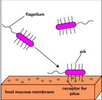
Fig: Formation of biofilm by P.  aeruginosa.

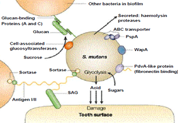
Two bacteria involved in dental caries, Streptococcus mutants and Streptococcus sobrinus, break down sucrose to glucose and fructose. Streptococcus mutans may use a dextransucrase enzyme in a sticky dextran polysaccharide, which forms a biopathic film that allows bacteria to bind to the tooth enamel and form the plaque. The pathogenicity of S bacteria. S. and mutans. In order to generate energy, sobrinus often ferments glucose. Glucose fermentations produce lactic acid, which is released on the tooth surface and causes decay. The mechanism is described in the given image.

Microbial Leaching:

  • Copper extraction from ore deposits was carried out over centuries using acidic solutions, but the involvement of bacteria was not confirmed in metal dissolution before the 1940s.
  •  Today about 10–20% of copper mined in the United States is extracted by low-grade microbial processing. In the expansion of microbial leaching, other elements, including Uranium, Silver, Gold, Cobalt and Molybdenum, are also invested considerably.
  • Most microbial liquidation relies on metal sulphides’ microbial oxidation. Aqueous environments combined with mineral waste create very harsh conditions with a low pH, high metal concentrations and high temperatures that select very specialized nutrient requirements for a microbial flora.
  • Heap leaching is the most common method in which copper and other minerals are extracted microbially from spent ore.
  •  The method involves arranging the spent ore fragments into a configuration of the packed bed to allow the passage of water. Acidized water (pH = 1.5-3.0) is sprayed on the porous ore bed in order to start the operation.
  • The solvent ferrous iron is actively oxidized, and sulphide minerals are attacked by acidophilic bacteria such as Thiobacillus iron, which can then be released from aquatic ions by releasing soluble cupric ions. In terms of the corrosion on metal surfaces, this oxidation is identical.

Biological reactions and mass transfer rates currently constrain the industrial applications of microbial fluid, but in recent years significant changes have been made to process design, and the mining industry sees the method as being promising.

Removal of Biofilms:

  • Traditional cleaning of biofilm has been accomplished by detergents, biocides, enzymes, and mechanical or physical methods of cleaning the biofilm.
  •  Biofilms in sensitive locations in the food and drink manufacturing industries have grown, leading to problems such as food spoilage, production quality and other nutrient supplementation and insufficient cleaning and disinfection.
  • Depending on the medium temperature and relative humidity of these microorganisms, they will live longer after application. The pulp- and paper-based agent for biofilm removal is classified into three groups: chlorine, chlorine dioxide, hydrogen peroxide (hydrogen peroxide), Ozone, antioxidants, and enzymes.
  • The cells adhering to the staphylococcus aureus are effectively used in sodium dichloro isocyanurate, hypochlorite, iodophore, hydrogen peroxide and peracetic acid. The most powerful method of extracting adhered large amounts of L was to eliminate peracetic acid.
  • Monocytogenes cells remaining on chips of stainless steel after sanitization. Scanning electron microscopy has shown that on chlorine and anionic acid-treated surfaces biofilm cells and extracellular matrices remain better than iodine and ammonium-quaternary detergent sanitizers from which a viable cell is not released.
  • Oxidative and antioxidant biocides have long been used, although enzyme use is being studied at present.
  • In order to develop a quality management programme in different industries, tanks, pipes, pasteurizers, coolers, membrane filtration units and fillers must be tested, because they help to avoid microbiological hazards and severe financial losses.

COMPONENTS OF BLOOD

BY: Reddy Sailaja M (MSIWM030)

BLOOD

Blood is a specialized body fluid, comprises of plasma and the cells that circulate throughout the human body. It supplies oxygen, glucose, antibodies, vitamins, electrolytes, heat, hormones, and immune cells across the body for life survival. It removes carbon dioxide and other waste generated from the cells in the body.

BLOOD COMPONENTS

Blood is made up of three main components: plasma, blood cells (red and white) and platelets.

Plasma: Plasma occupies 55% of blood fluid in human beings. Plasma comprises of 92% water and the remaining 8% contains carbon dioxide, glucose, hormones, proteins etc.

 Blood cells: Blood cells comprise of 45% of total blood fluid. These are produced in bone marrow by the process called ‘hematopoiesis’ from a common precursor cell (hematopoietic stem cells). Then these blood cells mature into red blood cells (RBCs), white blood cells (WBCs) and platelets. The organs like lymph nodes, liver and spleen regulate the generation, destruction and regulation of blood cells.

Figure 1: Blood cells production by hematopoiesis

Red blood cells: RBCs are also called erythrocytes. They are double concave shaped structures without nucleus. Approximately, 4.5 – 6.2 million/microliter are present in the blood. Their main function is to carry oxygen from lungs to all parts of the body. RBCs contain a special protein called ‘Hemoglobin’ that aids in oxygen transport. RBCs have a life span of 120 days.

White blood cells: WBCs are otherwise known as leucocytes. WBCs are vital in fighting against invading pathogens and infections. Approximately 3700 – 10500/microliter are present in the blood. Apart from fighting infection, WBCs also help heal wounds by ingesting dead cells and debris, protects against foreign entity that enter blood stream and fights against cancerous cells.

The following are the types of WBCs that are produced in response to the kind of infection (bacterial vs fungal vs viral vs parasitic).  Life span varies from hours to days to years depending upon the type of WBCs. WBCs are majorly divided into two subtypes: Granulocytes and agranulocytes. Granulocytes contains protein containing granules in their cytoplasm. Eosinophils, basophils and neutrophils constitute granulocytes. Monocytes and lymphocytes constitutes agranulocytes. The following table explains the types of WBCs’, their nature and function.

Type of white blood cellPercentage of abundance in WBCS (%)Function
Basophils0.5 – 1Basophils produce in response to parasite infections, allergy and bone marrow damage. It secretes histamine – involve in allergic reactions and heparin – an anticoagulant that aids blood clotting at the site of injury and subsequent wound healing.
Eosinophils2 – 4Eosinophils defend against bacterial and parasite infections by releasing toxic substances and results in immflamatory reaction.
Neutrophils60 – 70First line of defense against invading pathogens. They attack the pathogens, engulf and digest them by phagocytosis process and maintain normal health.
Monocytes3 – 8Maintains tidiness of the blood and other tissues by clearing the dead pathogen particles and damaged cells and their debris.
Lymphocytes20 – 25B – cells produce antibodies against bacterial, viral and fungal infection. T – cells are two types, cytotoxic T-cells kills the antigens and helper T-cells aid antibody production from B-cells. Natural killer cells attack any foreign object that comes in contact with the body.

Figure 2: Blood and its components

Blood platelets: These are also called thrombocytes. Approximately 1,50,000 – 4,00,000 platelets/microliter are present in the blood. Platelets help clot the blood to stop bleeding during injury by protects the wound against further infection.

In brief, blood functions include:

  • Oxygen supply to cells and tissues
  • Supply essential nutrients – glucose, aminoacids, fatty acids,
  • Removal of waste material – carbon dioxide, urea, lactic acid
  • Fighting against infections
  • Regulating body temperature and pH balance
  • Transport hormones and transmits neuromessages

Blood disorders:

Blood disorders often cause life threatening situations as the infection spreads out throughout the body by blood circulation. General blood disorders are as follows:

RBCs disorder – Anemia: Low number of RBCs in blood cause anemic situation. This results in low oxygen supply in the body, fatigue and pale skin.

WBCs disorder – Cancer: Lymphoma, myeloma and leukemia are the major blood related cancers.

Platelets disorder – Internal blood clots: these clots block blood supply and can be dislodged and spread through various organs like lungs, heart, brain etc, which can be fatal to the body.

CYTOKINES

BY: Ria Fazulbhoy (MSIWM031)

Cytokines are an important group of proteins or glycoproteins which play a major role in cell-to-cell communication between cells like lymphoid cells, inflammatory cells and hematopoietic cells. They are secreted by white blood cells (WBCs) and other cells of the body. They respond to stimuli and assist in the regulation of development of immune effector cells and sometimes have a direct effect of their own as well. The cytokine binds to the target cell by the presence of specific membrane receptors present on the target cell. (very high affinity-cytokines work at picomolar concentrations)

Mode of action

  1. Autocrine

The cytokine is released from a cell and binds to the membrane receptor present on the same cell.

  • Paracrine

Cytokine is released from the producer cell and binds to the target cell which is in close proximity

  • Endocrine

Cytokine binds to the target cell which is in a distant part of the body.

Four major groups of cytokines are Hematopoietic family (interleukins-ILs), Interferons (IFs) family, Tumor necrosis factors (TNF) family and chemokine family.

A variety of cells secrete cytokines, but the major principal producer cells are Th cells and macrophages. Cytokines secreted from these cells activate an entire network of interacting cells.

   Macrophage and Th cells are major producers of cytokines in the body.

Some biological functions of cytokines include:

  • Cellular and humoral immunity development
  • Inflammatory response induction
  • Control of cellular proliferation and differentiation
  • Healing of wounds
  • Development of innate/acquired immunity
  • Hematopoiesis

NOTE: Cytokines have a non-antigen specific mode of action and have very short half-lives.

Functions of some cytokines

Cytokine secretion by Tн cell subsets: Tн1 and Tн2

Difference in the pattern of cytokine secretion amongst Tн cell subsets determines the immune biological response made to a particular antigenic challenge. These two subsets are Tн1 and Tн2, which secrete different cytokines and mediate in different ways. Both these subsets secrete IL-3 and GM-CSF.

Tн1 and Tн2 have the following functional differences:

1) Tн1 subset:

  • It is responsible for mainly cell-mediated immune responses like activation of Tc cells and delayed hypersensitivity reactions.
  • Helps in promotion of excessive inflammation and tissue injury
  • Helps in production of opsonization-promoting IgG antibodies.
  • Effective in viral infections and intracellular pathogens.
  • IFN-४, IL-12 and IL-18 are responsible for the development of Tн1 cell response.
  • E.g.: IFN-४ and TNF-ß mediates inflammation and delayed hypersensitivity.
  • E.g.: IL-2 and IFN-४ promote differentiation of cytotoxic cells Tc from CD8 precursors.

2) Tн2 subset:

  • Responsible for secretion of antibodies for immune response.
  • Stimulates eosinophil activation and differentiation
  • Helps B cells
  • Promotes production of large amount of IgM and IgG
  • Supports allergic reactions
  • IL-4 is essential for the development of Tн2 response.
  • E.g.: IL-4 and IL-5 induce production of IgE and helps eosinophil attack on helminth or roundworm infections.

Tн2 development is favoured over Tн1. The cytokines produced by the two subsets are cross regulated. The cytokines produced by a subset (Tн1 or Tн2) promote the growth of their subset and simultaneously inhibit the activity and development of the opposite substrate (cross regulation). Two transcription factors known as T- Bet and GATA-3 are important in determining the cross regulation of the two subsets

  • T-Bet drives cells to differentiate towards Tн1 cells.
  • GATA-3 drives cells to differentiate along Tн2 cells.

EXTRAVASATION OF LYMPHOCYTES

BY: K. Sai Manogna (MSIWM014)

At inflammatory sites and secondary lymphoid glands, different subsets of lymphocytes exhibit directed extravasation. Therefore, lymphocyte recirculation is closely monitored to ensure that sufficient populations of B and T cells are recruited into various tissues. Extravasation of lymphocytes involves interactions between a variety of cell-adhesion molecules, as with neutrophils. The overall process is similar to what occurs during the extravasation of neutrophils and involves the same four stages of touch and rolling, activation, arrest and adhesion and, eventually, transendothelial migration.

Sites of Lymphocyte Extravasation:

1. Some regions of vascular endothelium consist of specialised cells with a plump, cuboidal (‘high’) form in the postcapillary venules of different lymphoid organs; such regions are referred to as high-endothelial venules or HEVs.

2. In appearance, their cells contrast strongly with the flattened endothelial cells that line the rest of the capillaries. Each of the secondary lymphoid organs comprises HEVs, except the spleen.

3. There are about 1.4 × 10⁴ lymphocytes extravasate into a single lymph node every second through HEVs.

4. Cytokines developed in response to antigen capture influence the production and maintenance of HEVs in lymphoid organs.

5. In order to prevent the antigen from entering the node, the role of lymphocyte antigenic activation in the preservation of HEVs has been demonstrated by surgical blocking of afferent lymphocyte vasculature of the node.

6. The HEVs demonstrate impaired function within a short period and gradually return to a more flattened morphology.

7. High-endothelial venules express several cell-adhesion molecules. HEVs, like other vascular endothelial cells, express CAMs from the selectin (E- and P-selectin) family, the mucin-like (GlyCAM-1 and CD34) family, and the superfamily of immunoglobulins (ICAM-1, ICAM-2, ICAM-3, VCAM-1, and MAdCAM-1).

8. In a tissue-specific way, some of these adhesion molecules are distributed. These tissue-specific adhesion molecules have been named vascular addressins (VAs) because they help to guide the extravasation to specific lymphoid organs of various populations of recirculating lymphocytes.

Receptor Profiles and Signals Guided by Lymphocyte Homing:

1. Related to neutrophil extravasation, the general lymphocyte extravasation mechanism is similar.

2. The fact that different subsets of lymphocytes migrate differently into different tissues is a significant aspect that separates the two processes. This method is known as trafficking or homing.

3. The numerous lymphocyte subset trafficking patterns are regulated by unique combinations of adhesion molecules and chemokines; homing receptors are called receptors that guide the circulation of different lymphocyte populations to specific lymphoid and inflammatory tissues.

Researchers have established several lymphocytes and endothelial cell adhesion molecules that are involved in lymphocyte interactions with HEVs and endothelium at tertiary sites or sites of inflammation.

Recirculating Naive Lymphocytes into Secondary Lymphoid Tissue:

Until it has been triggered to become an effector cell, a naive lymphocyte is not able to mount an immune response.

1. In specialised microenvironments within secondary lymphoid tissue (e.g., peripheral lymph nodes, Peyer patches, tonsils, and spleen), activation of a naive cell occurs.

2. Dendritic cells catch antigen inside these microenvironments and present it to the naive lymphocyte, resulting in its activation.

3. Naive cells do not display a preference for a specific form of secondary lymphoid tissue but instead circulate indiscriminately across the body to secondary lymphoid tissue through recognising HEV adhesion molecules.

4. The initial attachment to HEVs of naive lymphocytes is usually mediated by the binding of the L-selectin homing receptor to HEV adhesion molecules such as GlyCAM-1 and CD34.

5. The naive cell trafficking pattern is designed to keep these cells continuously recirculating across secondary lymphoid tissue, the primary purpose of which is to trap antigen transmitted by blood or tissue.

6. They are activated and enlarged into lymphoblasts until naive lymphocytes encounter antigen trapped in secondary lymphoid tissue. Activation takes approximately 48 h, and the blast cells are retained in the paracortical area of secondary lymphoid tissue during this time.

7. The antigen-specific lymphocytes cannot be identified in the circulation during this process, called the shutdown phase.

8. During the shutdown point, rapid proliferation and differentiation of naive cells occur. Then the effector and memory cells that this process produces leave the lymphoid tissue and begin to recirculate.

Lymphocytes of Effector and Memory follow distinct patterns of trafficking:

1. Effector and memory lymphocyte trafficking patterns vary from those of naive lymphocytes.

2. By recognising inflamed vascular endothelium and chemoattractant molecules produced during the inflammatory response, effector cells appear to be home to regions of infection.

3. On the other hand, memory lymphocytes selectively house the type of tissue in which antigen was first encountered.

4. This presumably ensures that a specific memory cell returns to the tissue where the antigen it recognises is most likely to re-encounter a subsequent threat.

5. Memory cells and effector cells express increased levels of specific molecules of cell adhesion, such as LFA-1, which interact with ligands present in additional tertiary lymphoid tissue (such as skin and mucosal epithelial) and at inflammation sites, allowing these sites to be accessed by effector and memory cells.

6. Naive cells lack the corresponding molecules of cell-adhesion and do not house these sites.

7. A variety of adhesion molecules, including E- and P-selectin and the Ig-superfamily molecules VCAM-1 and ICAM-1, are expressed in inflamed endothelium and bind to receptors expressed at high levels in the memory and effector cells.

8. Subsets of the memory and effector populations display tissue-selective homing activity, unlike naive lymphocytes.

9. Such tissue specificity is imparted by multiple combinations of adhesion molecules rather than by a single adhesion receptor.

10. A mucosal homing subset of memory/effector cells, for example, has high levels of LPAM-1 (4 7) and LFA-1 (Lb2) integrins that bind to MAdCAM and various ICAMs on venules of intestinal lamina propria.

11. However, since they have low levels of L-selectin that would promote their entry into secondary lymphoid tissue, these cells prevent direction to secondary lymphoid tissues.

12. Preferential homing to the skin is shown by the second group of memory/effector cells. Low levels of L-selectin are also expressed in this subset, however high levels of cutaneous lymphocyte antigen (CLA) and LFA-1, which bind to E-selectin and ICAMs on skin dermal venules, are seen.

13. While effector cells and memory cells that express decreased L-selectin levels do not appear to reach peripheral lymph nodes via HEVs, they may enter peripheral lymph nodes via afferent lymph vessels.

Adhesion-Molecule Interactions Play Extravasation Vital Roles:

1. A multi-stage mechanism involving a cascade of adhesion-molecule interactions is the extravasation of lymphocytes into secondary lymphoid tissue or regions of inflammation, similar to those involved in bloodstream neutrophil emigration.

2. This shows the usual interactions in the extravasation of naive T cells into lymph nodes through HEVs.

Mechanism:

1. In the first stage, a selectin-carbohydrate interaction similar to that seen with neutrophil adhesion.

2. L-selectin, which acts as a homing receptor which directs the lymphocytes to specific tissues expressing a corresponding mucin-like vascular addressin such as CD34 or GlyCAM-1, initially binds naive lymphocytes to HEVs.

3. The rolling of lymphocytes is less pronounced than neutrophil rolling.

4. Although the initial interaction of selectin-carbohydrate is minimal, the slow rate of blood flow in postcapillary venules, especially in regions of HEVs, reduces the possibility that the tethered lymphocyte can dislodge the sheer force of the flowing blood.

5. In the second stage, chemokines that are either localised on the endothelial surface or secreted locally mediate an integrin-activating stimulus.

6. To maintain these soluble chemoattractant variables on the HEVs, the thick glycocalyx covering of the HEVs can work.

7. If, as some have indicated, HEVs secrete lymphocyte-specific chemoattractants, it will clarify why, while they express L-selectin, neutrophils do not extravasate into lymph nodes at the HEVs.

8. As happens in neutrophil extravasation, chemokine binding to G-protein – coupled receptors on the lymphocyte contributes to the activation of integrin molecules on the membrane.

9. The integrin molecules interact with the adhesion molecules of the Ig superfamily (e.g., ICAM-1) once activated, so that the lymphocyte adheres tightly to the endothelium.

10. In the final stage, molecular mechanisms involved in the transendothelial migration are poorly understood.

ION EXCHANGE CHROMATOGRAPHY

BY: Ria Fazulbhoy (MSIWM031)

Principle

  • Ion exchange chromatography is based on the principle of reversible exchange of ions and polar molecules by retaining the sample molecules on a column (inert support medium).
  • It is based on electrostatic force of attraction or ionic interactions between the ions and the surface of the stationary phase which has ionic functional groups (R-X)
  • Ion exchange chromatography is carried out in columns packed in an ion exchanger, which is an inert insoluble support medium. Ions bound electrostatically to the column are known as counterions.
  • The stationary phase can be ion exchange resins that carry charged functional groups to interact with oppositely charged groups in the sample.
  • It can be employed on charged molecules like ions, amino acids, small nucleotides, large proteins, etc.
  • The sample containing the ionic species which has to be separated is allowed to percolate through the exchanger for a sufficient amount of time so that equilibrium can be achieved.

Types of exchanges in ion exchange chromatography

  1. Based on charge:
  1. Anion-exchange chromatography:

This uses ion exchange resins containing positively charged groups like diethyl-aminoethyl groups. In solution, resins are coated with positively charged counter ions, which have an affinity for molecules with net negative charges on the surface. Also known as “Basic ion exchange” materials.

  1. Cation-exchange chromatography

Cation exchange chromatography is a technique that uses a negatively charged ion exchange resin which has an affinity for molecules having net positive charges on their surface. Also known as “Acidic ion exchange” materials.

The total number of equivalents of replaceable protons per unit volume of resin determines the exchange capacity of the resin.

Based on this there are two kinds of exchangers:

  1. Strong exchangers

Strong ion exchangers show no variation in ion exchange capacity with changes in pH. They are prepared with a tertiary amine, yielding a strongly basic quaternary ammonium group.

  1. Weak exchangers

They are ionized over only a limited pH range. Weak anion exchanger is prepared with secondary amines which yield a weakly basic tertiary amine.

What is the Isoelectric point in ion exchange chromatography?

Ion exchange chromatography is based on the different charges of ions and the electrostatic force between the ionic charges and that of the column of chromatography.

Isoelectric point is the pH at which the overall number of negative and positive charges is equal to zero. Thus, no ion exchange takes place at the isoelectric point.

Resins used in ion exchange chromatography:

Applications of ion exchange chromatography

  1. Used in in amino acid analysis. Amino acids are known as “autoanalyzer” and this is based on ion exchange principle
  2. Ion exchange has also been extensively used to determine the base composition of nucleic acids. Treatment with DNAses and RNAses which results in a mixture of nucleotides can be readily separated by ion exchange chromatography.
  3. In Biological applications, ultrapure, metal ion free reagents are needed. This is commercially performed by ion exchange chromatography.
  4. Ion exchange chromatography has been used for the separations of many vitamins, other biological amines, and organic acids and bases.

MOLECULAR MARKERS

BY: Reddy Sailaja M (MSIWM030)

A molecular marker is a specific gene fragment present at a specific position called ‘locus’ (pleural loci) in the genome of a cell. These molecular markers are ‘phenotypically neutral’ i.e., they won’t exhibit any genotypic or phenotypic properties. Rather, they ‘flag’ or give ‘sign’ of a particular gene, its functions, variations and inheritance. They act as ‘tags’ if they are present in close proximity of a gene of interest.

These markers help to detect a particular character/trait by analyzing the variations that occur in a particular gene fragment over a period of time.

Ideal characteristics of a molecular marker:

  • Polymorphic
  • Co-dominant inheritance
  • Frequent and even incidence across the genome
  • Easy, cost and time effective to use
  • Consistency in results
  • Reliable data across globe
  • Reproducibility

There are three types of molecular markers as follows:

  1. Morphological markers: These markers are widely used in animal breeding and selection of superior quality farm animals. Morphological characters like skin color, body structure, coat color etc. were considered on visual observation and classify superior quality breeds. However, this technique is not always accurate.
  2. Cytological markers: These are used to identify proper location of a gene, its genetic diversity with respect to chromosome number and structure in the domesticated animals in comparison to their wild ancestors. Karyotypes, translocations, insertions, deletions, repeats etc. are the characteristics of these cytological markers to be investigated to understand the function of the gene of interest in inheritance, principally in the origin and phylogenetic classification of a species.
  3. Biochemical markers: Blood type and isozymes are the major biochemical markers that are being investigated at protein level with respect to amino acids composition. These are helpful in understanding phylogenetic relationships at intra and inter-species level. But these are not widely used as proteins are not genetic entities, but an end product of the expressed gene after many modifications.

Figure 1: Types of molecular markers

In short, molecular markers at gene level are the more reliable markers to understand variations and inheritance of a particular gene.  

In this session, four major types of molecular markers are discussed as follows:

  1. Restriction Fragment Length Polymorphism (RFLP): This is one of the early and widely used techniques for DNA analysis. Main principle of the technique is to generate restriction fragments with different sizes that were formed because of nucleotide base insertions, deletions, substitutions, inversions, and duplications etc. in the gene of interest that belongs to the same species. This technique helps scientists to generate gene map/ profile/ finger printing of a particular disease. This technique helps to understand the genetic disease inheritance within the family like hemophilia, a rare blood disorder. Autoradiography (using radioactive probes) or Chemiluminiscence (enzyme linked probe labeling) are the common methods used to visualize the RFLP results.

Figure 2: RFLP process illustration

Advantages:

  • Simple
  • Co-dominant markers expression
  • No PCR is requirement
  • Distinguish homozygous or heterozygous condition

Disadvantages:

  • Needs large amount of pure DNA
  • Identification of suitable markers is laborious
  • Time consuming
  • Requires trained technician to operate
  • Random Amplified Polymorphic DNA (RAPD):

RAPD is the most widely used technique to develop DNA markers. It uses short, random oligonucleotide primers (10 – mers) that amplify random sequences at various loci and the PCR to detect variations in the genome. This is a dominant marker selection system and detects polymorphism by analyzing difference in the primer binding site in the DNA sequence between closely arranged sequences of less than 2kb (kilobases).

Figure 3: RAPD process illustration

Advantages:

  • Requires small amount of DNA
  • Doesn’t require specific primers
  • Detects polymorphism effectively

Disadvantages:

  • It’s a dominant trait
  • Sensitive to PCR conditions
  • Generation of non-parental bands in the progeny of known pedigree warns its use
  • Not reproducible
  • Amplified Fragment Length Polymorphism:

AFLP is a combination of RFLP and polymerase chain reaction (PCR) techniques that detects variation in the DNA sequence from two individuals of a species. Whole genome is digested with the known and rare restriction endonuclease to generate DNA fragments. Adapters are linked to these DNA fragments and primers that are complimentary to these adapters are used to amplify the DNA fragments.

Fragments that are generated by PCR are analyzed using agarose gel electrophoresis. AFLP technique was majorly used in determining genetic variation in the population and has applicability in phenotyping, population genetics, DNA finger printing and quantitative trait loci (QTL) mapping.

Figure 4: AFLP process illustration

Advantages:

  • Sensitive – can distinguish homo and heterozygotes
  • Wide range of applicability
  • Gene mapping

Disadvantages:

  • Expensive
  • Require large amount of DNA
  • Needed trained personnel for sequencing the gels.
  • Simple Sequence Repeats (SSR)/Microsatellites:

SSRs are 1 – 6 nucleotides in length and are present throughout the genome as repeats in most of the eukaryotes and a few prokaryotes. They occur as di-, tri-, tetra nucleotide repeats that occur 5-20 times in the genome. The number of repeats varies among different alleles of a gene among population. SSR uses unique sequences as primers that act as flanking regions of a specific DNA fragment. These DNA fragments are further amplified by PCR to generate enough DNA for visualization on agarose or polyacrylamide gels.

Figure 5: SSR process illustration

Advantages:

  • Simple to use
  • Co-dominant marker
  • Map based cloning
  • Used to identify genetic distances between population, inbreeds and breeding material during evolution.

Disadvantages:

  • Development of proper primers for the satellite region is time consuming and costly.
  • Require DNA sequencing

Major applications of molecular markers:

  • DNA fingerprinting
  • Measure of genetic diversity among the species
  • Selection of a Genotype
  • Marker assisted selection of a particular trait

HUMAN INSULIN PRODUCTION

BY: Reddy Sailaja M (MSIWM030)

INSULIN

Insulin is a peptide hormone that plays a critical role in human metabolism. It is synthesized and secreted by beta cells of Islets of Langerhans in the pancreas. It is the first peptide hormone to be discovered (by Frederick Banting and Charles Herbert Best, 1921). It is the first protein to be sequenced in 1951 by Frederick Sanger. Dorothy Hodgkin has determined the crystal structure of insulin in 1969. Nevertheless, it is the first hormone to be synthesized by recombinant DNA technology.

Figure 1: Structure of insulin

STRUCTURE AND FUNCTION

 Human insulin is made up two polypeptide chains of 51 amino acids (A-chain- 21 amino acids and B-chain, 30 amino acids) with a molecular mass of 5808 Daltons.  Insulin is an anabolic hormone that plays a crucial role in the metabolism of carbohydrates and fats by converting the free glucose available in the blood into glycogen that can be stored in the muscles.

Figure 2: Functions of insulin

SYNTHESIS

 Insulin is synthesized as a single polypeptide called ‘preproinsulin’ along with a 24 residue signal peptide in the pancreatic beta cells. The signal peptide guides preproinsulin to endoplasmic reticulum (ER), where the signal peptide gets separated, resulting in ‘proinsulin’ formation. In the ER, the proinsulin is further processed and folded with the formation of three disulphide bonds and gets transported to golgi complex. In golgi, the folded proinsulin is converted to ‘active insulin’ by cellular endopeptidases, namely prohormone convertases 1 & 2 and exoprotease carboxypeptidase E. These endonucleases cleave at two positions in the proinsulin, resulting in the separation of a fragment called C-peptide. The active and mature insulin now consists of two chains: A-chain (21 amino acids) and B-chain (30 amino acids), both liked to each other by two disulphide bonds.

Figure 3:  Synthesis of active insulin from precursor

INSULIN MALFUNCTION AND THE ASSOCIATED DISEASES

Insulin helps maintain blood sugar level normal at all the times. When the blood sugar level is high, insulin directs liver to store glucose in the form of glycogen. In need, insulin directs the liver and muscles to release the stored glycogen in the form of glucose to boost energy to the body.

When the insulin production is less or uncontrollable, malfunction of the hormone results in the development of a condition called as diabetes mellitus (DM), where the body is unable to maintain balance between normal blood sugar levels and sysnthesis or breakdown of glycogen. Malfunction of Insulin hormone leads to two major types of diabetes milletus: Type 1 and Type 2.

Type 1 DM: It is an autoimmune disease, where one’s own immune system attacks the pancreatic cells and results in low or no insulin production. Environmental factors, genes and certain viruses trigger the immune system to damage the pancreatic cells.

Type 2 DM: The condition develops either by low insulin production by pancreatic cells or inability of the body to utilize the released insulin for glycogen synthesis. Insulin resistance is the condition developed when the major organs like muscles, body fat and liver starts ignoring the signals form insulin and fail in converting free glucose into glycogen. As more insulin is being produced, pancreatic cells get damaged and the free glucose (that was not being stored) affects the body with surge of energy. This, type 2 DM is a lifestyle disease that results majorly of  over body weight, lack of exercise, smoking, lower belly fat etc.

HUMAN INSULIN PRODUCTION BY RECOMBINANT DNA TECHNOLOGY

 Recombinant DNA technology is a revolutionary technique, where DNA molecules from two different organisms are joined together and inserted into a host organism in order to generate new genetic combination that adds value to varied fields like science, health care, agriculture, poultry and industry.

Human insulin is being produced by recombinant DNA technology using E.coli or Saccharomyces cerevisiae as host organisms in many ways. The popular one is the production of insulin A-chain and B-chain separately in two E.coli strains and then joined together by disulphide bonds to produce active insulin.

The mRNA sequence of A-chain (basically, mRNA is a blue print of functional protein after modifications) is fused with ß-galactosidase gene (lac Z) present in the pBR322 plasmid (now called recombinant plasmid) and inserted into E.coli by transformation process. The recombinant bacteria is allowed to grow in the presence of an antibiotic, so that only transformed E.coli with A-chain will be selected. The whole lacZ gene and the fused A-chain will synthesize ß-galactosidase enzyme and A-chain. Similarly the B-chain was also synthesized separately.

Both the chains are purified from bacteria, combined, oxidized and reduced to form disulphide bridges to produce active insulin.

Figure 4: Human insulin production using recombinant DNA technology

Recombinant human insulin was first approved in 1982 for human administration. Humulin, is the first human insulin that was released into market in 1986.

Major insulin manufacturers include: Novo Nordisk A/S (Denmark), Sanofi S.A. (France), Eli Lilly and Company (U.S.), Bioton S.A. (Poland), Wockhardt Ltd. (India) and Julphar (UAE).

ANTIGEN – ANTIBODY INTERACTIONS

BY: RIA FAZULBHOY (MSIWM 031)

Antigen- antibody interactions are used to detect a number of immune diseases, check for humoral immunity and identify biological molecules. There is noncovalent interaction between the epitopes/antigenic determinants of antigens and the variable region (Vh & Vl) domain of antibodies. Noncovalent bonds include ionic bonds, hydrophobic bonds, hydrogen bonds and van der Waals forces.

Noncovalent bonds between antigen and antibody

Different Antigen – Antibody interactions:

  1. PRECIPITATION REACTIONS:

Precipitation reactions occur between antibodies (Ab) and soluble antigens (Ag) present in aqueous solution. They bind by noncovalent bonds to give Ag-Ab complexes known as lattices, which are in turn seen as precipitate. The term precipitins is given to antibodies which aggregate with soluble antigens.

The formation of Ag-Ab lettuces depends on the valencies of both antigens and antibodies:

  • Antibodies must be bivalent.
  • Antigens must be bivalent or polyvalent.

Precipitation reactions in fluids form a precipitation curve:

  1. Constant amount of antibodies is taken in a series of test tubes.
  2. Soluble antigens are added in an increasing amount to each test tube.
  3. Precipitates are formed in each test tube, and this precipitate is then centrifuged in order to form a pellet. Amount of precipitate is measured by the pellet.
  4. By plotting the graph of the amount of precipitate against increasing antigen concentration, we get a precipitin curve.
  5. Maximum precipitation occurs in the zone of equivalence where ration of antigen : antibody is optimum.
  6. If there is an excess of antigens or antibodies, such extensive lattices are not formed and precipitation is not seen.

                                                                       Excess Antibodies   Equivalence   Excess Antigens

    2) AGGLUTINATION REACTIONS

Definition of agglutination states that it is the interaction between antibodies and particulate antigens which results in visible clumping known as agglutination. Antibodies participating in such reactions are called agglutinins. Agglutination reactions gave a principle similar to precipitation reactions (based on cross-linking of polyvalent antigens). Excess of antibodies inhibits agglutination and this effect is known as the prozone effect.

There are 2 types of agglutination reactions:

  1.  Active (natural) agglutination
  2. Epitopes of the antigen are naturally found on the test particle.
  3. Eg. Antigens found on RBCs, bacteria, and fungal cells
  • Passive (chemically fixed) agglutination
  • Epitopes and soluble antigens do not occur naturally on the surface of the cells or particles
  • They need to be chemically fixed onto either RBCs (with the help of tannins/ chromium chloride) or synthetic materials like latex beads and polystyrene.
  • The synthetic materials offer more stability, uniformity and consistency.
  • Eg: soluble antigens, viral diseases.

            Examples of agglutination reactions

  1. Hemagglutination in blood typing

This is done to detect the blood group of patients and carry out proper blood transfusion. In typing for ABO antigens, red blood cells are mixed on a slide with antisera to the A or B blood group antigens. If antigen is present on the cells, they visibly clump due to agglutination taking place.

  1. Agglutination inhibition

Agglutination inhibition is used to detect use of illicit drugs and also used in pregnancy tests. It is also used to detect viral infections in patients.

3) RADIOIMMUNOASSAY (RIA)

Radioimmunoassay is a technique used to detect the binding of antigen and antibodies in the given sample. It is based on the principle that there is a competition for binding between radio-labelled antigens and unlabelled antigens when they are in the same vicinity as high affinity antibodies. The antibody does not distinguish between labelled and unlabelled antigens, thus there is competitive binding between the two.

>The radio-labelled antigen is generally labelled with:

  • Gamma emitting isotope like I125
  • Beta emitting isotope like 3H (tritium)

>The test sample which contains unlabelled antigens is a complex mixture like serum or other body fluids.

How does Radioimmunoassay take place?

  1. First the radio-labelled antigen (Ag*) is mixed with the antibodies at a concentration such that the antigens saturate the antigen binding site of the antibody. This concentration of antibodies which should bind to labelled antigens should be anywhere between 50-70%.
  2. An increasing amount of unknown test sample of non labelled antigens is added to the mixture.
  3. As the amount of non labelled antigens increase and bind to the antibody, the number of radio labelled antigens which bind to the antibody decreases. They compete to bind to the samples.
  4. To determine the amount of labelled and non labelled antigens bound to the antibody, the Ag-Ab complex is precipitated to separate from unbound, free antigen.
  5. Unbound antigens are separated by various methods like use of formalin killed S.aureus, other antibodies which react with free antigens, use of solid-phase RIAs, etc.
  6. The precipitated Ag-Ab complex’s radioactivity is measured with the help of a radiation counter.
  7. A standard curve can be obtained in order to plot and determine the amount of antigen present in the test sample.

GLYCOLYSIS

BY- RIA FAZULBHOY (MSIWM031)

GLYCOLYSIS (glykys = sweet ; lysis = split/breakdown)

Other name: Embden Meyerhof Parnas pathway (EMP pathway)

Introduction:

A very important pathway in the body, glycolysis is the breakdown of sugar which is glucose (a molecule containing 6 carbons – hexose) into 2 pyruvate molecules, each containing 3 carbon molecules. This process releases energy for utilization by the body in the form of adenosine triphosphate (ATP) through a sequence of enzyme reactions. Glycolysis is a catabolic pathway, i.e a pathway which involves the breakdown of larger complexes through oxidative reactions. Catabolic pathways release energy and are exogenic in nature. Glycolysis is a very important part of the metabolism of glucose and takes place in aerobic as well as anaerobic organisms and does not require molecular oxygen. This takes place in the cytosol of the cell.

Glycolysis is carried out in a sequential 10 step reaction, which are enzyme catalysed. It is represented in the following manner:

C6H12O6 + 2ADP + 2Pi + 2NAD+   →   2C3H4O3 + 2H2O + 2ATP + 2NADH + 2H+

Thus, one molecule of glucose in the presence of phosphate and adenosine diphosphate gives two 3 carbon molecules of pyruvate, along with releasing water and energy in the form of ATP.

Enzymes involved in glycolysis

Each step of the glycolysis pathway requires the presence of an enzyme to continue the process. These enzymes include:

1) Hexokinase

2) Phosphohexose isomerase

3) Phosphofructokinase 1

4) Aldolase

5) Phosphotriose isomerase / Triose – P – isomerase

6) Glyceraldehyde 3-phosphate dehydrogenase

7) Phosphoglycerate kinase

8) Phosphoglycerate mutase

9) Enolase

10) Pyruvate kinase

Glycolysis takes place in two steps:

  1. Preparatory phase (energy invested)
  • This phase comprises steps 1-5 of the glycolysis pathway. 
  • It is called the preparatory phase as glucose is prepared for the conversion to pyruvate by the cleaving of the hexose chain – ringed structure to form a linear structure. 
  • Energy is invested in this phase in the form of 2 ATP molecules which helps to convert glucose into 2 three carbon sugar phosphates known as Glyceraldehyde-3-phosphate, which is the final product of the preparatory phase.
Preparatory phase of glycolysis
  1. Payoff phase (energy is released)
  • This phase is steps 5-10 of the glycolysis pathway
  • This phase is known as the payoff phase as energy is released in the form of 2 ATP molecules as glyceraldehyde-3-phosphate converts to 2 moles of pyruvate.
  • This is the final phase of glycolysis and consists of intermediates and there is a net gain of the energy-rich molecules ATP and NADH.
Payoff Phase of glycolysis

TO SUMMARISE:

RESULT OF GLYCOLYSIS:

  1. Pyruvate is oxidised from glucose
  2. NAD+ is reduced to NADH
  3. Phosphorylation of ADP into ATP

WHAT HAPPENS AFTER GLYCOLYSIS?

Pyruvate formed has different fates in the body, depending on the organism and also the metabolic fate, pyruvate has 3 different paths:

REGULATION OF GLYCOLYSIS:

The enzymes are the most important factors that help carry the pathway forward and thus play an important role in regulation of glycolysis.

The most important of these are the 3 enzymes which carry out irreversible kinase reactions:

  1. Hexokinase/glucokinase
  2. Phosphofructokinase
  3. Pyruvate kinase
Regulation of glycolysis

Enzymes are regulated by the following biological mechanisms:

1. Gene Expression

2. Allostery

3. Protein-protein interaction (PPI)

4. Post translational modification (PTM)

5. Localization

CONCLUSION:

In conclusion, Glycolysis is an extremely important pathway which is essential for many organisms for the formation and utilisation of energy (ATP). Pyruvate formed is utilised in many future pathways in the body.

Disease Associated With Viruses

BY- REDDY SAILAJA M (MSIWM030)

Viral disease:

A Virus is a genetic entity that comprises of either DNA or RNA, surrounded by a protein coat and require a host to survive. Viral disease is a condition, when a pathogenic virus attacks host, weakens its immune system and replicate inside host cells to further spread the infection.

Origin of virus:

Human infectious viruses have emerged from non-human reservoirs like – poultry, farm mammals, wild animals and rarely arthropods. About 75% of emerging human infectious viruses are due to zoonosis, which means spread of virus from animals or insects to humans. Viruses generally reside in nose, throat, upper or lower respiratory system and also attacks gastrointestinal, nervous and reproductive systems of the host. Common viral reservoirs include: deer, rat, pigs, bats, boar, mangoose, camel, goat, ferret, rabbit etc.

Figure 1: Animal to human viral transfer

Spread of virus:

  • Unprocessed foods
  •  Uncooked meat – zoonotic viral reservoirs
  • Infectious air droplets that spread through air
  • Poor personal sanitization and hospital environment
  • Unsterilized hospital equipment
  • Insect/animal bite
  • Sexual transmission
Figure 2: Common viral diseases symptoms.

Major types of viral diseases were stated in the following table with all the information like – symptoms, mode of transmission, treatment, prevention along with the examples.

Type of viral diseaseSymptomsMode of transmissionTreatmentPreventionExamples
Gastrointestinal viral disease: Virus attacks digestive system of the host and lead to inflammation of stomach and small intestine called gastroenteritis. .-Abdominal cramps -Diarrhea -Vomiting-Food/water contaminated with virus containing feces. -sharing objects infected with the virus-Intake of lots of water to prevent dehydration because of vomiting/diarrhea-Proper sterilization of cooking utensils. -Disinfection of surroundings -Proper washing of hands before food intake and after toilet usage-Rotavirus -Norvovirus – Astrovirus
Respiratory viral disease: Virus that invades nose, throat, upper air ways and deep lungs-Cough -Cold -Runny nose -Fever Body pains-Droplets of cough and sneeze. -Contaminated objects with contagious droplets like door knobs etc-Over the counter medication like pain relievers, cough suppressants, decongestants, antiviral drugs  -Personal hygiene -Social distance -Covering mouth face during cough and sneeze-Influenza virus -Common cold -Respiratory syncytial virus -Severe acute respiratory syndrome (SARS) – SARS-CoV-2  
Hepatic viral disease: Virus that infects liver and cause inflammation -Bodily fluids – Viral infected objects -Contaminated food/water-Anti viral drugs -Intake of fluids-Vaccination – No sharing of blades/razors – Safe sex-Hepatitis A – Hepatitis B -Hepatitis C
Neurologic viral disease: Virus that infects brain and associated systems-Fever -Drowsiness -Seizures -Confusion-Infected animal/insect -Contaminated objects-Rest -Intake of fluids -Anti inflammatory drugs-Proper rest -Intake of fluids – Anti-inflammatory drugs-Polio -Rabies -Viral Encephalitis -Viral meningitis
Exanthematous viral disease: Virus that causes skin rashes  -Fever -Body pains-Droplets of cough and sneeze. -Contact with infectious skin lesions -Mosquito bite (Chickun gunya virus)-Fever reducing medication -Pain relievers-Vaccination – Protecting from viral vectors (mosquitoes)-Measles – Rubella -Chicken pox -Chikun gunya

Treatment to viral diseases:

Most viral symptoms are mild and go away in a few days in most of the people. Only a few, with weak immune system suffers. Treatment against viral diseases comprise mainly of over-the-counter medications to relieve symptoms, subside cold or cough irritations. Drinking plenty of fluids – to keep hydrated, self isolation – to prevent spread of the virus, act as effective as a drug. Not a single antiviral medication is effective against viruses. Anti viral medicines act on viruses by preventing – viral entry into host cells, viral DNA/RNA replication, viral machinery assembly and spread. However, anti viral drugs are effective when taken during the early onset of infection or during outbreak of virus in a particular season.

Viral vaccines:

Vaccines against viruses stimulate host’s immune system to be defensive against the invading virus. Viral vaccines include: measles/mumps/rubella, polio, hepatitis A, hepatitis B, chicken pox, human papillomavirus, rotavirus, yellow fever and other common viruses.

Over all, prevention is better than cure. Maintaining personal hygiene, minimal social contact, intake of healthy food, fully cooked meet are the best way to prevent the spread of viruses, both existing and the emerging ones.