OXIDATIVE AND NON-OXIDATIVE DEAMINATION

INTRODUCTION:

The removal of amino group from the amino acid as ammonia (NH3) is called deamination.

A chemical reaction that is catalysed by the deaminase class of enzymes which results in the liberation of ammonia is called deamination and this liberated ammonia is used for urea synthesis. 

These reactions occur in the liver and kidney of humans. In the kidney, the ammonia which is a result of the conversion of the amine group (that is removed) is excreted from the human body.

Deamination can be either oxidative or non-oxidative.

OXIDATIVE DEAMINATION:

When an amine group is removed from a molecule by the process of oxidation, the reaction is called an oxidative deamination reaction

These types of reactions largely occur in liver and kidney.

These reactions lead to the production of alpha-keto acids etc. via the amine groups.

This reaction is very important in the catabolism of amino acids as it forms a catabolized product from amino acids.

The by-product of this reaction is ammonia which is a toxic. Here, the amine group converts into ammonia. The ammonia that is formed is the transformed in urea which is an excretion product of the body.

Image result for non-oxidative deamination

The primary reactants of such a reaction are glutamic acid or glutamate. This is because usually the end product of most transamination reactions is glutamic acid.

Glutamate dehydrogenase is the enzyme involved that catalyzes the transfer of an amino group to an alpha-keto acid group. 

Another enzyme involved in these reactions is the monoamine oxidase enzyme that catalyzes the deamination via the addition of oxygen.

  • Glutamate dehydrogenase (GDH):

Glutamate dehydrogenase is a mitochondrial enzyme. It also contains the element zinc.

It contains six identical units and has a molecular eight of 56,000 each.

GDH is controlled by allosteric regulation where GTP, ATP, steroid and thyroid hormones inhibit GDH whereas GDP and ADP activate it.

  • Glutamate dehydrogenase and its roles in the process of oxidative deamination:

Glutamate serves as a “collection centre” for amino groups in biological systems because the amino groups of most amino acids are transferred to glutamate.

Rapid oxidative deamination of glutamate leads to the production of ammonia. This is catalysed by glutamate dehydrogenase.

The importance if this GDH catalysed reaction lies within the reversibility of linking up glutamate metabolism with the tricarboxylic acid cycle. This reversibility is credited to the enzyme of alpha-ketoglutarate that is involved.

GDH is unique because it can utilize either NAD+ or NADP+ as a coenzyme.

The intermediate that is formed during the conversion of glutamate to alpha-ketoglutarate is iminoglutarate.

  • Regulation of GDH activity:

The glutamate levels are increased in the body after the consumption of a protein-rich meal and glutamate is converted to alpha-ketoglutarate with the liberation of ammonia.

Further, the degradation of glutamate is increased when the cellular energy levels are low to provide alpha-ketoglutarate which enters the TCA cycle to liberate energy.

  • Process of oxidative deamination by amino acid oxidases:

Alpha-keto acids and ammonia are produced by L-amino acid oxidase and D-amino acid oxidase flavoproteins which possess FMN and FAD respectively.

The result of this reaction is a reduced form of oxygen, which is H2O2

This H2O2 then undergoes a decomposition reaction for which the enzyme is catalase.

L-amino acid oxidase does not act on glycine and dicarboxylic acids and therefore the activity of L-amino acid oxidases is much lower than that of D-amino acid oxidases.

  • Fate of D-amino acids:

D-amino acids are found in plants and microorganisms but are not found in mammalian cells. 

However, they are taken regularly in the diet and metabolised in the body by D-amino acid oxidases to produce respective alpha-keto acids by oxidative deamination.

The first step for the conversion of unnatural D-amino acids to L-amino acids is catalysed by D-amino acid oxidase and is therefore of value within the body.

NON-OXIDATIVE DEAMINATION:

The process of removal of amine groups from a molecule via reactions, all except the oxidation reactions, is called a non-oxidative deamination reaction

The main types of reactions that are involved in this process are: 

1. Reduction

2. Hydrolysis

3. Intramolecular reactions.

However, this reaction also involves the production of toxic by-product ammonia from amino acids. 

Moreover, the most common amino acids that undergo this type of reactions are hydroxy amino acids (serine, threonine, cysteine and histidine), sulphur amino acids (cysteine and homocysteine). Similarly, the most common enzymes involved in this reaction are dehydratases, desulphahydrases, lyase, histidase etc.

The examples of non- oxidative deamination are:

(a) Amino acid hydrases: The hydroxy amino acids undergo deamination by PLP-dependent dehydrases to produce respective alpha-keto acids with the release of ammonia.

(b) Amino acid desulphhydrases: Form keto acids by undergoing a coupled reaction of sulfur amino acids undergoing deamination along with desulphhydration.

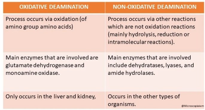
DIFFERENCE BETWEEN OXIDATIVE AND NON-OXIDATIVE DEAMINATION:

1. Oxidative deamination: Process occurs via oxidation (of amino group amino acids).

2. Non-oxidative deamination: Process occurs via other reactions which are not oxidation reactions (mainly hydrolysis, reduction or intramolecular reactions).

1. Oxidative deamination: Main enzymes that are involved are glutamate dehydrogenase and monoamine oxidase.

2. Non- oxidative deamination: Main enzymes that are involved include dehydratases, lyases, and amide hydrolases.

BY- Shaily Sharma (MSIWM041)

Cholesterol: Sources, Structure and Biosynthesis.

Cholesterol present in the body is derived from dietary foods, hydrolysis of cholesteryl esters and synthesis of cholesterol (more than half of the cholesterol is present due to synthesis).

fig:

                     Figure: Chemical structure of cholesterol

The biosynthesis of cholesterol takes place in the endoplasmic reticulum and the cytosol of the cell. Majorly, the process takes place in nucleated cells of the liver, like hepatic cells. The biosynthesis takes place in 5 major steps:

  1. Biosynthesis of mevalonate:
  • Mevalonate, a conjugate base of mevalonic acid, is formed from acetyl CoA during the synthesis of cholesterol.
  • This takes place in the cytosol of the cell.
  • Aceto-acetyl CoA is formed by the condensation of 2 molecules of Acetyl CoA, catalyzed by thiolase.
  • This product further condenses with another acetyl CoA molecule to give HMG-CoA, a reaction catalysed by HMG-CoA synthase.
  • This HMG-CoA is reduced to mevalonate by NADPH. This is catalysed by HMG-CoA reductase.
  • This is an important regulatory step as well in the biosynthesis of cholesterol, where HMG-CoA reductase plays a key role.

Figure: Biosynthesis of Mevalonate

  1. Formation of isoprenoid units
  • Isoprenoid units are formed when mevalonate is phosphorylated sequentially in the presence of ATP and Mg2+ and 3 kinases to give mevalonate-3-phospho-5-diphosphate.
  • The final kinase of the phosphorylation, mevalonate-3-phospho-5-diphosphate undergoes decarboxylation to give an active isoprenoid unit.

Figure: Biosynthesis of isoprenoid

  1. Formation of Squalene
  • Six molecules of Isopentenyl Pyrophosphate (C5) undergo condensation to form a 30 carbon molecule known as Squalene.
  • It is a sequential pathways which proceeds as: C5——>C10——->C15——>C30
  • Intermediates that are observed are geranyl diphosphate (C10), farnesyl diphosphate (C15)  and finally which goes on to form squalene (C30).

Figure: Biosynthesis of Squalene

  1. Formation of Lanosterol
  • Squalene is converted to squalene-2,3-epoxide in the endoplasmic reticulum by squalene epoxidase.
  • After this, ring closure occurs in the presence of oxidosqualene: lanosterol cyclase  to give lanosterol, which is a freshly formed cyclic structure.

Figure: Biosynthesis of Lanosterol

  1. Cholesterol formation
  • Finally, cholesterol is formed from lanosterol in the membranes of the endoplasmic reticulum.
  • This takes place through a number of changes in the side chains and the steroid nucleus.
  • An intermediate known as desmosterol is also formed due to the shift of a double bond.
  • This double bond of the side chain is reduced, which ultimately forms cholesterol.

Figure: Formation if cholesterol from lanosterol

Cholesterol is thus synthesized by nucleated cells in the body through a long pathway involving numerous enzymes and steps. It is an essential molecule as it has various biological functions such as it acts as the precursor for steroid hormones and bile salts, it is required for nerve transmission and is a major constituent of plasma membrane and lipoproteins.

BY- Shaily Sharma (MSIWM041)

Enzyme Inhibition

BY- Ezhuthachan Mithu Mohan (MSIWM043)

Enzyme Inhibitor: Enzyme inhibitor decreases the rate of reaction by binding to the substrate or decreasing the turnover number. It can be organic or inorganic. 

The process which decreases the rate of reaction, by either binding to enzymes or making configurationally changes is known as enzyme inhibition.

Allosteric Inhibition:

When enzyme poses allosteric side other than active site, allosteric inhibitors bind to the allosteric site causing the configuration change in enzyme, making it less feasible for enzyme to bind with substrate. This type of inhibition is partially reversible, when excess of substrate is added.  Km increases and Vmax reduces.

://www.qui.html

Image source: socratic.org

Allosteric inhibition is mainly of two types

  • Positive allosteric inhibition: Which increases the Enzyme activity
  • Negative allosteric inhibition: Which decrease the Enzyme activity

Examples of allosteric inhibition

PhosphofructokinaseActivator: AMPInhibitor: ATP and citrate
Glycogen phosphorylaseActivator: AMPInhibitor: ATP

End Point inhibition: This type of inhibition is also known as Negative feedback inhibition .It is a specialized form of allosteric inhibition, which controls metabolic pathway by regulating various cellular functions. In this type of inhibition end product when formed in excess, it regulate the pathway by binding to the enzymes in reaction, thus controlling production rate.


Reversible Inhibition:

Weak interaction of inhibitor to enzyme causes reversible inhibition, which can be reverted back to normal by adding excess substrate to compete with inhibitor or by removing inhibitor. This type of inhibition follows Michaelis- Menten rate equation. It has rectangular hyperbolic curve.

Competitive inhibitorNoncompetitive inhibitorUncompetitive inhibitor
When inhibitor binds to the site where substrate binds it causes competitive inhibitorWhen inhibitor binds with other site except active siteWhen inhibitor binds with Enzyme substrate complex, It causes structural distortions
These inhibitors are mainly analogues to substrate These inhibitors are not  analogues to substrateThese inhibitors are not  analogues to substrate
Vmax not changed Km increased and Velocity decreasesVmax decreases and Km remains unchangedVmax and Km decreases
Example : Malonate binds  Succinate dehydrogenase and competes with SuccinateExample : Ethanol binds with Acid phosphataseExample: inhibition of placental alkaline phosphatase by phenylalanine

Irreversible inhibition: 

Strong interaction of inhibitor to enzyme causes irreversible inhibition (mostly Covalent interaction). The conformation change caused in Enzyme due to inhibitor is irreversible. Enzyme activity is not regained even by adding or increasing substrate concentration. Vmax decreases and Km is not changed. Suicide inhibition is a specialized form of inhibition, which is also known as mechanism based inactivation. When inhibitor binds to enzyme, it inactivates or complete degradation of enzyme occurs. This type of inhibition  does not follows Michaelis- Menten rate equation. It has sigmoidal curve.

Examples:  Idoacetate , oxidizing agents etc.

Significance of Enzyme inhibition: 

  • To study Drug action
  • To study efficiency of enzyme
  • The interaction of enzyme with substrate can be clearly understood
  • Elucidating cellular reactions by accumulation of intermediates
  • Identification of catalytic site
  • Various drugs used are inhibitors of reaction, so the efficiency of drug and its catalytic function can be clearly known.

.

PRIONS


By: N. Shreya Mohan (MSIWM042)

Prions are a misfolded pathogenic form of proteins that have the ability to transfer its misfolded form to the normal shape of the same protein thereby, folding abnormally. The word “prions” was coined in 1982 by Stanley B. Prusiner and it is a derivative name from “proteinaceous infectious particle”. They are responsible for transmitting several lethal neurodegenerative diseases, particularly in human and few other animals. It is believed that the abnormal 3D configuration of the misfolded protein contains infectious properties resulting in turning the surrounding normal proteins into abnormal ones. These infectious agents are very different from other known infectious agent such as virus, bacteria, fungi as, all of them contain a genetic material (either DNA or RNA).

STRUCTURE

Prions are made up of proteins called as PrP that is generally found throughout the body of healthy humans and animals. But, PrP found in of the infectious prion has a different structure and is very resistant to proteases. Proteases are those enzymes that usually breakdown the proteins in the body. The healthy PrP is known as PrPc while the infectious one is known as PrPSC meaning ‘scrapie’, a disease of prion occurring in sheep. The structure of PrPc is well defined while the structure of PrPSC is poorly defined.

REPLICATION-

There were two hypotheses explaining the replication of prions. First one was a heterodimer model in which an assumption was made that a single PrPSC molecule binds to PrPc and further catalyzes its conversion to PrPSC. These PrPSC can go and further convert PrPc. But it was difficult to explain the spontaneity of the two molecules, hence it was disproved. Second model assumes that PrPSC exists as fibrils and its ends bind to the PrPc and further convert it into PrPSC. But an exponential growth was observed along with quantity of infectious particles thereby, explaining a breakage in the fibril.

THEIR ROLE IN NEUROGENERATIVE DISEASE

Prions cause neurogenerative disease by aggregating from outside inside the nervous system. This forms plaque known as amyloids that completely changes the tissue structures. This is visualized by “holes” in the tissue resulting in a spongy-like structure in the neurons. The incubation period of prion-related disease is (5-20 years), but once symptoms appear, they progress very quickly. The result might be brain damage or death. The structure of infectious prions usually is similar to every species and hence there is a little chance that it will get transmitted. However, the human prion disease Creutzfeldt- Jakob is said to be the prion that usually infects cattle, knowans as mad cow disease which severely infects the cattle and is transmitted through contaminated meat. Also, these proteins have been implicated in the ontogeny of neurodegenerative diseases such as Alzheimer’s disease, Parkinson’s disease, Hunington disease, and frontotemporal lobar degeneration with ubiquitin-positive inclusions (FTLD-U). 

TREATMENT

All prion-related diseases do not have a cure and are fatal. The clinical trials for the same have not produced successful results yet because of the rarity of disease. Although, some experiments built genetically engineered cattle and mice which lacked the gene which is necessary prion production, therefore, building research on the same. 

 Symptoms of prion diseases are:

  • Rapidly developing dementia
  • Ataxia
  • Hallucinations
  • Muscle stiffness
  • Confusion
  • Fatigue
  • Difficulty speaking

REFRENCES-

https://en.wikipedia.org/wiki/Prion

life sciences fundamentals and practice I – Pranav Kumar and Usha Mina.

lehninger principle of biochemistry

DETECTION OF PEPTIDE HORMONES

BY: Reddy Sailaja M (MSIWM030)

HORMONES

A hormone is a signaling molecule secreted by endocrine glands in response to physiological stimuli in multi cellular organisms. These hormones circulate in blood and reach its destination to exert specific function. Hormones help maintain physiological and behavioral functions in the organisms.

Hormones are classified into three main classes:

  • Steriod hormones – Lipid soluble and move across plasma membrane of the targeted cells.
  • Peptide hormones – Water soluble and act through cell surface receptors present on the targeted cells.
  • Aminoacid derivatives

Table 1: Three classes of hormones

Peptide hormones

Peptide hormones are made up of small amino acid chains called, peptides. Peptide hormones are synthesized in the cells from amino  acids based on mRNA sequence that is derived from DNA template within the nucleus.

Peptide hormones can’t navigate across plasma membrane of the cell. Hence, they exert their function by binding to the receptor present on the cell surface of the target cell that in turn trigger signal transduction and cellular response. Some peptide hormones like parathyroid hormone-related protein, angiotensin II etc interact with intracellular components within cytoplasm or nucleus by an intracrine mode of interaction.

Some of the examples of peptide hormones are as follows:

  • Adrenocorticotrophic hormone
  • Thyroid stimulating hormone
  • Vasopressin
  • Angiotensin II
  • Antrial natriuretic peptide
  • Calcitonin
  • Follicle stimulating hormone
  • Insulin
  • Growth hormone
  • Parathyroid hormone
  • Prolactin

Table 2: Main peptide hormones – details and functions

Detection of peptide hormones

As peptide hormones circulate in blood to reach their destination, serum generally acts as the source of detection and measurement.

The following are the main detection methods used to detection peptide hormones:

  1. Sandwich ELISA technique:
  2. In sandwich ELISA method, two antibodies are used to detect hormone of interest. One of the antibodies is attached to the solid support on the micro titer plate, called as capture antibody.
  3. Second antibody labeled with a signal molecule (enzyme or radioisotope or chemilumiscent) acts as detector.
  4. When the analyte containing mixture is loaded onto the micro titer plate, capture antibodies bind to the analyte via epitope that is present on the analyte surface and catch hold of analytes.
  5. When the detector antibodies are added to the plate, they bind to different location on the analyte.
  6. In the enzyme based reaction, when substrate is added, it reacts with enzyme that is attached to the detector body and shows response in the form of color change.

Figure 1: Sandwich ELISA to detect analytes in the blood

  • Radioimmunoassay (RIA):
  • RIA is an in vitro detection technique that detects and measures antigens (like hormones and other foreign substances) in the blood. RIA technique is discovered by Berson and Yalow in 1960 to analyze insulin levels in blood.
  • RIA method is based on the radioactivity measurement associated with antigen-antibody interactions in the reaction.
  • A known antigen that is radiolabelled is incubated with antibody at known concentrations.
  • When the analyte containing solution is added to the labelled antigen-antibody mixture, antigen of interest replaces labelled antigen and bind to the antibody.
  • More the antigen of interest present in the solution, more labelled antigen will be displaced and replaced with antigen of interest.

Figure 2: Radioimmunoassay

  • Enzyme multiplied Immunoassay Technique (EMIT):
  • EMIT is more easy and equivalent detection method, both qualitatively and quantitatively to measure wide-range of analytes from the serum.
  • EMIT is based on the principle that the amount of analyte present in the solution is directly proportional to the inhibition of enzyme-substrate reaction complex.
  • In this technique, initially a known analyte is labelled with an enzyme and antibody specific to drug is allowed to bind drug-enzyme complex. This results in inhibition of enzyme activity.
  • When the solution containing analyte is added to the above mixture, the analyte releases the antibody from the drug-enzyme complex, thereby increasing enzymatic activity.
  • Therefore, enzyme activity is proportional to the analyte present in the sample added and is measured by absorbance value changes of the enzyme.

Figure 3: Enzyme multiplied Immunoassay Technique

  • Immunoradiometric assay (IRMA)
  • IRMA utilizes radiolabeled antibodies to detect analytes of interest.
  • In this technique, antibody is directly labeled with radioisotopes rather than using two antibodies as in other immune assays.
  • When an analyte containing solution is added to micro titer plateradiolabeled antibodies bind to the specific epitopes of the anlytes and forms the antigen-antibody complex.
  • I125 and I131 radioisotopes used in general for this assay.
  • Unbound radiolabeled antibodies are removed from the plate by second wash during the process.

Figure 4: Immunoradiometric assay

The other peptide hormone detection methods are listed below:

  • Ultrafiltration
  • Chromatography
  • Time resolved fluorescence
  • Mass spectrometry
  • Two site immunometric technique

GALACTOSE TO GLUCOSE SYNTHESIS

BY: Ria Fazulbhoy (MSIWM031)

Introduction

Galactose is a sweet tasting monosaccharide sugar which is a carbohydrate, which is an aldohexose molecule. Galactose is a very important molecule in the body as it is needed for the synthesis of lactose, glycolipids, glycoproteins as well as proteoglycans. It is also known as “brain sugar” as it is useful in formation of the glycolipids which occur in the brain and the myelin sheaths of the nerve cells. Galactose can be hydrolysed from interstitial disaccharide lactose, which is known as the ‘sugar of milk’. (lactose = glucose + galactose). Glucose and galactose are stereoisomers of each other, differing in stereochemistry at carbon 4.

Conversion of galactose to glucose

Galactose can be readily converted to glucose in the liver. This is done in the following steps:

  1. The first step is the conversion of galactose to galactose-1-phosphate on the action of galactokinase. It is a phosphorylation reaction, in which ATP acts as the donor of the phosphate group.
  2. Galactose-1-phosphate reacts with UDP glucose i.e. uridine diphosphate glucose (UDPGlc) to form uridine diphosphate galactose (UDPGla). Glucose-1-phosphate is also formed and given out in this step.

This reaction is catalysed by an enzyme called galactose-1-phosphate uridyl transfer

  1. After this, UDP galactose which is formed is converted to UDP glucose by an epimerization reaction.

This is catalysed by UDP-4 glucose epimerase.

  1. The epimerization reaction will involve the oxidation and reduction of carbon 4, using NAD+ as coenzyme.
  2. Finally, from the so formed UDP glucose, glucose is released in the form of glucose 1 phosphate

NOTE: The epimerization reaction is freely reversible, thus glucose can convert back to                 galactose as and when required. This is the reason that galactose is not a dietary essential.

To yield lactose in the mammary gland, UDPGal condenses with glucose. This reaction is catalysed by lactose synthase

Galactosemia is a disease related to this pathway:

  • Galactosemia is the disruption of galactose metabolism.
  • Classic galactosemia is the most common type, in which a deficiency of galactose 1 phospahte uridyl transferase activity is inherited.
  • Due to this, galactose 1 phosphate is accumalated and as a consequence, the inorganic phosphate of the liver is depleted.
  • This results in the ulitimate liver failure and mental detoriation.
  • Infants who are afflicted seriously fail to thrive. They go through symptoms of vomitting and diarhhea after consuming dairy products, enlargement of the liver, and jaundice (which can progress to cirrhosis)
  • Cataract, lethargic developement and retarded mental developement is also very common amongst children. Cataract is observed due to deposition of a galactose metabolite, galactitol, in the lenses of the eyes. Galactitol is osmotically active, due to which water diffuses in the eyes and instigates cataract formation.
  • Other side effects include increase in level of blood galactose level, which is then found in urine.
  • Females can also display ovarian failure.
  • A stirct limitation of galactose consumption in diet can greatly reduce these symptoms.
  • An important diagnostic criterion for this the absence of transferae in the red blood cells.

Conclusion:

Thus, galactose is a very important carbohydrate present in the body which can be easily converted to glucose ( in the form of glucose-1-phosphate) for energy. This is a  reversible reaction, as glucose and galactose are stereoisomers. This is why galactose is not an essential need for the body.

Deficiency in the enzymes that carry out this conversion may result in a vast range of symptoms under galactosemia.

CELL WALL POLYSACCHARIDES: BACTERIAL PEPTIDOGLYCAN

BY: Ria Fazulbhoy (MSIWM031)

Introduction

Bacteria require a thick, rigid extracellular wall that protects them from osmotic lysis. There is a structure known as the peptidoglycan (also known as muerin) which protects the bacteria and gives the bacterial envelopes their strength and rigidity. Peptidoglycan is a polymer made up of amino acids and sugars which forms a mesh like layer. It is a linear, alternating copolymer of N Acetylglucosamine (GlcNAc) and N-acetylmuramic acid (Mur2Ac), linked by beta 1->4 glycosidic bonds, which is crosslinked by short peptides attached to the Mur2Ac. Both GlcNAc

and Mur2Ac are activated by attachment of a uridine at their anomeric carbons during the assembly of the polymer of this complex macromolecule.

Bacterial peptidoglycan synthesis

The peptidoglycan outside the plasma membrane of bacteria is synthesised in the following manner:

  1. In the first step, GlcNAc 1-phosphate condenses with UTP to form UDP-GlcNAc.
  2. UDP-GlcNAc then reacts with phosphoenolpyruvate to form UDP-Mur2Ac, with NADPH present.
  3. To this, 5 amino acids are added to form Mur2Ac pentapeptide moiety.
  4. The Mur2Ac pentapeptide moiety is made from amino acids L alanine, D glutamate, L lysine, D ala D alanine.
  5. This pentapeptide moiety is then transferred to membrane lipoid dolichol, a long chain isoprenoid alcohol, from the uridine nucleotide.
  6. UDP GlcNAc also donates a GlcNAc.
  7. In a number of bacteria, five glycines are then added in peptide linkage to the amino group of the Lys residue of the pentapeptide.
  8. After this, the disaccharide decapeptide which is formed, is added to the non reducing end of an already existing peptidoglycan molecule.  Dolichol leaves the macromolecule in this step.
  9. A transpeptidation reaction occurs in which there is crosslinking of the adjacent polysaccharide chain. This step is catalysed with the help of the enzyme transpeptidase.
  10. Thus, a huge, strong macromolecule is formed which contributes to the macromolecular wall surrounding the bacterial cell.
  11. Most of the effective antibiotics that are used today against  bacteria, have a mode of action by inhibiting the reactions which are involved in the synthesis of bacterial peptidoglycan.
  12. Numerous other oligosaccharides and polysaccharides are synthesized by similar routes, where sugars are activated for subsequent reactions by attachment to nucleotides

Difference of peptidoglycan in gram positive and gram negative bacteria

Importance of peptidoglycan in bacteria

The peptidoglycan which surrounds the bacteria is very important, and sometimes is essential for their survival. They have 2 main functions:

  1. To counteract and maintain the osmotic pressure of the cell. If the peptidoglycan is absent, the bacteria undergoes very abrupt osmotic lysis. Hence, a lot of antibiotics target the peptidoglycan of the bacteria, since it plays a crucial part in the maintenance of osmotic pressure and protection of the bacterial cell. These antibiotics inhibit the synthesis of peptidoglycan in bacteria and initiate osmotic lysis of it.
  2. Another very important function of the peptidoglycan is the regulation of the molecules entering and leaving the bacterial cell. The peptidoglycan regulates the diffusion of cells which play important roles like division of the cell and anchoring the cell wall (eg : teichoic acid)
  3. Some bacteria also release peptidoglycan fragments, which play an important role in cell to cell communication.

BIOAEROSOLS

BY: K. Sai Manogna (MSIWM014)

Bioaerosols are airborne particles created by biological materials and generate a great deal of energy to distinguish the small particles from the larger particles. Based on their sizes, bioaerosols are graded and often range in diameter from 0.02-100µm. The name of these bioaerosols is given to microorganisms distributed in the atmosphere by the transport and deposition process followed by the launch process.

Launching: 

Launching is the process in which the particles filled by microbes are suspended in the earth’s atmosphere. It is achieved predominantly by aquatic and terrestrial sources. For example, the sneeze exposes the atmosphere to bioaerosols. 

Three considerations are included in this process: 

(a) Air turbulence caused by the human, animal, and machine movement; 

(b) The production, storage, processing, and disposal of waste materials; 

(c) natural mechanical processes, such as the movement of water and wind on solid or liquid surfaces that are contaminated; and 

(d) As a result of regular fungal life cycles, the production of fungal spores. Any other examples may be a passing aircraft releasing a biological warfare agent or a passenger jet releasing unburnt carbon particles’ source as an instantaneous linear source. 

Conveyance of bioaerosols: 

Transport or dispersion is the mechanism by which a viable particle travels at wind speed from one point to another or when it is released by force into the air. The airborne particle’s force depends on its kinetic energy derived from the force at which it is launched into the atmosphere and the speed of the wind. Bio-aerosol transport can be described in terms of time and distance. Inside buildings or other enclosed spaces, this method of transport is standard. 

Deposition of Bio Aerosols: 

The deposition is the last pathway involving the distribution of bioaerosols in the atmosphere. It is then split into the other three forms. 

1. Settling Gravity 

2. Effect on the Surface 

3. Deposition of Rain 

Settling Gravity: 

The action of gravity on particles is the primary mechanism associated with deposition. Strength works more intensely on the particles than air, dragging them down. Larger particles would have higher speeds and settle more rapidly down the aero microbiology pathways However it should be noted that gravitational deposition may be negligible for particles of microbiological interest exposed to winds above 8103 m/hr. 

Impacting the surface: 

It is the mechanism in which the particles of bioaerosols have contact with surfaces such as leaves, trees, walls, with the effect of kinetic energy loss. The potential for impact allows a particle to collide with the surface and encourages its binding to it. However, after a collision, depending on the nature of a particle’s surface, it will bounce. 

Bouncing off a surface allows the particle at a lower rate to re-enter the air current, which can have one of two effects: 

1. It allows for subsequent molecular downward diffusion and gravitational settling, resulting in deposition on or on another nearby surface. 

2. It will cause the particle to escape from the surface and re-enter the air current once more. 

Deposition of Rain: 

The deposition also impacts rainfall and electrostatic charges. It occurs as the condensation reaction between two particles, which combine and produce a massive mass bioaerosol, making it settle faster. The overall efficiency of the deposition of rain also depends on the particle plume’s distribution area. Massive, more diffuse plumes have a substantial impact than smaller, more diffuse plumes. The rainfall rate also influences rain deposition. On the other hand, electrostatic deposition still operates the same way, condenses bioaerosols, but is based on electrovalent particles’ attraction. Both particles appear to have an associated charge of some kind. Usually, microorganisms have an overall negative charge at neutral pH associated with their surfaces. Such negatively charged particles may interact with other airborne particles of positively charged, leading to electrostatic condensation.

Mechanisms for Laboratory Regulation of Bioaerosols: 

Two such indoor conditions are hospitals and microbiology laboratories that fall under intramural aero microbiology, with probably the highest potential for pathogenic microbe aerosolization. The centers for the care of immense numbers of patients with a range of diseases are hospitals. It accounts for a high percentage of individuals being the active carriers of several contagious airborne pathogens or microorganisms, including workers and patient visitors. In this respect, microbiological laboratories are just as important as they also serve as a breeding ground for pathogenic species. 

Physical Bioaerosol Removal through filtration: 

Technologies that tackle the bioaerosol threat fall into two categories: 

(1) capture or physical elimination from the air stream of bioaerosols, and 

(2) inactivation on-line or airborne. 

Technologies that make up the former group have typically not been established explicitly for bioaerosols but aerosols’ general regulation. In the latter case, to make airborne microbes non-infectious and exclusively target bioaerosols, technologies apply external stress such as heat or ultraviolet light. Since bioaerosols are physically identical to non-biological particles of the same aerodynamic size and composition, it is possible to apply standard aerosol control devices (air filters, electrostatic precipitators. that physically extract particles from the airstream for bioaerosol control. Filtration is the most successful method for particle removal, both viable and nonviable. For example, high-performance particulate air (HEPA) filters have a 99.97 percent removal efficiency of 0.3-μm sized particles by definition. 

Mechanically, filters extract particles by integrating four simple filtration components. 

Mechanisms: inertial effect, gravitational settling, interception, and diffusion. Impact occurs with larger aerosols that do not adjust to changes in a flow streamline induced by a collector (fiber, granule.) due to their inertia. Gravity, especially when the flow velocity is insufficient, may also cause larger particles to contact a collector. For particles in the submicrometric scale, the two dominant mechanical collection mechanisms are diffusion and interception. As they deviate from a flow streamline by Brownian motion, aerosols are collected by diffusion and eventually deposited on a collector. Aerosols follow a streamline during interception and contact a collector when the streamline distance from the collector is equal to the particle’s radius. 

Disinfection by Air Filter: 

Due to the risks associated with bioaerosols sustained viability, many technologies for disinfecting filter media have been developed. These include photocatalytic oxidation (PCO), UV illumination, and other technologies, as well as anti-microbial filters. A brief overview of several unique technologies for filter disinfection are described below:  

UV light: Irradiation with UV light of bioaerosols (without the presence of UV light, photocatalyst) may cause inactivation. This procedure, known as ultraviolet germicidal irradiation (UVGI), creates thymine dimmers in DNA and inhibits replicating the targeted microbe. 

Anti-microbial filters: Bioaerosols have also been tested against air filters, which have been treated with biocidal chemicals such as iodine. For iodine treated filters, inactivation is hypothesized through the penetration of iodine molecules through the cell wall of microbe and subsequent damage to the capsid protein. In addition to killing microbes obtained from the filter, it is speculated that microbes passing through the filter can be inactivated by iodine species, leading to a decrease in viable bioaerosols’ penetration. A benefit of anti-microbial filters is that additional equipment (e.g., UV light) is not required and can therefore be readily integrated into respirators. 

Technologies for Airborne-Inactivation: 

Besides the physical elimination by filtration of bioaerosols from an airstream, air 

With airborne-inactivation technologies, it can be disinfected. Technologies may be mounted in an on-line system, e.g., ventilation and cooling of heating systems) 

System) to process air that is polluted. Descriptions of several airborne-inactivation technologies are given below: 

UVGI: UVGI lamps may be placed before or after an air filter by direct irradiation of the suspended microbes to minimize the amount of penetrating infectious bioaerosols. The implementation of UVGI is relatively complicated because several factors must be taken into account in the engineering design: airflow patterns, residence time (dose) of the microbe, relative humidity, different resistance of bioaerosols to UV light, ray-tracing optics, power consumption, lamp dust, shielding effect of the material surrounding the bioaerosol, and ozone production from UV lamps. 

Microwave irradiation: By direct irradiation, bioaerosols may be inactivated. Microwave radiation at a frequency of 2.45-GHz decreases the concentration of laboratory-generated and atmospheric bioaerosols. Electron microscopy of irradiated cells revealed that cell death could be responsible for structural damage. 

Cold plasma: Inactivation of plasma has been used in surface disinfection and disinfection. 

Sterilization, however, on-line bioaerosol inactivation has recently been implemented. 

Dielectric barrier discharge (DBD) – a non-thermal technique that uses electrical discharge between electrodes separated by a dielectric material – can generate plasma for disinfection purposes. DNA and cell membrane damage likely cause microbe death. 

Toxic vapours: It has been shown that chemicals such as chlorine dioxide (ClO2) reduce the concentration of culturable airborne bacteria and fungi effectively and thus decontaminate buildings. ClO2 is an oxidizing agent suspected of causing microbes’ death through membrane damage or protein synthesis destruction. Unlike the systems discussed above, due to the vapor’s toxicity, harmful vapours cannot be incorporated in an on-line environment and cannot be used with human occupants. 

Ultra-high temperature (UHT) treatment: UHT methods have historically been used to sterilize or disinfect liquids (e.g., milk) in order to destroy resistant bacterial spores (applying temperatures of > 125 °C for several seconds). However, recent studies have shown their effectiveness against bioaerosols. Airflow was heated to temperatures greater than 1,000 °C for less than a second for inactivation in UHT bioaerosol tests. 

GLYCOLYSIS

BY- RIA FAZULBHOY (MSIWM031)

GLYCOLYSIS (glykys = sweet ; lysis = split/breakdown)

Other name: Embden Meyerhof Parnas pathway (EMP pathway)

Introduction:

A very important pathway in the body, glycolysis is the breakdown of sugar which is glucose (a molecule containing 6 carbons – hexose) into 2 pyruvate molecules, each containing 3 carbon molecules. This process releases energy for utilization by the body in the form of adenosine triphosphate (ATP) through a sequence of enzyme reactions. Glycolysis is a catabolic pathway, i.e a pathway which involves the breakdown of larger complexes through oxidative reactions. Catabolic pathways release energy and are exogenic in nature. Glycolysis is a very important part of the metabolism of glucose and takes place in aerobic as well as anaerobic organisms and does not require molecular oxygen. This takes place in the cytosol of the cell.

Glycolysis is carried out in a sequential 10 step reaction, which are enzyme catalysed. It is represented in the following manner:

C6H12O6 + 2ADP + 2Pi + 2NAD+   →   2C3H4O3 + 2H2O + 2ATP + 2NADH + 2H+

Thus, one molecule of glucose in the presence of phosphate and adenosine diphosphate gives two 3 carbon molecules of pyruvate, along with releasing water and energy in the form of ATP.

Enzymes involved in glycolysis

Each step of the glycolysis pathway requires the presence of an enzyme to continue the process. These enzymes include:

1) Hexokinase

2) Phosphohexose isomerase

3) Phosphofructokinase 1

4) Aldolase

5) Phosphotriose isomerase / Triose – P – isomerase

6) Glyceraldehyde 3-phosphate dehydrogenase

7) Phosphoglycerate kinase

8) Phosphoglycerate mutase

9) Enolase

10) Pyruvate kinase

Glycolysis takes place in two steps:

  1. Preparatory phase (energy invested)
  • This phase comprises steps 1-5 of the glycolysis pathway. 
  • It is called the preparatory phase as glucose is prepared for the conversion to pyruvate by the cleaving of the hexose chain – ringed structure to form a linear structure. 
  • Energy is invested in this phase in the form of 2 ATP molecules which helps to convert glucose into 2 three carbon sugar phosphates known as Glyceraldehyde-3-phosphate, which is the final product of the preparatory phase.
Preparatory phase of glycolysis
  1. Payoff phase (energy is released)
  • This phase is steps 5-10 of the glycolysis pathway
  • This phase is known as the payoff phase as energy is released in the form of 2 ATP molecules as glyceraldehyde-3-phosphate converts to 2 moles of pyruvate.
  • This is the final phase of glycolysis and consists of intermediates and there is a net gain of the energy-rich molecules ATP and NADH.
Payoff Phase of glycolysis

TO SUMMARISE:

RESULT OF GLYCOLYSIS:

  1. Pyruvate is oxidised from glucose
  2. NAD+ is reduced to NADH
  3. Phosphorylation of ADP into ATP

WHAT HAPPENS AFTER GLYCOLYSIS?

Pyruvate formed has different fates in the body, depending on the organism and also the metabolic fate, pyruvate has 3 different paths:

REGULATION OF GLYCOLYSIS:

The enzymes are the most important factors that help carry the pathway forward and thus play an important role in regulation of glycolysis.

The most important of these are the 3 enzymes which carry out irreversible kinase reactions:

  1. Hexokinase/glucokinase
  2. Phosphofructokinase
  3. Pyruvate kinase
Regulation of glycolysis

Enzymes are regulated by the following biological mechanisms:

1. Gene Expression

2. Allostery

3. Protein-protein interaction (PPI)

4. Post translational modification (PTM)

5. Localization

CONCLUSION:

In conclusion, Glycolysis is an extremely important pathway which is essential for many organisms for the formation and utilisation of energy (ATP). Pyruvate formed is utilised in many future pathways in the body.

VITAMIN B6 AND ITS PRODUCTION

BY: SAI MANOGNA (MSIWM014)

Introduction:

An essential compound for general cellular metabolism is vitamin B6 (vitB6), or pyridoxine. Vit B6 has been involved in over 140 biochemical reactions in the cell as a cofactor. This vitamin was first discovered in 1934 by György and colleagues. While most of the co-catalyzed vitB6 reactions are linked to amino acid biosynthesis and catabolism, vitB6 also contributes to the biosynthesis of fatty acids and animals’ breakdown and plants of certain storage compounds, as well as in animals and plants.

Plant hormones, neurotransmitters, and organelle-specific compounds such as chlorophyll are biosynthesized. Moreover, reactive oxygen species ( ROS) can be quenched by vitB6. VitB6 is advantageous for photosynthesis because of its function in ROS scavenging and chlorophyll synthesis and is discussed as a potential factor to relieve biotic and abiotic stress.

Components of Vit B6:

A group of six chemically related compounds, all of which contain a pyridine ring as their center, constitute the vitamin. They differ from each other in the 4 ‘location of the pyridine variable group, which can either be an aminomethyl group (pyridoxamine (PM)), a pyridoxine (PN) hydroxyl methyl group, or an aldehyde group (pyridoxal (PL). Once the various derivatives are phosphorylated, they can serve as cofactors, with vitB6 being the biologically active form of pyridoxal 5’-phosphate (PLP).

Fig: a. Pyridoxal, b. Pyridoxal 5’-phosphate

       c. Pyridoxamine, d. Pyridoxamine 5’-phosphate

       e. Pyridoxine, f. Pyridoxine 5’-phosphate

Production of Vit B6:

Several studies on the production of vitamin B6 and vitamin B6 are known to be provided by various microorganisms belonging to the Saccharomyces, Pichia, Klebsiella, Achromobacter, Bacillus, and Flavobacterium genera. It is possible to manufacture high-yielding vitamin B6. Microorganisms belonging to the genus Rhizobium will accumulate in the culture broth a significant amount of vitamin B6 that can be recovered from it in the desired purity or under aerobic conditions by extracting the resulting vitamin B6 from the fermentation broth in an aqueous culture medium.

1. Saccharomyces carlsbergensis will assay the quality of vitamin B6 in a fermentation broth.

2. High-performance liquid chromatography may also separately calculate the quality of vitamin B6 components such as pyridoxine, pyridoxal, and pyridoxamine in a fermentation broth.

3. The aqueous culture medium, which contains assimilable sources of carbon, digestible sources of nitrogen, inorganic salts, and other nutrients necessary for microorganism development, microorganisms belonging to the genus Rhizobium are incubated.

4. Glucose, fructose, sucrose lactose, galactose, maltose,  dextrin, starch, or glycerol may be used as carbon sources.

5. Peptone, soybean powder, steep corn liquor, meat extract, ammonium sulfate, ammonium nitrate, urea, or mixtures can be used as a source of nitrogen.

6. Also, calcium, magnesium, zinc, manganese, cobalt, and iron may be used in inorganic salts, sulfates, hydrochlorides, or phosphates. Moreover, it is also possible to incorporate conventional nutrient factors or an anti-foaming agent such as vegetable oil, animal oil, or mineral oil.

7. Appropriately, the pH of the culture medium is about 5.0-9.0, ideally about 6.5-7.5.

8. The temperature of cultivation is appropriate from about 10 °- 40 ° C., ideally from about 26 ° to about 30 ° C.

9. The cultivation time is appropriate for 1 to approximately 14 days, ideally for 2 to approximately seven days.

10. Aeration and agitation usually give favorable results in cultivation.

11. The presence in the medium of a compound chosen from pyruvate, D-glyceraldehyde, glycolaldehyde, glycine, 1-deoxy-D-threopentulose, 4-hydroxy-L-threonine, and a suitable combination thereof gives the vitamin B6 titer with more desirable results and is therefore favored.

12. As a supplement for the development of vitamin B6, the combination of 1-deoxy-D-threopentulose and 4-hydroxy-L-threonine is particularly significant.

13. The synthesis of vitamin B6 can also be accomplished by incubating pyruvate, D-glyceraldehyde, glycolaldehyde, glycine, 1-deoxy-D-threopentulose and 4-hydroxy-L-threonine cells of microorganisms belonging to the genus Rhizobium, separated from the culture broth in a buffer of the correct pH value.

14. Vitamin B6 produced may be separated from the culture broth after cultivation and purified.

15. For this reason, by using different vitamin B6 properties, a method used to separate the product from the culture broth can be applied. Thus, for instance, the filtrate’s desired material is purified using an ion exchange resin or similar means after the cells have been separated from the culture broth.

16. The ideal substance is recrystallized from alcohol upon elution.

The microorganism is used in conjunction with all strains belonging to the genus Rhizobium that are capable of producing vitamin B6 and are deposited for availability in a public depository, i.e., culture collection.

First Oxygen producing Species:

According to researchers, vitamin B6 may have given birth to the Earth’s first oxygen-producing species. The earth witnessed a significant increase in atmospheric oxygen levels around 2.4 billion years ago. Scientists have long held that this surge in oxygen, called the Great Event of Oxygenation, was related to the first photosynthetic organisms. Oxygen is a by-product of photosynthesis that converts carbon dioxide into sugar foods using sunlight. Nevertheless, no one understood why these species producing oxygen appeared in the first place.

Researchers found that about 2.9 billion years ago, at the same time that the enzyme manganese catalase appeared, the oldest oxygen-based method involved the manufacture of pyridoxal, a type of vitamin B6. Manganese catalase splits hydrogen peroxide into water and oxygen. When attempting to cope with environmental hydrogen peroxide, early species may have come across this enzyme, which some geochemists claim was plentiful at the time in Earth’s glaciers and was released by the bombardment of solar radiation. By breaking down the glacial hydrogen peroxide with manganese catalase, the species ultimately obtained the oxygen they required to create pyridoxal.