MICROBIOLOGY OF WATER INDICATOR ORGANISMS

BY: K. Sai Manogna (MSIWM014)

For life, water is essential. There must be sufficient, stable, and usable supplies available to everyone. Improving access to clean drinking water will lead to substantial health benefits.  Most individuals fail to gain access to clean water. The supply of clean and filtered water to each house may be the standard in Europe and North America, but access to clean water and sanitation is not the rule in developing countries, and water-borne infections are common. There is no access to better sanitation for two and a half billion people, and more than 1.5 million children die from diarrheal diseases each year. According to the WHO, the mortality of water-related illnesses exceeds 5 million individuals per year; more than 50 percent of these are microbial intestinal diseases, with cholera standing out first and foremost.

A prominent public health concern in developing countries is acute microbial diarrheal diseases. Those with the lowest financial resources and the worst hygiene services are those affected by diarrheal diseases. Children under five, especially in Asian and African countries, are the most affected by water-borne microbial diseases. Often affecting developing countries are microbial water-borne diseases—the most severe water-borne bacterial illnesses.

Table 1: Some bacterial diseases transmitted through drinking water.

DiseaseBacterial agent
CholeraVibrio cholerae, serovarieties O1 and O139
Typhoid fever and other serious salmonellosisSalmonella enterica subsp. enterica serovar Paratyphi Salmonella enterica subsp. enterica serovar Typhi Salmonella enterica subsp. enterica serovar Typhimurium
Bacillary dysentery or shigellosisShigella dysenteriae Shigella flexneri Shigella boydii Shigella sonnei
Gastroenteritis – vibrioMainly Vibrio parahaemolyticus

Indicator Organisms:

1. Microorganisms such as viruses and bacteria in water bodies, which are used as a proxy to determine the existence of pathogens in that area, are indicator organisms.

2. It is preferred that these microorganisms are non-pathogenic, have no or limited water growth, and are consistently detectable at low concentrations.

3. In larger populations than the related pathogen, the indicator species should be present and preferably have comparable survival rates instead of the pathogen.

4. In the monitoring of water quality, different indicator species can be used, and the efficacy of predicting pathogens depends on their detection limit, their tolerance to environmental stresses and other pollutants.

Cholera:

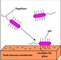
Tiny, curved-shaped Gram-negative rods with a single polar flagellum are Vibrio. Vibrios are optional anaerobes capable of metabolism that are both fermentative and respiratory. For all animals, sodium promotes the development and is an absolute prerequisite for most.

1. The majority of plants are oxidase-positive, and nitrate is reduced to nitrite.

2. Pili cells of some microbes, such as V. cholerae, V. parahaemolyticus, and V. vulnificus, have structures consisting of TcpA protein.

3. The production of TcpA is co-regulated with cholera toxins expression and is a primary determinant of in vivo colonization.

4. Several species of Vibrio can infect humans. The most significant of these species is, by far, V. cholerae.

5. Several forms of soft tissue infections have been isolated from V. alginolyticus.

6. The cells of Vibrio cholerae will expand at 40°C at pH 9-10.

7. The presence of sodium chloride stimulates growth. Vibrio cholerae is a bacterial genus that is very diverse.

8. It is split into 200 serovarieties, distinguished by the lipopolysaccharide (LPS) structure (O antigens). Only O1 and O139 serovarieties are involved in true cholera.

9. Gastroenteritis may be caused by many other serovarieties, but not cholera.

10. Biochemical and virological features are the basis for the differentiation between Classical and El Tor biotypes.

Disease characterization:

1. The incubation period for cholera is 1-3 days.

2. Acute and severe diarrhea, which can reach one liter per hour, characterises the disease.

3. Patients with cholera feel thirsty, have muscle pain and general fatigue, and display anuria symptoms accompanied by oliguria, hypovolemia, and hemoconcentration.

4. In the blood, potassium decreases to deficient levels. With cyanosis, there is circulatory collapse and dehydration.

Several factors depend on the seriousness of the illness:

(a) personal immunity: both previous infections and vaccines can confer this immunity;

(b) inoculum: disease arises only after the absorption of a minimum quantity of cells, approx. 108.

(c) Gastric barrier: V. cholera cells like simple media and the stomach is also an adverse medium for bacterial survival, usually very acidic. Patients that take anti-acid drugs are more vulnerable than healthy patients to infection.

(d) Blood group: persons with O-group blood are more vulnerable than others for still unexplained causes.

5. In the absence of treatment, the cholera-patient mortality rate is approx—fifty percent.

6. The lost water and the lost salts, mostly potassium, must be replaced.

7. Water and salts can be administered orally during light dehydration, but rapid and intravenous administration is mandatory under extreme conditions.

8. Presently, doxycycline is the most effective antibiotic. In some instances, if no antibiotic is available for treatment, the administration of salt and sugar water will save the patient and help with recovery.

9. Two significant determinants of infection exist:

(a) the adhesion of bacterial cells to the mucous membrane of the intestine. It depends on the presence on the cell surface of pili and adhesins;

(b) development of a toxin from cholera.

Salmonellosis:

1. Gram-negative motile straight rods include the genus Salmonella, a member of the family Enterobacteriaceae.

2. Cells are oxidase-negative and positive for catalase, contain D-glucose gas, and use citrate as a sole source of carbon. There are many endotoxins in Salmonellae: O, H, and Vi antigens.

3. S. Subsp enterica. enterica serovar Enteritidis is the most widely isolated serovariety worldwide from humans. Other serovarieties can, however, be prevalent locally.

4. A fermented juice historically extracted from the palm-tree was the source of insulation.

Disease Characterization:

1. Two forms of salmonellosis can be pathogenic to humans:

(a) typhoid and paratyphoid fever (not to be confused with rickettsia-induced typhus disease);

(b) Gastroenteritis.

2. Low infection doses are sufficient to cause clinical symptoms (less than 1,000 cells).

3. There are different clinical signs of salmonellosis in newborns and children, from a severe typhoid-like disease with septicemia of a range to a mild or asymptomatic infection.

4. The infection is commonly spread through the hands of staff in pediatric wards.

5. Ubiquitous Salmonella serovars, such as Typhimurium, are often caused by food-borne Salmonella gastroenteritis.

6. Symptoms such as diarrhea, vomiting, and fever occur about 12 hours after consuming infected food and lasts 2 to 5 days.

7. Spontaneous healing typically happens. All kinds of food can be associated with Salmonella.

8. The prevention of food-borne Salmonella infection is focused on the prevention of contamination, the prevention of food-borne Salmonella multiplication (persistent storage of food at 4oC), and where possible, the use of pasteurization (milk) or sterilization (other foods).

9. When infected with fertilizers of the fecal origin or washed with polluted water, vegetables and fruits can carry Salmonella.

10. The incidence of typhoid fever decreases as a country’s development level grows, such as pasteurization of milk, dairy products, and controlled water sewage systems.

11. The risk of fecal contamination of water and food remains high where these hygienic conditions are absent, and so is the occurrence of typhoid fever.

Bacillary Dysentery or Shigellosis:

Shigella are members of the Enterobacteriaceae family that are Gram-negative, non-spore-forming, non-motile, straight-rod-like. Without gas production, cells ferment sugars. There is no fermentation of salicin, adonitol, and myo-inositol. Cells do not use citrate, malonate, and acetate as the primary source of carbon and do not create H2S. It is not decarboxylated with lysine. Cells are oxidase-negative and positive for catalase. Members of the genus Shigella have a complex antigenic sequence, and their somatic O antigens are the basis of taxonomy.

Disease Characterization:

1. The incubation time for shigellosis is 1-4 days.

2. Typically, the illness starts with fever, anorexia, tiredness, and malaise. Patients exhibit irregular, low-volume, sometimes grossly purulent, bloody stools, and abdominal cramps.

3. Diarrhea progresses to dysentery after 12 to 36 hours, with blood, mucus, and pus appearing in feces that decrease in volume (no more than 30 mL of fluid per kg per day).

4. Even though the molecular basis of shigellosis is involved, the colonic mucosa’s penetration is the initial phase in pathogenesis.

5. Degeneration of the epithelium and acute inflammatory colitis in the lamina propria define Shigella infection’s resulting concentration.

6. Desquamation and ulceration of the mucosa eventually contribute to leakage into the intestinal lumen of blood, infectious elements, and mucus.

7. The colon’s water absorption is hindered under some circumstances, and the amount of stool depends on the flow of ileocecal blood.

8. As a consequence, normal, scanty, dysenteric stools can move through the patient.

9. The bacterium must first adhere to its target cell in order for Shigella to penetrate an epithelial cell.

10. The bacterium is usually internalized into an endosome, which is then lysed to obtain entry to the cytoplasm where replication occurs.

AERO MICROBIOLOGY

BY: K. Sai Manogna (MSIWM014)

Introduction:

The study of living microbes that are suspended in the air is known as Aero microbiology. Such microbes are known as bioaerosols. There are significantly fewer microorganisms in the atmosphere than in the oceans and in the soil; there are still many microorganisms that can impact the atmosphere. With the help of wind and precipitation, these microbes have a chance to migrate long distances and increase the rate of infectious diseases caused by these microbes. In humans, animals, and plants, these aerosols are ecologically important because they can be associated with the disease. Microbes can suspend themselves in the atmosphere, where they can communicate and precipitate with the clouds and create specific shifts in the clouds. 

The air has two microbial ecosystems. 

A. Atmosphere 

B. Clouds 

A. Atmosphere: 

1. High light intensities, extreme temperature fluctuations, low amount of organic matter, and a lack of water availability; characterize the atmosphere as a habitat, making it a non-hospitable environment for microorganisms and a generally inadequate habitat development. 

2. In the lower regions of the atmosphere, however, large numbers of microbes are contained. 

B. Clouds: 

1. In the atmosphere, an apparent mass of concentrated watery vapor floating, usually well above the general ground level. 

2. Clouds, with a pH ranging from 3 to 7, are also an acidic environment. 

Sources of airborne microorganisms: 

1. Air is not a favourable microbial growth environment because it does not provide adequate moisture and nutrients to sustain growth and reproduction, and there is also no indigenous flora growth in the air. 

2. Quite a range of sources responsible for introducing microbes into the air have been identified and researched. 

3. The most popular of these is dirt. Microbes are suspended in the air with wind flow and remain there and often accumulate. 

4. Microbes are often released into the air by human activities such as digging, sloughing, and running. 

5. Microbes are often released into the air through air currents and splashes of water. 

6. Besides, air currents strip plant and animal pathogens from their surfaces and disperse them across the atmosphere. 

7. In contrast to animal pathogens, plant pathogens can spread more quickly. For example, a gamine flies over a thousand kilometers with Puccini spores.

Examples of airborne plant pathogens:

Examples of airborne animal pathogens:

8. Human beings are the primary cause of the introduction of bacteria into the air.

Examples of airborne human pathogens:

9. The most comprehensive source is human activity. The pathogenic bacteria in the human respiratory tract and the mouth’s microbes are continuously released into the air, but they cough, sneeze, and laugh. 

Depending on the size and moisture content, the microbes released into the air come in three forms. Those are the three forms: 

1. Droplets 

2. Nuclei Droplets 

3. Dust that is contagious 

Droplets: As we sneeze, millions of droplets are released, and mucus is expelled from about 200 miles away. Such droplets are water droplets that hold microorganisms if a diseased individual releases them. Saliva and mucus comprise these droplets. Most of the microorganisms they transport are from the respiratory tract. The droplet size determines how long microorganisms live on the droplet. Large-sized droplets settle quickly in the air. The source of these droplets carrying the microorganism can be a source of infectious disease. 

Nuclei Droplets: 

Water particles emitted 1 to 5 micrograms in diameter during sneezing and coughing. For respiratory disorders, droplet nuclei are known to be the raw material. On its surface, it contains saliva and mucus. They are stuck in the air for a more extended period because of their small scale. Droplet nuclei, if the bacteria are the constant source of bacterial infections, are known to be 

The present remains viable on its surface. The viability of bacteria depends on physical conditions, such as humidity, sunlight, moisture, and droplet size. 

Dust Particles: 

By bed making, holding a handkerchief, working with a patient with dried secretion, digging and ploughing, these dust particles are released into the air. Microorganisms adhere to these droplets’ surface and are then suspended by the above techniques to dry them. There is a more significant size of dust particles laden with bacteria and settle down in the air. Two forms of droplets cause airborne diseases. 

a. Droplet infection due to droplets with a diameter greater than 100-micron meters. 

b. Any dried droplet residues cause airborne infections. 

Infection with droplets remains localized and concentrated, while airborne infection may be long-distance. Microorganism can grow on dust particles for a more extended period. It is proven harmful in hospitals and laboratories when closed bottles of dried specimens are opened, and cotton plugs are removed from the bottles. 

Factors influencing airborne microbes:

Factors that influence microbial survival in the atmosphere are 

a. Temperature

b. Moisture/Humidity

c. Content of Nutrients 

d. pH and Acidity 

1. The main factor in regulating the growth of microbes in the air is temperature. 

2. High temperatures hinder the production of microbes and often denature the microbes’ structural conformation. 

3. Very few microbes can live and withstand high temperatures, i.e., extremophiles. 

4. Likewise, when ice crystal formation occurs, shallow temperatures are also not ideal for microbial growth. 

5. Humidity has a role in preserving the development of airborne microbes. 

6. Gram-Bacteria associated with aerosols tend to live in low humidity for a more extended period. 

7. The abundance of nutrients in the atmosphere is lower, so it does not help microbial growth. 

HABITATS (2)

BY : K. Sai Manogna (MSIWM014)

SOIL HABITATS:

For microorganisms, soils are widespread and essential ecosystems that play crucial roles in providing plants with nutrients. If you dig a hole in the earth, you will find that the ground has a structure with various levels of evidence. These include the top organic horizon (O horizon), which includes freshly fallen litter on top by partially decomposed organic matter lower down, followed by horizon A, which comprises a range of minerals. The horizon B where humus, clays, and other materials transported live, and finally, the weathered parent material horizon C. With thin soils overlying calcareous regions of the world, such as the Yucatan, and dense soils occurring in some of the rich farmlands, such as those found in the Midwestern United States, the depth of these layers can differ drastically. Areas where heavy rainfall occurs, have nutrient-poor soils, such as the tropics, and over time the rains leach nutrients from the soils. The degree to which nutrients and microorganisms can travel can influence the permeability of the soil. Light does not penetrate beneath the first centimetre or two, eliminating phototrophy as a means of energy acquisition. The rhizosphere, the region around plant roots, is a habitat where abundant microbial populations occur.

Microbial Food Webs:

One of the greatest treasures of novel species is antibiotics and insights into how populations are organized in soil ecosystems on earth. On average, there are 109 bacterial cells in one gram of soil representing up to 5000 [or even 10,000] bacterial species by some estimates. The nature of soil food web and its inhabitants are more complicated than you might think. Plants play a significant role in physically structuring the under-ground ecosystem through their roots and the impact of their aboveground canopy, depending on the aridity of the aboveground environment. The larger organisms in the soil, such as earthworms, mites, springtails, nematodes, protists, and other invertebrates, were the subject of much of the soil biota research. These species help ‘engineer’ the soil conditions through their absorption and excretion of soil sections. On the scale of the microenvironment, bacteria and fungi even serve as engineers.

Soil microbial abundance varies with the microenvironment’s physical and chemical characteristics, including the content of moisture, organic matter abundance, and the size of soil aggregates. Although seasonal changes contribute to the dynamics of microbial soil populations, discussions of soil microorganisms typically refer to the topsoil sampled during the growing season. The most abundant microorganisms in the soil follow this sequence, as determined by plate count methods:

Algae < Fungi < Actinomycetes = Anaerobic bacteria < Aerobic bacteria

Microorganisms are most common on the surface and decrease as the depth increases in colony-forming units. In the variety of physiological forms of bacteria in various soil environments, soil types, including different organic content and related microbial processes, are seen. There is a substantial genetic diversity of bacteria in soil, and many of the physiological classes still have to be cultivated in the laboratory. Thus, molecular techniques produce more knowledge than conventional plating exercises for the study of the soil culture.

Soils comprise almost all the main microbial groups: bacteria, viruses, fungi, and archaea. Progress has been made in delineating which groups are most prevalent using culture-independent approaches in soil societies. Thirty-two different libraries of sequences from different soils were studied. Their results are impressive: 32 different phyla were present in the tests, but nine phyla were dominant: Proteobacteria, Acidobacteria, Actinobacteria, Verrucomicrobia, Bacteroidetes, Chloroflexi, Planctomycetes, Gemmatimonadetes, and Firmicutes. Proteobacteria account for the highest percentage of soils (39 percent on average). The majority of the sequences were new, and the results of this study vary significantly from the cultivation studies found in previous decades.

Symbiotic nitrogen fixers and mycorrhizae, which provide 5-20 percent of grassland and savannah nitrogen and 80 percent of nitrogen in temperate and boreal forests, are two main classes of bacteria in soil have been extensively studied. Nitrogen and phosphorus derived from symbiotic microorganisms are dependent on at least 20,000 plant species. Plants require nitrogen, and they are unable to fix atmospheric nitrogen in a beneficial form without their symbiotic partners. Examples of essential nitrogen-fixing bacteria are Frankia, an actinomycete that is important in forest growth, and Rhizobium, a key player in the health of crop legumes. These trees may grow in more marginal areas where nitrogen is restricted by interactions between plants, such as the alder tree (Alnus) and Frankia. More than 25 different genera of trees and shrubs have been recorded for cultivation in association with Frankia. Chemoheterotrophs, free-living in the soil or associated with a broad range of legumes, including alfalfa, clover, lupines, and soybeans, are rhizobia, including Azorhizobium, Bradyrhizobium, and Rhizobium. In order to infect them, plants release chemical compounds to attract soil rhizobia. On root hairs, most of the nodules form, but some form on stems. Nodules can use 7-12 percent of the plant’s photosynthetic production when active, but the expense is well worth the return in the form of fixed nitrogen available to the plant.

Despite the progress in understanding soil food webs, our understanding of soil food webs’ mechanisms is hindered by significant challenges. Different feeding classes are generally aggregated because, in their feeding patterns, most soil species are very “flexible,” muddling the distinctions between trophic stages. Relatively unknown are the diets of tiny species. New molecular techniques such as fluorescent in situ hybridization are exciting instruments that can help to expose the dynamic relationships in the soil population of who eats whom. Another instrument that is used to expose feeding relationships and energy sources is stable-isotope analysis.

HABITATS (1)

BY: K. Sai Manogna (MSIWM014)

For a human being who is 890,000 times larger than an E.coli cell, it is difficult to think of microbial environments on the order of micrometres to thousands of meters. Conditions like oxygen or pH will drastically change over this time. It creates microenterprises, and ecosystems are therefore more patchy than stable. Different abiotic factors influence and help establish these microenvironments in microbial communities in these habitats. Any disruption can lead over time to changes in microbial populations in habitats.

Table: Effects of abiotic factors:

Abiotic factorsRange of States
O2 LevelAnoxic-microoxic-oxic
SalinityHypersaline-marine-freshwater
Moisture levelArid-moist-wet
pHAcidic-neutral-alkaline
TemperatureHot-warm-cold
Light levelAphotic-low level-bright-UV

The Niche:

The ‘niche’ turning to the general ecological literature shows that there is what is known as the ‘fundamental niche,’ which reflects all environmental factors. In the ecosystems, the environment affects a species’ ability to survive and reproduce in the environment. The ‘realized niche’ is also the proper niche when biotic interactions (i.e., competition) restrict a species’ growth and reproduction. The definition of niche has been extended to microscopic organisms, enabling bacteria and archaea to exploit new niches not available to parentage by acquiring new genes through horizontal transmission. This niche definition for bacteria and archaea focuses more on the organism’s acquisition of new functional ability by transmitting horizontal genes, which implies a more complex character for niche boundaries. The survival and best reproduction niche of Ferroplasma are characterized by acidic, stable, rich in iron and heavy metals, and moderate temperatures. These conditions distinguish Ferroplasma’s niche space. Species also may change their climate so that other species have a more or less habitable environment.

Aquatic Habitats:

The oceans and flowing water bodies, for example, rivers and streams, range in aquatic ecosystems. Water, more than 97% found in the world’s oceans, covers nearly 71 percent of the earth’s surface. In streams, rivers, and lakes, less than 1 percent of water is contained. Water is continuously renewed through the hydrological cycle in all those various marine ecosystems. The scale of aquatic environments and their diversity suggests the significance for microorganisms of aquatic habitats. Key microbial players in aquatic environments include primary production phototrophs and the heterotrophs involved in carbon cycling in aquatic habitats.

In these marine settings, the environmental and physicochemical conditions vary greatly. Water movement is one of the apparent factors; streams and rivers will flow quickly, with lakes moving less. Winds produce surface water movement in the seas, create ocean waves, and create upwelling areas. These winds transfer nutrients, organisms, oxygen, and heat worldwide, in addition to deep-water currents. As in the seas, water circulation in all marine environments determines different properties of water. Physicochemical factors, such as the pH, the abundance and availability of macro-and micronutrients, salinity, phosphorus, nitrogen, sulphur, and carbon, can vary significantly within the various ecosystems.

Table: Characteristics of different aquatic habitats:

Aquatic habitatTemperature rangeSalinity (%)
Oceans-1.5 to 27oC at surface3.5
Rivers0-30oC0.001-0.05
Lakes
a. freshwater
b. great salt lake

4-50oC

0.01
avg. 12%

Storms are very fluid, have significant variations in physical and chemical environments, are greatly affected by their drainage range, and have a single water flow. In contrast, the lakes, particularly the stream’s headwaters, have more stable conditions and primary productivity. Lakes can be acidic or alkaline (e.g., Mono Lake, California), but often they can be saltier than freshwater, like the Great Salt Lake, Utah.

Aquatic microbial ecology has been advanced from descriptive research on who’s home” to hypothesis-driven studies of interactions and environmental and biological controls on the diversity and population distributions. A broad range of anti-predating mechanisms, including the secretion of exopolymer substances and capsules made up of polysaccharides and morphologic adaptations, are two of the exciting features and the subject of several studies. They are gram-positive. Studies have focused on how predators evade microbial communities, like predation, particularly by protists, and virus lysis is a significant mortality factor. Viruses in various aquatic environments are standard, with a difference of between one or two orders in size in these different habitats, while in freshwater, the abundance of the virus is more seasonal. In aquatic settings, what governs viral abundance is still under review.  In aquatic environments, viruses have a vital role in recovering organic matter dissolved by lysing their presence into their bodies, converting the carbon and other nutrients.

a. Fresh Water

The word wetlands for freshwater generally applies to rivers, streams, reservoirs, lakes, and groundwater. Freshwater that contains less than 1.000 mg/ l dissolved solids is classified under the United States Geological Survey (USGS). As noted above, freshwater microorganisms vary greatly from marine environments in their phylogenetic diversity. Typical freshwater bacterial classes include beta-proteobacteria (e.g., the relative of Rhodoferax and Polynucleobacter necessarius), Actinobacteria, Cytophaga/Flexibacter/Flavobacterium hydrolysis relatives.

b. Lakes:

Lakes are aquatic lakes, initially formed by glaciation, volcanism, or tectonics. The Great Lakes in North America, and Lake Baikal, Siberia, comprise approximately 40% of the world’s freshwater at a few vast lakes.

There are many gradients within water bodies that affect microbial distribution populations. The oxygen gradient is one of the most critical. In lakes where upper waters can be oxic and warmer, the lower gradient is colder and often anoxic. The thermocline is separated by these two layers, which is a transition region between the two layers. Seasonal changes in atmospheric temperature and water temperature can result in changes in density that turn the water over and allow oxygenated water to enter the lake’s lower reaches. It influences the microbial communities of the lake.

The vegetation around lakes supplies some nutrients that have been found in lakes. Low nutrient quantity lakes are oligotrophic, while high nutrient quantities, productivity, and oxygen depletion can affect species that can survive under such conditions. Lakes are eutrophic. The transformation of contaminants such as sulphur dioxide and nitrous oxide (NO3) into acid rain causes some lakes to be naturally acidic while other acidic. Lakes in North-East America recover from acid rain impacts. The pH of the water in lake also influences the population of microbes.

c. Rivers and Streams:

During and after a rainstorm, the water will change and get a water movement force in rivières. Water is moving in streams and rivers through vast material, soil, trees, rocks, and other substances. It ensures a steady supply of nutrients to biotic communities and a great deal of trouble during floods. Many rivers cross cities and thus are exposed to human wastewater and other contaminants that can directly affect the river’s population. As the metabolic diversity of microorganisms is such that specific contaminants are potential energy sources in microorganisms. Since high organic loading can result in high productivity that diminishes oxygen levels, areas of urban rivers can be anoxic, limiting microorganisms in such regions.

The ecosystem of the river consists of many components like horizontal

(1) the active channel that can go dry part of the year in some rivers and streams and

(2) the transitional zone between the marine and the terrestrial habitats, in the riparian zone.

Vertically, streams and rivers are marked by

(1) Waters of the surface;

(2) the sub-surface water region of the hyporheic zone;

(3) the phreatic groundwater field.

The physicochemical properties of these ecosystems differ. Rivers and streams have many suspended organic and inorganic particles, restricting how much light penetrates the water column. At least partly shaded by trees that hang over the streams, the parts of the reaches have extensive vegetation. The extent of photosynthesis in the streams is restricted by both turbidity and shading. Desert streams are much higher than in tropical and temperate regions and have no shading of microbial photosynthesis. Rivers and rivers differ in their salinity by order of magnitude; desert rivers have the highest amounts.

d. Hot Springs:

Springs are springs of geothermal water, groundwater which comes into contact with hot rocks or magma from the world’s earth’s crust in volcanically active regions. Some impressive examples are found in Yellowstone’s national park in Wyoming, Iceland, Japan, and New Zealand.  Hot springs reflect extreme temperature conditions and, in some cases, pH. There is a high concentration of anaerobic or microaerophilic hot spring that suggests low oxygen concentrations. In hot springs where temperature limits are photosynthesis, they were suggested to be primordial producers. Hyperthermophiles, who use carbon dioxide as their carbon source, are also chemoautotrophs and serve as primary producers within hot spring ecosystems. Hot springs contain various gases, including molecular hydrogen and a reduced number of iron and sulphur compounds, dissolved and provide electron donors. It implies that Yellowstone’s primary productivity results from molecular hydrogen oxidation, which can happen to levels above 300 nM. in hot springs. Hot springs are the prime habitat for archaeological animals.

Marine habitats:

a. Oceans: 

In addition to the fact that it is a saline ocean rather than a terrestrial habitat. It is one of several environmental parameters that influence the existence of marine habitat microorganisms. Furthermore, temperature, light, food supply, and pressure vary from the surface to the ocean’s depths. The ecosystems of the ocean shift from shore to vertical depth. Traveling further into the water, from the surface or epipelagic region to the mesopelagic zone (200–1000m), you move into the bathypelagic zone (1000–4000m), the abyss area (4000–6000m), and eventually the Hadean area (<6000m).

b. Food and Microbial Aquatic Habitats:

The marine food web is typically characterized by the low nutrient abundance and patchy nature of the gradients, as mentioned above, and high salinity.

Although the food web has been studied on the ocean for more than a hundred years, several recent findings have led us to believe that the classical description of a chain from diatoms through copepods and to fish and whales can only be a small part of the energy flow. Recent studies of microorganisms, organic dissolved matter, and organic particles in the sea have shown other mechanisms by which a significant share of the energy available will flow. For decades, marine scientists have been cautiously approaching this food web view, and care should be taken when a paradigm is challenged.

INTRODUCTION TO MICROBIAL ECOLOGY

BY : K. Sai Manogna (MSIWM014)

The microbial ecology research covers subjects ranging from individual cells to complex structures and includes several different types of microbes. In studying pure cultures and unique microbial ecosystems, there is not only a visual difference, but there is a difference in the approach of analysis in each of these images.

Fig: Soil bacterium showing multiple flagella in SEM

Fig : Fruiting bodies of various molds.

Studies by scientists from many different research fields discussing ecosystems around the globe have benefited from microbial ecology. The great interest lies in understanding the structure of microbial communities in the ecosystem at this period. It is essential to classify microbes present to attain this understanding; this can be done by using molecular methods even if the microbes have not been cultivated in the laboratory. Microorganisms’ enzymatic processes and microbial adaptations to the environment contribute to our understanding of microorganisms’ physiological ecology.

Roots of Microbial Ecology:

For millennia and long before bacteria were identified, people from various regions around the world used selective procedures to control the production of desired foods.

1. To make fermented milk, starter cultures were passed within a population, and traditional processes were used for fruit juice fermentation.

2. For food preservation, pickling processes involving natural fermentations are customary.

3. Increased rice production has resulted from unique practises in different regions of the world that we now understand are chosen for the growth of nitrogen-fixing cyanobacteria.

4. Some claim that microbiology began in 1675 with the reports by Anton van Leeuwenhoek (1632-1723) describing “very tiny animalcules” in the form of bacteria, yeast, and protozoa.

5. Saliva, dental plaque, and polluted water were the conditions that van Leeuwenhoek investigated.

6. As scientists in different countries studied the environment through direct observations or experimentation, knowledge of microorganisms gradually emerged.

7. Scientists’ contributions to disproving the “spontaneous generation theory” had a significant influence on microbiology, and the presentation by Louis Pasteur at the Sorbonne in Paris in 1864 was particularly important.

8. Pasteur emphasised the significance of microorganisms in fermentation by studying the function of microorganisms in diseases and their effect on our lives.

9. Many consider that Sergei Winogradsky and Martinus Beijerinck were the pioneers of microbial ecology, they were the first people to demonstrate the role of bacteria in nutrient cycles and to formulate concepts of soil microbial interactions.

10. Beijerinck invented the enrichment culture technique to isolate many bacterial cultures, including those now known as Azotobacter, Rhizobium, Desulfovibrio, and Lactobacillus.

11. The early studies of Beijerinck also contributed to the tobacco mosaic virus demonstration and offered insight into virology concepts.

12. Winogradsky was a Russian soil microbiologist who while working with nitrifying bacteria, developed the principle of chemolithotrophy. Winogradsky developed the notion of nitrogen fixation resulting from his experiment with Clostridium pasteurianum, and also demonstrated that bacteria could grow autotrophically with CO2 as the carbon source.

With the prominent interest in microbiology, it became evident that the relationship between microorganisms and also between microorganisms with their environment was highly complex. The study of microbial ecology today covers many different areas.

Current perspectives:

The study of microbial ecology covers the effect of the environment on microbial production and growth. Not only are microorganisms selected for physical and chemical changes in the climate, but biological adaptation helps bacteria and archaea to maximise the use of available nutrients to sustain development.

1. For early life forms, the prokaryotic cell was the ideal device because it had the facility for rapid genetic evolution.

2. Horizontal gene transfer between prokaryotes serves as the mechanism for the cellular evolution of early life forms to generate progeny with different genotypes and phenotypes, as we now understand.

3. While fossils provide evidence of the evolution of plants and animals, they also provide evidence of extinct early animal forms.

4. It is an irony in biology that dinosaurs and other prehistoric forms have engaged in the decomposition of the same prokaryotic organisms that evolved to create eukaryotic organisms. Not only does the prokaryotic mode of life survive today, but it prospers and continues to evolve.

5. It has been calculated that the top one inch of soil has more living microbial cells than several eukaryotic species living above ground.

6. It was estimated by William Whitman and colleagues that there are 5 × 1030 (five million trillion) prokaryotes on earth, and over half of the living protoplasm on earth is made up of these cells.

7. In the human body, the amount of bacteria that develop exceeds the number of human cells by a factor of 10. Although the function of each of these prokaryotic cells cannot be assessed, collectively, groups of prokaryotic cells can have a significant impact on eukaryotic life.

8. Human microbiome analysis shows that although the microbial flora of the skin is identical, each human being has a unique bacterial biome for that individual.

9. Not only are microorganisms involved in nutrient cycling, but they play an essential role in the organisation of the community and interactions with other types of life.

10. Without microorganisms, it would be difficult to imagine life on earth. It is useful to focus on the production of microbes on earth before discussing important divisions in microbial ecology.

TIMELINE:

The formation of the earth took place around 4.5 billion years ago, followed by the creation of the crust and oceans of the earth.

1. The Earth’s volcanic and hydrothermal activities have emitted different gases into the atmosphere. Dinitrogen (N2), carbon dioxide (CO2), methane (CH4), and ammonia (NH3) were the primary greenhouse gases in addition to water vapour, while hydrogen (H2), carbon monoxide (CO), and cyanide hydrogen (HCN) were present at trace amounts.

2. Scientists have critically examined the chemical discoveries of prebiotic earth that are important to life evolution. The anaerobic climate on earth gave decreasing power for the formation of the first organic compounds.

3. Early life forms were anaerobes that included chemolithotrophs, methanogens, and various microbes using thermophilic H2-showing dissimilar mineral reduction.

4. One of the earliest life forms is suggested to have been hyperthermophilic prokaryotes, and has gathered over 1500 strains of these species from hot terrestrial and submarine environments.

5. There is a significant abundance of these microorganisms in the environment, with 107 Thermoproteus cells found in a gram of boiling mud near active volcanoes, 108 Methanopyrus cells found in a gram of hot chimney rock, and 107 Archaeoglobus and Pyrococcus cells found in deep subterranean fluids below the North Sea per millilitre.

6. Although hyperthermophiles typically grow at 80o-113o C with a pH range of 0-9.0, one archaeal cell, Pyrolobus fumarii, resists in an autoclave with a temperature of 121o C for one hour.

7. Around 90 species of microorganisms are hyperthermophiles at present. The majority of hyperthermophiles are chemolithotrophic species that use molecular hydrogen (H2) as an energy-yielding electron source for reactions.

8. Although S0 is used as an electron acceptor by hyperthermophilic archaea, some hyperthermophiles may combine growth with the use of Fe3+, SO42-, NO3-, CO2 or O2 as electron acceptors.

9. For a few hyperthermophilic archaea, molecular oxygen (O2) is an appropriate electron acceptor and in these situations, only under microaerophilic conditions.

10. To sustain their anaerobic or aerobic growth, hyperthermophilic bacteria typically need organic material. Many anaerobes have active structures that use H2 as the donor of electrons.

The biological synthesis of methane is considered an ancient mechanism and the following reaction would have been attributed to catalysing prokaryotes:

4H2 + CO2 → CH4 + 2H2O

Methanogens might have developed methane from methanol, formate, or acetate if organic compounds such as acetate had accumulated in the environment. Only members of the Archaea domain are capable of the development of methane.

Based on the following reaction, chemoautotrophic microbes may have evolved to expand on energy from molecular hydrogen oxidation and carbon dioxide reduction:

2H2 + CO2 → H2O + [CH2OH]

Ultraviolet radiation may have produced H2 according to the following reaction, in addition to the output of H2 from geological formations:

2Fe2+ + 4H+ → 2H2 + 2Fe3+

The radiolysis of water due to alpha radiation will be another cause of H2. Heterotrophic prokaryotes metabolising organic carbon materials would have emerged sometime after the chemoautotrophs were formed with the accumulation of diverse organic compounds in the ecosystem.

Approximately 3 billion years ago, anaerobic photo-driven energy activities may have been present, using light to cause bacteriorhodopsin-like proteins to pump ions through cell membranes. The bacteriorhodopsin type of photo-driven energetics would have accompanied Chlorophyll-containing anoxygenic bacterial photosynthesis involving purple and green photosynthetic bacteria where H2S was the electron source. Although microbial evolution was initially in the aquatic world, about 2.75 billion years ago, microorganisms may have migrated to dry land. Cyanobacteria formed the aerobic atmosphere with oxygenic photosynthesis, and this was called the “great oxidation case.” Since the photocatalytic process produced O2 from water, the rate of O2 released was not limited by water availability.

Once molecular oxygen was released into the atmosphere, it reacted by microbial and abiotic processes with reduced iron and sulphur compounds (i.e. FeS and FeS2) to create oxidised inorganic compounds. The O2 level in the earth’s atmosphere steadily increased and oxygen respiration may have been used to help the development of the first single-cell eukaryotes by around 1.78-1.68 billion years ago.

The production of ozone (O3) from O2 due to an ultraviolet light reaction was another significant development in the aerobic atmosphere. Ozone absorbs ultraviolet light and forms a protective layer in the atmosphere to shield the earth from ultraviolet radiation’s harmful activities. Microorganisms may have been developing only in subsurface areas or habitats covered by rocks before the creation of the ozone layer.

THE PROCESS OF THE INFLAMMATION

BY : K. Sai Manogna (MSIWM014)

Inflammation is a systemic reaction for several causes, such as tissue damage and infections. An acute inflammatory response typically has a fast onset and lasts for a short time. In general, acute inflammation is followed by a systemic reaction known as an acute-phase response, marked by a rapid shift in the concentrations of many plasma proteins. Persistent immune activation in certain diseases can lead to chronic inflammation, which often has pathological implications.

An essential role of neutrophils in inflammation:

1. The predominant cell type infiltrating the tissue is the neutrophil at the early stages of an inflammatory response.

2. Within the first six hours of an inflammatory response, neutrophil penetration into the tissue peaks, with development of neutrophils in the bone marrow growing to meet this need.

3. An average adult produces more than 1010 neutrophils per day, but during a time of acute inflammation, neutrophil production can increase by as much as tenfold.

4. The bone marrow is left by the neutrophils and circulates inside the blood.

5. Vascular endothelial cells increase their expression of E- and P-selectin in response to the mediators of acute inflammation.

6. Increased P-selectin expression is caused by thrombin and histamine; cytokines like IL-1 or TNF-induce increased E-selectin expression. The circulating neutrophils express mucins such as PSGL-1 or the tetrasaccharides Lewisa sialyl and Lewisx sialyl bind to E- and P-selectin.

7. This binding mediates the attachment or tethering of neutrophils to the vascular endothelium, enabling the cells to roll in the direction of blood flow.

8. Chemokines such as IL-8 or other chemoattractants function on the neutrophils during this time, causing an activating signal mediated by G-protein that leads to a conformational shift in the molecules of integrin adhesion, resulting in neutrophil adhesion and subsequent transendothelial migration.

9. When in tissues, activated neutrophils also express elevated levels of chemoattractant receptors and thus show chemotaxis, migrating up the chemoattractant gradient.

10. Several chemokines, complement split products (C3a, C5a, and C5b67), fibrinopeptides, prostaglandins, and leukotrienes are among the inflammatory mediators that are chemotactic to neutrophils.

11. Furthermore, microorganism-released compounds, such as formyl methionyl peptides, are also chemotactic to neutrophils.

12. Increased levels of Fc antibody receptors and complement receptors are expressed by activated neutrophils, allowing these cells to bind more efficiently to antibody- or complement-coated pathogens, thereby increasing phagocytosis.

13. The triggering signal also activates the metabolic pathways into a respiratory burst, creating intermediates of reactive oxygen and intermediates of reactive nitrogen.

14. In the killing of different pathogens, the release of some of these reactive intermediates and the release of mediators from neutrophil primary and secondary granules (proteases, phospholipases, elastases and collagenases) play a significant role.

15. The tissue damage that can result from an inflammatory reaction also leads to these substances. The aggregation, along with accumulated fluid and different proteins, of dead cells and microorganisms, makes up what is known as pus.

Inflammatory Responses:

A complex cascade of non-specific events, known as an inflammatory response, is caused by infection or tissue injury, which provides early protection by minimising tissue damage to the site of the infection or tissue injury. Both localised and systemic responses are involved in the acute inflammatory response.

LOCALISED INFLAMMATORY RESPONSE:

Redness, swelling, pain, heat, and loss of function are the hallmarks of a localised acute inflammatory response first identified almost 2000 years ago. There is an increase in vasodilation within minutes of tissue injury, resulting in an increase in the area’s blood volume and a decrease in blood flow. The increased volume of blood heats the tissue and causes it to turn red. Vascular permeability also increases, leading to fluid leakage, especially in postcapillary venules, from the blood vessels. This results in the fluid deposition in the tissue and, in some cases, leukocyte extravasation, which leads to the area’s swelling and redness. The kinin, clotting, and fibrinolytic processes are triggered when fluid exudes from the bloodstream. The direct effects of plasma enzyme mediators like bradykinin and fibrinopeptides, which induce vasodilation and increased vascular permeability, are responsible for many of the vascular changes that occur early in the local response. Some of these vascular changes are due to the indirect effects of histamine-released complement anaphylatoxins (C3a, C4a, and C5a) that induce local mast-cell degranulation.

1. Histamine is a potent inflammatory mediator, inducing vasodilation and contraction of smooth muscle.

2. Prostaglandins may also contribute to the acute inflammatory response associated with vasodilation and increased vascular permeability.

3. Neutrophils bind to the endothelial cells within a few hours of the initiation of these vascular changes and move from the blood into the tissue areas.

4. These phagocytose neutrophils invade pathogens and release mediators that lead to the inflammatory reaction.

5. The macrophage inflammatory proteins (MIP-1 and MIP-1), chemokines which attract macrophages to the inflammation site, are among the mediators. Around 5-6 hours after an inflammatory response starts, macrophages arrive.

6. These macrophages are activated cells that show increased phagocytosis and increased release of mediators and cytokines that contribute to the inflammatory response.

7. Three cytokines (IL-1, IL-6, and TNF-𝛼) that induce activated tissue macrophages secrete many of the localised and systemic changes, which observed in the acute inflammatory response.

8. All three cytokines function locally, causing coagulation and vascular permeability to increase.

9. Both TNF-𝛼 and IL-1 induce increased expression of adhesion molecules on vascular endothelial cells. For example, TNF-𝛼 stimulates the expression of E-selectin, a molecule of endothelial adhesion that binds adhesion molecules to neutrophils selectively. IL-1 induces increased ICAM-1 and VCAM-1 expression, which binds to lymphocyte and monocyte integrins.

10. Neutrophils, monocytes, and lymphocytes circulating identify these adhesion molecules on the walls of the blood vessels, bind to them and then pass into the tissue spaces via the vessel wall.

11. IL-1 and TNF-𝛼 also act on macrophages and endothelial cells to induce the development of chemokines that, by increasing their adhesion to vascular endothelial cells and by acting as potent chemotactic factors, contribute to the influx of neutrophils.

12. Besides, macrophages and neutrophils are activated by IFN-𝞬 and TNF-𝛼, facilitating increased phagocytic activity and increased release of lytic enzymes into tissue areas.

13. Without the overt intervention of the immune system, a local acute inflammatory response may occur.

14. Cytokines released at the inflammation site also promote both the adherence of immune system cells to vascular endothelial cells and their migration into tissue spaces through the vessel wall.

15. This results in an influx of lymphocytes, neutrophils, monocytes, eosinophils, basophils, and mast cells to the tissue damage site, where these cells are involved in antigen clearance and tissue healing.

To monitor tissue damage and promote the tissue repair processes that are important for healing, the length and strength of the local acute inflammatory response must be carefully controlled. TGF-β has been shown to play an essential role in limiting the response to inflammation. It also encourages fibroblast aggregation and proliferation and the deposition of an extracellular matrix necessary for proper tissue repair. Clearly, in the inflammatory response, the leukocyte adhesion processes are of great importance. As exemplified by leukocyte-adhesion deficiency, a failure of proper leukocyte adhesion may result in disease.

SYSTEMIC ACUTE-PHASE RESPONSE:

The systemic response is known as the acute-phase response accompanies the local inflammatory response. This response is characterised by fever induction, increased hormone synthesis such as ACTH and hydrocortisone, increased white blood cell development (leukocytosis), and the production of a large number of liver acute-phase proteins.

1. The rise in body temperature prevents a variety of pathogens from rising and tends to strengthen the immune response to the pathogen.

2. A prototype acute-phase protein whose serum level increases 1000-fold during an acute-phase response is a C-reactive protein.

3. It is made up of five similar polypeptides by noncovalent interactions kept together.

4. The C-reactive protein binds and activates complements to a wide range of microorganisms, resulting in the accumulation of opsonin C3b on the surface of microorganisms.

5. The C3b-coated microorganisms can then readily phagocytose phagocytic cells, which express C3b receptors.

6. The combined activity of IL-1, TNF-𝛼 and IL-6 is linked to several systemic acute-phase effects. To cause a fever response, each of these cytokines works on the hypothalamus.

7. Increased levels of IL-1, TNF- and IL-6 (as well as leukaemia inhibitory factor (LIF) and oncostatin M (OSM)) induce hepatocyte development of acute-phase proteins within 12–24 h of the onset of acute-phase inflammatory response.

8. To induce colony-stimulating factors (M-CSF, G-CSF, and GM-CSF) secretion, TNF-𝛼 also acts on vascular endothelial cells and macrophages.

9. These CSFs induce hematopoiesis, causing the number of white blood cells required to combat the infection to increase temporarily.

10. Redundancy in the capacity of at least five cytokines (TNF-𝛼, IL-1, IL-6, LIF, and OSM) to induce liver acute-phase protein development results from the induction of NF-IL6, a common transcription factor, after the receptor interacts with each of these cytokines.

11. Amino-acid sequencing of cloned NF-IL6 showed that it has a high degree of sequence identity with C / EBP, a liver-specific transcription factor.

12. NF-IL6 and C / EBP both contain a leucine-zipper domain and a simple DNA-binding domain, and in the promoter or enhancer of the genes encoding different liver proteins, both proteins bind to the same nucleotide sequence.

13. C / EBP, which stimulates albumin and transthyretin production, is hepatocyte-constitutively expressed.

14. Expression of NF-IL6 increases and that of C / EBP decreases as an inflammatory response arises, and the cytokines interact with their respective receptors on liver hepatocytes.

15. The inverse relationship between these two transcription factors reflects the observation that serum protein levels such as albumin and transthyretin decrease during an inflammatory response while those of acute-phase proteins increase.

BACTERIAL ENDOSPORES – FORMATION, STRUCTURE AND FUNCTIONS

BY: Reddy Sailaja M (MSIWM031)

Introduction

  • Microorganisms have the ability to adapt themselves to the changing conditions prevailing in the environment. Factors that influence microorganism’s survival could be physical, chemical or environmental.
  • Some microorganisms go in search of favorable conditions for survival, while some will become dormant till the favorable conditions arrive.
  •  One such mechanism adapted by certain gram-positive bacteria is the development of ‘endospores.  Gram positive bacteria, especially genera, Bacillus and Clostridium have the ability to form endospores in response to harsh conditions, nutrient deprivation in particular.
  •  When there is starvation due to nutrient deprivation, these bacteria produce most resistant and dormant ‘endospore ‘structures that preserve cell’s genetic composition to with stand the harsh assaults like high temperature, desiccation, UV radiation, chemical and enzymatic damage.
  •  Moreover, endospores are the most resistant form of “spores” or “cysts” produced by many bacteria and are resistant to most of the antibiotics. Altogether, endospores are resistant and dormant structures of life survival forms of bacteria and fight against harsh environments.


Formation of endospore

Figure 1: Development of Endospore

  • Bacillus subtilis is the model organism used to study and understand the development of endospore during the process is called sporulation.
  •  It takes many hours to complete endospore formation. Morphological changes that occur during this process are used as markers to classify stages of endospore development. Stage I is that when the bacterial cell is under favorable conditions.
  • Under unfavorable conditions, bacterial cell initiates endospores formation by asymmetric cell division and is called Stage II. Asymmetric cell division results in the formation of a larger mother cell and a smaller forespore (or pre-spore) with septum in between them.
  • Even though, these two cell types in stage II has varied developmental fates, intercellular communication system harmonize cell specific gene regulation by influencing specialized sigma factors in the cells.
  •  In stage III, peptidoglycan present in the septum gets dissolved and the mother cell engulfs forespore, which becomes a cell within a cell.
  • In the stages IV+V, cortex and the spore coat layers are formed around the forespore, leading to the production of endospore specific compounds.
  •  In the stages VI+VII, further dehydration and the maturation of the endospore happens. Finally, the mother cell dies by apoptosis (also called programmed cell death) and the endospore is release into the environment and remains dormant until favorable conditions prevail.

Endospore structure

Endospore structure comprises of multiple layers of coats that resist against harsh surrounds. The following table details various layers (from outer to inner), their compositions and functions.

Endospore layerCompositionFunction
ExosporiumCarbohydrates, proteins  and lipidsGives hydrophobic character to the endospore and is responsible for endospore pathogenicity
Spore coatCoat proteins cross-linked with disulfide bondsActs as primary permeability barrier and allows only smaller molecules like germinants
Outer membraneNot known
CortexPeptidoglycan without teichoic acids with low cross linkingStructural differences in the peptidoglycan of cortex and germ cell wall allow selective degradation of outer protection, germination of endospore and transformation of germ cell wall into vegetative cell.
Germ cell wallPeptidoglycan
Inner membraneSimilar to cell membrane composition. Germinant receptorsVaried fluidity and permeability and decreased mobility of the membrane lipids make the structure highly impermeable to the molecules including water, protecting core. Germinant receptors allow binding of germinants and begin germination and vegetative growth.
CoreBacterial DNA, RNA, ribosomes, essential enzymes, small acid-soluble spore proteins (SASPs), Dipicolinic acidDehydrated state protects enzymes and heat resistance. SASPs protect DNA from destructive chemicals and enzymes by forming shield. SASPs also function as carbon and energy source during germination into vegetative cell. Dipicolinic acid also protects endospore’s DNA against harsh environment.

Figure 2: Structure of endospore

Mechanism of sporulation

  • Sporulation of endospores is under the control of five kinases, namely KinA, KinB, KinC, KinD and KinE that act under phosphorelay signal transduction mechanism.
  • Each of these kinases gets activated based on specific environmental stimuli. Under a specific kind of environmental stimulus, one of the five sensor kinases undergoes autophosphorylation at conserved histidine residue by an ATP dependent reaction through a protein called Spo0F. Then the Spo0F transfers the phosphate to Spo0B, that act as a mediator and delivers signal to Spo0A.
  •  Spo0A further positively regulate genes necessary for sporulation and negatively regulate genes required for vegetative growth.

Figure 3: Mechanism of sporulation

Functions of endospores

  • Endospores mainly resist harsh conditions like high temperatures, disinfectants, radiation, etc.
  •  Endospores are reported to survive for millions of years. For example, viable endospores were isolated from gastrointestinal tract of a bee that was embedded in amber around 25-40 years ago.
  • Dipicolinic acid and SASPs are crucial in protecting core of the endospore that contains genetic material.

Infectious diseases caused by endospores

In spite of defensive mechanism, endospores also transmit some infectious diseases as follows:

i)Anthrax – caused by Bacillus anthracis endospores when inhaled, ingested will germinate under suitable conditions and spread the infection

ii)Botulism – Caused by Clostridium botulinum. Spreads through unprocessed food and infect

iii) Tetanus – caused by Clostridium tetani. Spread through anaerobic wounds and cause infection.

 Other infectious diseases like gas gangrene and pseudomembranous colitis are also popular.

CHEMOKINES

BY: K Sai Manogna (MSIWM014)

1. Chemokines are the superfamily of small polypeptides, most of which contain residues of 90-130 amino acids are chemokines. 

2. They regulate the adhesion, chemotaxis, and activation of several kinds of leukocyte populations and sub-populations selectively, and sometimes explicitly. 

3. Consequently, they are significant leukocyte traffic regulators. Some chemokines are predominantly involved in inflammatory processes; others are expressed constitutively and play essential roles in the homeostatic or developmental activity. 

4. Housekeeping chemokines are manufactured in lymphoid organs and tissues or in non-lymphoid sites such as the skin where normal lymphocyte trafficking is directed, such as deciding the correct placement of newly-generated hematopoiesis leukocytes arriving from the bone marrow. 

5. Chemokines are constitutively expressed by the thymus, and normal B cell lymphopoiesis often depends on suitable chemokine expression. 

6. Effects mediated by chemokines are not confined to the immune system. 

7. Mice missing either the CXCL12 chemokine (also referred to as SDF-1) or its receptor display significant defects in brain and heart production. 

8. In the formation of blood vessels and wound healing, members of the chemokine family have also been shown to play regulatory roles. 

9. In response to infection, inflammatory chemokines are usually induced. 

10. The expression of inflammatory cytokines as inflammation sites is regulated by interaction with pathogens or the action of pro-inflammatory cytokines, such as TNF-𝛼. 

11. By inducing, adherence of these cells to the vascular endothelium, chemokines induce leukocytes to migrate into different tissue locations. 

12. Leukocytes are drawn to high localised concentrations of chemokines after migration into tissues, resulting in the selective recruitment of phagocytes and lymphocyte effector populations to inflammatory sites. 

13. The assembly of leukocytes at infection sites, coordinated by chemokines, is an integral part of mounting an infection response that is correctly oriented. 

Of more than 50 chemokines and at least 15 chemokine receptors have been identified. The chemokines have four residues of conserved cysteine and almost all fall into one or the other of two distinctive subgroups based on the position of two of the four invariant cysteine residues: 

■ Chemokines of the C-C subgroup, in which conserved cysteines are contiguous; 

■ Chemokines of the C-X-C subgroup, in which several other amino acids (X) distinguish the conserved cysteines. 

Receptors whose polypeptide chain traverses the membrane seven times mediate chemokine action. CC receptors (CCRs), which recognise CC chemokines, and CXC receptors (CXCRs), which recognise CXC chemokines, are two subgroups of receptors. 

As with cytokines, there is a high affinity (Ka > 109) and high specificity for the interaction between chemokines and their receptors. For instance, at least six different chemokines are recognised by CXCR2, and several chemokines can bind to more than one receptor. 

It activates heterotrimeric large G proteins when a receptor binds an appropriate chemokine, activating a signal-transduction method that generates such potent second messengers as cAMP, IP3, Ca2 +, and activated small G proteins. Chemokine-initiated activation of these signal transduction pathways carries out drastic changes. The addition of adequate chemokine to leukocytes induces sudden and widespread changes in shape within seconds, the promotion of more outstanding adhesion to endothelial walls by activating leukocyte integrins, and the development of phagocyte microbicidal oxygen radicals. These signal-transduction pathways facilitate other changes, such as granular material release, neutrophil and macrophage proteases, basophil histamine, and eosinophil cytotoxic proteins.  

Chemokine-Receptor Profiles Mediate Activity of leukocytes: 

1. Neutrophils express CXCR1, -2, and -4 among the central populations of human leukocytes; eosinophils have both CCR1 and CCR3. 

2. Some activated T cells have CCR1, -2, -3, and -5, CXCR3 and -4, and possibly others, whereas resting naive T cells show a few types of chemokine receptors. 

3. Consequently, variations in the expression of chemokine receptors by leukocytes coupled with the development by destination tissues and sites of distinctive chemokine profiles provide rich opportunities for the differential control of the activities of the various populations of leukocytes. 

4. Indeed, variations in chemokine-receptor expression patterns occur both within and between different populations of leukocytes. 

Fig : Patterns of expressions on some principle chemokine receptors on the human leukocytes.

Note: Their various cytokine output patterns can distinguish the TH1 and TH2 subsets of TH cells. Different profiles of chemokine receptors also show these subsets. CCR3 and -4 are expressed by TH2 cells, and a variety of other receptors are not expressed by TH1 cells. TH1 cells, on the other hand, express CCR1, -3, and -5, but most TH2 cells do not. 

The Other Inflammatory Mediators: 

Several other mediators released by cells of the innate and acquired immune systems activate or improve particular aspects of the inflammatory response in addition to chemokines. Tissue mast cells, blood platelets, and several leukocytes, including neutrophils, monocytes/macrophages, eosinophils, basophils, and lymphocytes, release them. 

The plasma comprises four interconnected mediator-producing processes in addition to these sources: the kinin system, clotting system, fibrinolytic system, and the complement system. The first three systems share the Hageman factor, a common intermediate. These four systems are triggered when tissue damage occurs, to form a network of interacting systems that produce several inflammation mediators. 

The Tissue Injury Stimulates the Kinin System: 

1. The kinin mechanism is an enzymatic cascade that starts with tissue injury, a plasma clotting factor, called Hageman factor, is activated. 

2. In order to form kallikrein, the activated Hageman factor then activates prekallikrein, which cleaves kininogen to generate bradykinin. 

3. This inflammatory mediator is a potent fundamental peptide that enhances vascular permeability, causes vasodilation, pain, and induces smooth muscle contraction. 

4. By cleaving C5 into C5a and C5b, kallikrein also works directly on the complement mechanism. 

5. An anaphylatoxin that induces mast-cell degranulation, resulting in the release of several inflammatory mediators from the mast cell, is the C5a complement portion. 

The Clotting System Yields Inflammation Mediators Produced by Fibrin: 

1. Another enzymatic cascade caused by blood vessel disruption yields significant amounts of thrombin. 

2. To generate insoluble strands of fibrin and fibrinopeptides, thrombin works on soluble fibrinogen in tissue fluid or plasma. 

3. Clot formation acts as a barrier to the spread of infection, for which the insoluble fibrin strands cross each other. 

4. After tissue damage, the clotting mechanism is activated very quickly to avoid bleeding and limit the spread into the bloodstream of invading pathogens. 

5. As inflammatory mediators, the fibrinopeptides act, inducing increased vascular permeability and neutrophil chemotaxis. 

The Fibrinolytic System Yields Inflammation Mediators produced by Plasmin: 

1. The fibrinolytic method completes the elimination of the fibrin clot from the damaged tissue. 

2. The end product of this pathway is the plasmin enzyme, which is generated by plasminogen conversion. 

3. Plasmin breaks down fibrin clots into degradation products that are chemotactic for neutrophils, a potent proteolytic enzyme via activating the classical complement pathway.

4. Plasmin also contributes to the inflammatory response. 

Anaphylatoxins Formed by the Complement System: 

1. Activation by both classical and alternative pathways of the complement system results in the development of several complement split products that serve as essential inflammation mediators. 

2. Binding of anaphylatoxins such as C3a, C4a, and C5a to receptors on the membrane of tissue mast cells induces degranulation with histamine release and other pharmacologically active mediators.

3. Such mediators cause contraction of smooth muscles and increase vascular permeability. 

4. C3a, C5a, and C5b67 function together to induce the adherence of monocytes and neutrophils to vascular endothelial cells, extravasate through the capillary endothelial lining, and migrate to the tissue site of complement activation. 

5. Thus, activation of the complement system results in fluid inflows carrying antibody and phagocytic cells to the entry site of the antigen. 

Lipids as Inflammatory Mediators: 

1. Phospholipids in the membrane of many cell types (e.g., macrophages, monocytes, neutrophils, and mast cells) are degraded into arachidonic acid and lyso-platelet-activating factor following membrane disturbances. 

2. Subsequently, the latter is transformed into a platelet-activating factor (PAF) that induces platelet activation and has several inflammatory consequences, including eosinophil chemotaxis, neutrophil and eosinophil activation and degranulation. 

Arachidonic acid metabolism:

a. Arachidonic acid metabolism produces prostaglandins and thromboxanes through the cyclooxygenase pathway. 

b. Various cells produce various prostaglandins: 

i. monocytes and macrophages produce large quantities of PGE2 and PGF2; 

ii. neutrophils produce moderate amounts of PGE2, which is released by mast cells. 

There are various physiological effects of prostaglandins, including increased vascular permeability, increased vascular dilation, and neutrophil chemotaxis induction. Thromboxanes cause platelet aggregation and blood vessel constriction. 

The lipoxygenase pathway also metabolises arachidonic acid to yield four leukotrienes: LTB4, LTC4, LTD4, and LTE4. Three of these (LTC4, LTD4, and LTE4) together make up what was formerly referred to as a slow-reacting anaphylaxis material (SRS-A); these mediators cause contraction of smooth muscle. LTB4 is a potent neutrophil chemoattractant. Several cells, including monocytes, macrophages, and mast cells, make leukotrienes.

Some cytokines are essential mediators of inflammation:

1. In the formation of an acute or chronic inflammatory response, several cytokines play a significant role. 

2. There are redundant and pleiotropic effects of IL-1, IL-6, TNF, IL-12, and several chemokines that together lead to the inflammatory response. 

3. Besides, IFN-contributes to the inflammatory response, functioning later in the acute response and by attracting and stimulating macrophages, leading in a significant way to chronic inflammation. 

4. The differentiation of the pro-inflammatory TH1 subset is caused by IL-12.

CENTRIFUGATION

BY: Reddy Sailaja M (MSIWM031)

Centrifugation is one of the most extensively used technique in research and development fields of biochemistry, molecular biology, biochemistry and pharmaceutical industries for varied applications like isolation of cells, fractionation of sub cellular particles and other macromolecules for analytical and clinical applications.

Definition

Centrifugation is a process of separation (or concentration) of particles from suspended medium based on their size, shape, density, viscosity of the medium, rotor speed etc. Centrifugal force is a key in this technique to separate the particles in less time and it acts against gravitational force. Figure 1 gives overview of centrifugation process from muscle tissue.

Figure 1: Centrifugation overview

Principle:

In general, when a liquid suspension is placed idle for some time, particles of bigger size/density will start to settle at the bottom of the container because of gravitational force and so on. But, this is a slow process and can’t be applied practically. Centrifugation works on centrifugal force to separate the particles in a suspension in less time with more efficiency.

When a body with mass ‘m’ is rotating in a circle with radius ‘r’ and velocity ‘v’, the force acting on the body is measured using the following formula 1.

F = mv2/r

Where,

F = centrifugal force,

m = mass of body,

v = velocity of the body,

 r = radius of circle of rotation.

The gravitational force acting on the body ‘m’ is calculated using the formula 2: G = mg

Where,

G = gravitational force

g = acceleration due to gravity

U

The centrifugal force is further calculated using the formulae 1 and 2 as follows:

C = F/G = mv2/mgr = v2/gr

Since, v = 2π r n

Where,

 n = speed of rotation

C = F/G = (2π r n) 2/g r = 4π2 r2n2 = 2π2/g D n2 = kD n2

where,

k = 2π2/g = constant

D = maximum diameter of the centrifuge

D is able to measured either from centrifuge center to the free surface of the liquid or to the tip of the centrifuge tube.

From the equation C = kDn2 it was evident that,

Centrifugal effect ∝ diameter of centrifuge

Centrifugal effect ∝ (speed of rotation)2.

When a liquid suspension containing container is rotated at a certain speed called revolutions per minute (RPM), particles will move at a certain speed away from the axis of rotation. The force that’s being generated on the particles to move away from the centre is called relative centrifugal force (RCF). RCF depends mainly on the rotational speed (measured in RPM) and the distance of the particles from the centre of the rotation (rotor).

RCF = 11.2 × r (RPM/1000)2

Where,

r – Distance in centimeters

More the density of the particles, faster is the settlement at the bottom of the tube, while less dense particles will be floating in the liquid. The rate of sedimentation depends on the size and density of the particles and can be explained by Stokes equation (explains movement of a sphere in a gravitational field).


Where,

V = viscosity of the medium

d = diameter of the sphere

p = particle density

L = medium density

n = viscosity of medium

g = gravitational force

Stokes equation explains behavior of particles based on the rate of particle sedimentation as follows:

  • directly proportional to the size of the particle
  • directly proportional to the difference between the particle and the medium densities
  • zero when the particles and medium exhibits same density values
  • decreases when the medium viscosity increases
  • increases as the gravitational force increases

Table 1: Densities of cells and sub cellular fractions

The particles that gets settled at the bottom forms ‘pellet’ while the liquid suspension with lighter particles or no particles is called ‘supernatant’.  Therefore, centrifugation is a process that utilizes centrifugal force for the sedimentation of particles.

Figure 2: Densities and sedimentation coefficients of biomolecules, cell organelles and viruses

For example, ‘m’ is a particle in a centrifuge tube suspended in a liquid. During centrifugation process, the particle is influenced by three kinds of forces: FC– the centrifugal force, FB – the buoyant force and Ff – the frictional force between the particle and the liquid.

Figure 3: Centrifugal force

Centrifuge

Centrifuge is a tool designed to separate particles in the liquid suspension based on the centrifugation principle. It is operated using an electric motor that enables an object to move around in a fixed axis when a perpendicular force is applied to the axis.

Figure 4a: Front view of a typical centrifuge

Figure 4b: Rear view of a typical centrifuge

Centrifuge comprises of three major components:

  1. Rotor – Holds containers (tubes/bottles etc) containing liquid suspension to be centrifuged. Rotors of different types and sizes are available
  2. Fixed angle rotor – Requires short time to sediment particles as they need to travel only a little distance.

Figure 5a: Fixed angle rotor

  • Swinging bucket rotor – Allows better separation of particles as the particles have to move long distance. Stronger pellet is formed and supernatant can be easily removed.

Figure 5b: Swinging bucket rotor

  1. Drive shaft – Helps hold rotors which in turn connect to motor.
  2. Motor – Helps to rotate the rotor based on the input speed by providing power.

All the major centrifuge components are surrounded by a protective cabinet with operating controls and indicator dials for speed and time mounted on it. Centrifuges come with brake system to control rotor and allow it to come to standstill when the centrifugation run gets completed. Refrigerated centrifuges allow option to control temperature so that the delicate biological samples won’t be degenerated during the process.

Types of centrifugation techniques

There are two major types of centrifugation techniques to separate particles.

  1. Differential centrifugation
  2. Density gradient centrifugation
  3. Isopycnic centrifugation
  4. Rate-zonal centrifugation
  5. Differential centrifugation:

This is the simplest type of particle separation form, also called ‘pelleting’ down the particles. Sedimentation occurs at different rates based on the density of the particles. More dense particles will sediment fast and the lighter particles will be floating in the suspension. Sedimentation of the particles also depends on the centrifugal force applied. As the centrifugal force increased, pellet with decreased sedimentation rate will be formed and vice versa.

Differential centrifugation is applied during cell harvest or sub cellular fractionation from a tissue homogenate. When lower centrifugal force is applied, dense particles like nuclei, membrane vesicles etc gets pelleted first. To further pellet next order particles like mitochondria etc, more centrifugal force is applied. Greater than or equal to four differential centrifugation cycles are applied for sub cellular fractionation of the tissue homogenate. However, this process faces carry over contamination of the particles from previous fraction and not purity is less.

Figure 6: Differential centrifugation process overview

  • Density gradient centrifugation: This method is mainly applicable to separate sub cellular particles and other macromolecules with more purity.

In this process, gradient media of different density are layered one above the other, more dense at the bottom of the tube and the lightest at the top. The cell fractionate that need to be separated is placed on the top of the gradient layer and the centrifugal force is applied.

Figure 7: Density gradient process overview

Density gradient method is further classified into two types as follows:

  1. Isopycnic centrifugation:

This process is also known as buoyant or equilibrium separation. In this process, particles are separated base on density. Particle size plays a role when the density of the particles and the surrounding medium is same. When the centrifugal force is applied, initially the sample and gradient gets mix uniformly and the particles move through the gradient until the density of the particles and gradient medium becomes same. Now, the gradient is called as ‘isopycnic’ and the particles get separated based on their buoyancy. Therefore, it is important to make sure that the gradient medium is always dense than the particles to be separated. Particles get separated in the gradient medium in different layers, but never settle to the bottom of the tube. Gradient medium varies depending upon the kind of material being separated. Continuous gradient method is good for analytical separation while discontinuous gradient is more suitable for biological applications (E.g.: separation of lymphocytes from blood).

Table 2: Common density gradient media used for isopycnic centrifugation process

  • Rate zonal centrifugation:

The carryover contamination of particles in differential centrifugation is prevented by implementing rate zonal centrifugation. In this process, sample was layered in a narrow zone on the top of the density gradient and the centrifugal force is applied. Particles segregate based on their size and mass rather than density, and also on the centrifugal force. As a result, narrow load zone prevents less sample volume (≤10%) that can be accommodated on the density gradient and it further stabilizes the bands and allows medium of increasing density and viscosity. Centrifugation is applied for a short time at a low speed. As the density of the particles is more than the density of the gradient, there is a chance that all the particles form pellet if the centrifugation continues for a long time.

Figure 8: Rate zonal and isopycnic centrifugation processes overview

Common applications of centrifugation

  • Production of drugs and other biological products
  • Separation of subcellular particles
  • Separation of blood and urine components in forensic analysis
  • Protein purification
  • Clarification and stabilization of wine
  • Fat removal from milk

BIOFILMS

BY: K. Sai Manogna (MSIWM014)

  • Layer-like microorganism aggregations and the extracellular polymers bound to a solid surface are biofilms.
  •  Biofilms, innate cells, are ubiquitous and are becoming more and more important in processes that are designed for pollution control including philtres that drip, rotating biological contactors and anaerobic philtres.
  • Biofilm processes are simple, efficient, and sustainable because natural immobilization allows excellent retention and accumulation of the biomass without the need for separate solids.
  • We should consider a fundamental problem concerning biofilms (indeed all aggregated systems) before designing mathematical instruments for predicting the removal of substrates by biofilm:

Here are a few possibilities:

1. Due to the advection of substrates after the film, these biofilms are exposed continuously to the fresh substrate in space. It means that if the biofilm is attached near the source of the substratum supply, the substrate concentration is higher.

2. In mandatory substratum transportation consortia or other synergistic relationships, different types of bacteria must be co-existent; for the exchange, the near juxtaposition between cells is required in a biofilm.

3. The biofilms build a more friendly internal atmosphere (for example, pH, O2, or products) than the bulk liquid. In other words, the biofilm creates special and self-created cell-beneficial micro-environments.

4. The surface itself produces a single micro-environment, for example by removing contaminants or by corrosive releases of Fe2+, a donor of electrons.

5. The surface allows the bacteria to change physiologically.

6. The cell’s strict packing modifies the physiology of the cells.

Biofilm formation:

The development of biofilms can be divided into five phases:

1. Surface relation

2. Monolayer formation

3. Training in microcolony

4. Biofilm mature

5. Unlocking

  1. The initial step, which is still reversible, is the initial touch of the moving planktonic bacteria on the surface.
  2. The bacteria then begin to form a monolayer and create a protective “slime,” an extracellular matrix.
  3. The matrix contains extracellular polysaccharides, structural proteins, cell scrap, and nucleic acids known as EPS.
  4. Extracellular DNA (eDNA) predominates the initial steps of matrix development, and polysaccharides and structural proteins later take over.
  5. At these points, microcolonies are produced that display significant growth and contact between cells such as quorum sensing.
  6. During the last step of a new cycle of biofilm formation, some cells in mature biofilm begin to detach itself into the environment as planktonic cells.

The biofilm allows for:

a. tolerates antibiotic attacks;

b. Trap bacterial growth nutrients and stay in a favourable niche;

c. Adhere and avoid flushing to environmental surfaces;

d. live in close collaboration and associate with other biofilm bacteria;

e. Stop phagocytosis and attack the complementary routes of the body.

For example, Planktonic Pseudomonas aeruginosa uses polar flagella to travel by water or mucus and to reach a firm surface as mucous membranes of the body. The cell wall and pil adhesives can then be used to bind the mucous membrane epithelial cells. Attachment stimulates signalling and quorum sensing genes so that a polysaccharide alginate biofilm synthesization will eventually begin with the population of P. aeruginosa. As the biofilm forms, the bacteria lose flagellum and secrete different enzymes that allow the population to obtain nutrients from the host cells. Finally, the biofilm pillows and establishes water canals for all bacteria in the biofilm to provide water and nutrients. As the biofilm gets too crowded with bacteria, the sensing of quorum helps a few Pseudomonas to develop flagella again, escape the biofilm, and colonize a new spot.

Fig: Formation of biofilm by P.  aeruginosa.

Two bacteria involved in dental caries, Streptococcus mutants and Streptococcus sobrinus, break down sucrose to glucose and fructose. Streptococcus mutans may use a dextransucrase enzyme in a sticky dextran polysaccharide, which forms a biopathic film that allows bacteria to bind to the tooth enamel and form the plaque. The pathogenicity of S bacteria. S. and mutans. In order to generate energy, sobrinus often ferments glucose. Glucose fermentations produce lactic acid, which is released on the tooth surface and causes decay. The mechanism is described in the given image.

Microbial Leaching:

  • Copper extraction from ore deposits was carried out over centuries using acidic solutions, but the involvement of bacteria was not confirmed in metal dissolution before the 1940s.
  •  Today about 10–20% of copper mined in the United States is extracted by low-grade microbial processing. In the expansion of microbial leaching, other elements, including Uranium, Silver, Gold, Cobalt and Molybdenum, are also invested considerably.
  • Most microbial liquidation relies on metal sulphides’ microbial oxidation. Aqueous environments combined with mineral waste create very harsh conditions with a low pH, high metal concentrations and high temperatures that select very specialized nutrient requirements for a microbial flora.
  • Heap leaching is the most common method in which copper and other minerals are extracted microbially from spent ore.
  •  The method involves arranging the spent ore fragments into a configuration of the packed bed to allow the passage of water. Acidized water (pH = 1.5-3.0) is sprayed on the porous ore bed in order to start the operation.
  • The solvent ferrous iron is actively oxidized, and sulphide minerals are attacked by acidophilic bacteria such as Thiobacillus iron, which can then be released from aquatic ions by releasing soluble cupric ions. In terms of the corrosion on metal surfaces, this oxidation is identical.

Biological reactions and mass transfer rates currently constrain the industrial applications of microbial fluid, but in recent years significant changes have been made to process design, and the mining industry sees the method as being promising.

Removal of Biofilms:

  • Traditional cleaning of biofilm has been accomplished by detergents, biocides, enzymes, and mechanical or physical methods of cleaning the biofilm.
  •  Biofilms in sensitive locations in the food and drink manufacturing industries have grown, leading to problems such as food spoilage, production quality and other nutrient supplementation and insufficient cleaning and disinfection.
  • Depending on the medium temperature and relative humidity of these microorganisms, they will live longer after application. The pulp- and paper-based agent for biofilm removal is classified into three groups: chlorine, chlorine dioxide, hydrogen peroxide (hydrogen peroxide), Ozone, antioxidants, and enzymes.
  • The cells adhering to the staphylococcus aureus are effectively used in sodium dichloro isocyanurate, hypochlorite, iodophore, hydrogen peroxide and peracetic acid. The most powerful method of extracting adhered large amounts of L was to eliminate peracetic acid.
  • Monocytogenes cells remaining on chips of stainless steel after sanitization. Scanning electron microscopy has shown that on chlorine and anionic acid-treated surfaces biofilm cells and extracellular matrices remain better than iodine and ammonium-quaternary detergent sanitizers from which a viable cell is not released.
  • Oxidative and antioxidant biocides have long been used, although enzyme use is being studied at present.
  • In order to develop a quality management programme in different industries, tanks, pipes, pasteurizers, coolers, membrane filtration units and fillers must be tested, because they help to avoid microbiological hazards and severe financial losses.