THE PROCESS OF THE INFLAMMATION

BY : K. Sai Manogna (MSIWM014)

Inflammation is a systemic reaction for several causes, such as tissue damage and infections. An acute inflammatory response typically has a fast onset and lasts for a short time. In general, acute inflammation is followed by a systemic reaction known as an acute-phase response, marked by a rapid shift in the concentrations of many plasma proteins. Persistent immune activation in certain diseases can lead to chronic inflammation, which often has pathological implications.

An essential role of neutrophils in inflammation:

1. The predominant cell type infiltrating the tissue is the neutrophil at the early stages of an inflammatory response.

2. Within the first six hours of an inflammatory response, neutrophil penetration into the tissue peaks, with development of neutrophils in the bone marrow growing to meet this need.

3. An average adult produces more than 1010 neutrophils per day, but during a time of acute inflammation, neutrophil production can increase by as much as tenfold.

4. The bone marrow is left by the neutrophils and circulates inside the blood.

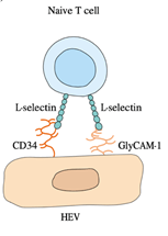
5. Vascular endothelial cells increase their expression of E- and P-selectin in response to the mediators of acute inflammation.

6. Increased P-selectin expression is caused by thrombin and histamine; cytokines like IL-1 or TNF-induce increased E-selectin expression. The circulating neutrophils express mucins such as PSGL-1 or the tetrasaccharides Lewisa sialyl and Lewisx sialyl bind to E- and P-selectin.

7. This binding mediates the attachment or tethering of neutrophils to the vascular endothelium, enabling the cells to roll in the direction of blood flow.

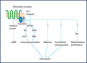
8. Chemokines such as IL-8 or other chemoattractants function on the neutrophils during this time, causing an activating signal mediated by G-protein that leads to a conformational shift in the molecules of integrin adhesion, resulting in neutrophil adhesion and subsequent transendothelial migration.

9. When in tissues, activated neutrophils also express elevated levels of chemoattractant receptors and thus show chemotaxis, migrating up the chemoattractant gradient.

10. Several chemokines, complement split products (C3a, C5a, and C5b67), fibrinopeptides, prostaglandins, and leukotrienes are among the inflammatory mediators that are chemotactic to neutrophils.

11. Furthermore, microorganism-released compounds, such as formyl methionyl peptides, are also chemotactic to neutrophils.

12. Increased levels of Fc antibody receptors and complement receptors are expressed by activated neutrophils, allowing these cells to bind more efficiently to antibody- or complement-coated pathogens, thereby increasing phagocytosis.

13. The triggering signal also activates the metabolic pathways into a respiratory burst, creating intermediates of reactive oxygen and intermediates of reactive nitrogen.

14. In the killing of different pathogens, the release of some of these reactive intermediates and the release of mediators from neutrophil primary and secondary granules (proteases, phospholipases, elastases and collagenases) play a significant role.

15. The tissue damage that can result from an inflammatory reaction also leads to these substances. The aggregation, along with accumulated fluid and different proteins, of dead cells and microorganisms, makes up what is known as pus.

Inflammatory Responses:

A complex cascade of non-specific events, known as an inflammatory response, is caused by infection or tissue injury, which provides early protection by minimising tissue damage to the site of the infection or tissue injury. Both localised and systemic responses are involved in the acute inflammatory response.

LOCALISED INFLAMMATORY RESPONSE:

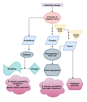
Redness, swelling, pain, heat, and loss of function are the hallmarks of a localised acute inflammatory response first identified almost 2000 years ago. There is an increase in vasodilation within minutes of tissue injury, resulting in an increase in the area’s blood volume and a decrease in blood flow. The increased volume of blood heats the tissue and causes it to turn red. Vascular permeability also increases, leading to fluid leakage, especially in postcapillary venules, from the blood vessels. This results in the fluid deposition in the tissue and, in some cases, leukocyte extravasation, which leads to the area’s swelling and redness. The kinin, clotting, and fibrinolytic processes are triggered when fluid exudes from the bloodstream. The direct effects of plasma enzyme mediators like bradykinin and fibrinopeptides, which induce vasodilation and increased vascular permeability, are responsible for many of the vascular changes that occur early in the local response. Some of these vascular changes are due to the indirect effects of histamine-released complement anaphylatoxins (C3a, C4a, and C5a) that induce local mast-cell degranulation.

1. Histamine is a potent inflammatory mediator, inducing vasodilation and contraction of smooth muscle.

2. Prostaglandins may also contribute to the acute inflammatory response associated with vasodilation and increased vascular permeability.

3. Neutrophils bind to the endothelial cells within a few hours of the initiation of these vascular changes and move from the blood into the tissue areas.

4. These phagocytose neutrophils invade pathogens and release mediators that lead to the inflammatory reaction.

5. The macrophage inflammatory proteins (MIP-1 and MIP-1), chemokines which attract macrophages to the inflammation site, are among the mediators. Around 5-6 hours after an inflammatory response starts, macrophages arrive.

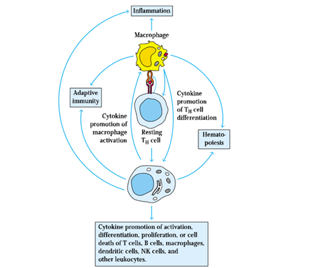
6. These macrophages are activated cells that show increased phagocytosis and increased release of mediators and cytokines that contribute to the inflammatory response.

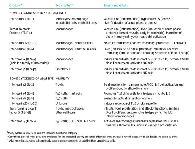
7. Three cytokines (IL-1, IL-6, and TNF-𝛼) that induce activated tissue macrophages secrete many of the localised and systemic changes, which observed in the acute inflammatory response.

8. All three cytokines function locally, causing coagulation and vascular permeability to increase.

9. Both TNF-𝛼 and IL-1 induce increased expression of adhesion molecules on vascular endothelial cells. For example, TNF-𝛼 stimulates the expression of E-selectin, a molecule of endothelial adhesion that binds adhesion molecules to neutrophils selectively. IL-1 induces increased ICAM-1 and VCAM-1 expression, which binds to lymphocyte and monocyte integrins.

10. Neutrophils, monocytes, and lymphocytes circulating identify these adhesion molecules on the walls of the blood vessels, bind to them and then pass into the tissue spaces via the vessel wall.

11. IL-1 and TNF-𝛼 also act on macrophages and endothelial cells to induce the development of chemokines that, by increasing their adhesion to vascular endothelial cells and by acting as potent chemotactic factors, contribute to the influx of neutrophils.

12. Besides, macrophages and neutrophils are activated by IFN-𝞬 and TNF-𝛼, facilitating increased phagocytic activity and increased release of lytic enzymes into tissue areas.

13. Without the overt intervention of the immune system, a local acute inflammatory response may occur.

14. Cytokines released at the inflammation site also promote both the adherence of immune system cells to vascular endothelial cells and their migration into tissue spaces through the vessel wall.

15. This results in an influx of lymphocytes, neutrophils, monocytes, eosinophils, basophils, and mast cells to the tissue damage site, where these cells are involved in antigen clearance and tissue healing.

To monitor tissue damage and promote the tissue repair processes that are important for healing, the length and strength of the local acute inflammatory response must be carefully controlled. TGF-β has been shown to play an essential role in limiting the response to inflammation. It also encourages fibroblast aggregation and proliferation and the deposition of an extracellular matrix necessary for proper tissue repair. Clearly, in the inflammatory response, the leukocyte adhesion processes are of great importance. As exemplified by leukocyte-adhesion deficiency, a failure of proper leukocyte adhesion may result in disease.

SYSTEMIC ACUTE-PHASE RESPONSE:

The systemic response is known as the acute-phase response accompanies the local inflammatory response. This response is characterised by fever induction, increased hormone synthesis such as ACTH and hydrocortisone, increased white blood cell development (leukocytosis), and the production of a large number of liver acute-phase proteins.

1. The rise in body temperature prevents a variety of pathogens from rising and tends to strengthen the immune response to the pathogen.

2. A prototype acute-phase protein whose serum level increases 1000-fold during an acute-phase response is a C-reactive protein.

3. It is made up of five similar polypeptides by noncovalent interactions kept together.

4. The C-reactive protein binds and activates complements to a wide range of microorganisms, resulting in the accumulation of opsonin C3b on the surface of microorganisms.

5. The C3b-coated microorganisms can then readily phagocytose phagocytic cells, which express C3b receptors.

6. The combined activity of IL-1, TNF-𝛼 and IL-6 is linked to several systemic acute-phase effects. To cause a fever response, each of these cytokines works on the hypothalamus.

7. Increased levels of IL-1, TNF- and IL-6 (as well as leukaemia inhibitory factor (LIF) and oncostatin M (OSM)) induce hepatocyte development of acute-phase proteins within 12–24 h of the onset of acute-phase inflammatory response.

8. To induce colony-stimulating factors (M-CSF, G-CSF, and GM-CSF) secretion, TNF-𝛼 also acts on vascular endothelial cells and macrophages.

9. These CSFs induce hematopoiesis, causing the number of white blood cells required to combat the infection to increase temporarily.

10. Redundancy in the capacity of at least five cytokines (TNF-𝛼, IL-1, IL-6, LIF, and OSM) to induce liver acute-phase protein development results from the induction of NF-IL6, a common transcription factor, after the receptor interacts with each of these cytokines.

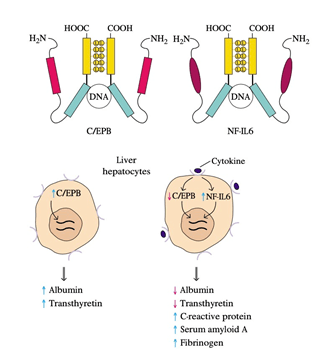
11. Amino-acid sequencing of cloned NF-IL6 showed that it has a high degree of sequence identity with C / EBP, a liver-specific transcription factor.

12. NF-IL6 and C / EBP both contain a leucine-zipper domain and a simple DNA-binding domain, and in the promoter or enhancer of the genes encoding different liver proteins, both proteins bind to the same nucleotide sequence.

13. C / EBP, which stimulates albumin and transthyretin production, is hepatocyte-constitutively expressed.

14. Expression of NF-IL6 increases and that of C / EBP decreases as an inflammatory response arises, and the cytokines interact with their respective receptors on liver hepatocytes.

15. The inverse relationship between these two transcription factors reflects the observation that serum protein levels such as albumin and transthyretin decrease during an inflammatory response while those of acute-phase proteins increase.

BACTERIAL ENDOSPORES – FORMATION, STRUCTURE AND FUNCTIONS

BY: Reddy Sailaja M (MSIWM031)

Introduction

  • Microorganisms have the ability to adapt themselves to the changing conditions prevailing in the environment. Factors that influence microorganism’s survival could be physical, chemical or environmental.
  • Some microorganisms go in search of favorable conditions for survival, while some will become dormant till the favorable conditions arrive.
  •  One such mechanism adapted by certain gram-positive bacteria is the development of ‘endospores.  Gram positive bacteria, especially genera, Bacillus and Clostridium have the ability to form endospores in response to harsh conditions, nutrient deprivation in particular.
  •  When there is starvation due to nutrient deprivation, these bacteria produce most resistant and dormant ‘endospore ‘structures that preserve cell’s genetic composition to with stand the harsh assaults like high temperature, desiccation, UV radiation, chemical and enzymatic damage.
  •  Moreover, endospores are the most resistant form of “spores” or “cysts” produced by many bacteria and are resistant to most of the antibiotics. Altogether, endospores are resistant and dormant structures of life survival forms of bacteria and fight against harsh environments.


Formation of endospore

Figure 1: Development of Endospore

  • Bacillus subtilis is the model organism used to study and understand the development of endospore during the process is called sporulation.
  •  It takes many hours to complete endospore formation. Morphological changes that occur during this process are used as markers to classify stages of endospore development. Stage I is that when the bacterial cell is under favorable conditions.
  • Under unfavorable conditions, bacterial cell initiates endospores formation by asymmetric cell division and is called Stage II. Asymmetric cell division results in the formation of a larger mother cell and a smaller forespore (or pre-spore) with septum in between them.
  • Even though, these two cell types in stage II has varied developmental fates, intercellular communication system harmonize cell specific gene regulation by influencing specialized sigma factors in the cells.
  •  In stage III, peptidoglycan present in the septum gets dissolved and the mother cell engulfs forespore, which becomes a cell within a cell.
  • In the stages IV+V, cortex and the spore coat layers are formed around the forespore, leading to the production of endospore specific compounds.
  •  In the stages VI+VII, further dehydration and the maturation of the endospore happens. Finally, the mother cell dies by apoptosis (also called programmed cell death) and the endospore is release into the environment and remains dormant until favorable conditions prevail.

Endospore structure

Endospore structure comprises of multiple layers of coats that resist against harsh surrounds. The following table details various layers (from outer to inner), their compositions and functions.

Endospore layerCompositionFunction
ExosporiumCarbohydrates, proteins  and lipidsGives hydrophobic character to the endospore and is responsible for endospore pathogenicity
Spore coatCoat proteins cross-linked with disulfide bondsActs as primary permeability barrier and allows only smaller molecules like germinants
Outer membraneNot known
CortexPeptidoglycan without teichoic acids with low cross linkingStructural differences in the peptidoglycan of cortex and germ cell wall allow selective degradation of outer protection, germination of endospore and transformation of germ cell wall into vegetative cell.
Germ cell wallPeptidoglycan
Inner membraneSimilar to cell membrane composition. Germinant receptorsVaried fluidity and permeability and decreased mobility of the membrane lipids make the structure highly impermeable to the molecules including water, protecting core. Germinant receptors allow binding of germinants and begin germination and vegetative growth.
CoreBacterial DNA, RNA, ribosomes, essential enzymes, small acid-soluble spore proteins (SASPs), Dipicolinic acidDehydrated state protects enzymes and heat resistance. SASPs protect DNA from destructive chemicals and enzymes by forming shield. SASPs also function as carbon and energy source during germination into vegetative cell. Dipicolinic acid also protects endospore’s DNA against harsh environment.

Figure 2: Structure of endospore

Mechanism of sporulation

  • Sporulation of endospores is under the control of five kinases, namely KinA, KinB, KinC, KinD and KinE that act under phosphorelay signal transduction mechanism.
  • Each of these kinases gets activated based on specific environmental stimuli. Under a specific kind of environmental stimulus, one of the five sensor kinases undergoes autophosphorylation at conserved histidine residue by an ATP dependent reaction through a protein called Spo0F. Then the Spo0F transfers the phosphate to Spo0B, that act as a mediator and delivers signal to Spo0A.
  •  Spo0A further positively regulate genes necessary for sporulation and negatively regulate genes required for vegetative growth.

Figure 3: Mechanism of sporulation

Functions of endospores

  • Endospores mainly resist harsh conditions like high temperatures, disinfectants, radiation, etc.
  •  Endospores are reported to survive for millions of years. For example, viable endospores were isolated from gastrointestinal tract of a bee that was embedded in amber around 25-40 years ago.
  • Dipicolinic acid and SASPs are crucial in protecting core of the endospore that contains genetic material.

Infectious diseases caused by endospores

In spite of defensive mechanism, endospores also transmit some infectious diseases as follows:

i)Anthrax – caused by Bacillus anthracis endospores when inhaled, ingested will germinate under suitable conditions and spread the infection

ii)Botulism – Caused by Clostridium botulinum. Spreads through unprocessed food and infect

iii) Tetanus – caused by Clostridium tetani. Spread through anaerobic wounds and cause infection.

 Other infectious diseases like gas gangrene and pseudomembranous colitis are also popular.

CHEMOKINES

BY: K Sai Manogna (MSIWM014)

1. Chemokines are the superfamily of small polypeptides, most of which contain residues of 90-130 amino acids are chemokines. 

2. They regulate the adhesion, chemotaxis, and activation of several kinds of leukocyte populations and sub-populations selectively, and sometimes explicitly. 

3. Consequently, they are significant leukocyte traffic regulators. Some chemokines are predominantly involved in inflammatory processes; others are expressed constitutively and play essential roles in the homeostatic or developmental activity. 

4. Housekeeping chemokines are manufactured in lymphoid organs and tissues or in non-lymphoid sites such as the skin where normal lymphocyte trafficking is directed, such as deciding the correct placement of newly-generated hematopoiesis leukocytes arriving from the bone marrow. 

5. Chemokines are constitutively expressed by the thymus, and normal B cell lymphopoiesis often depends on suitable chemokine expression. 

6. Effects mediated by chemokines are not confined to the immune system. 

7. Mice missing either the CXCL12 chemokine (also referred to as SDF-1) or its receptor display significant defects in brain and heart production. 

8. In the formation of blood vessels and wound healing, members of the chemokine family have also been shown to play regulatory roles. 

9. In response to infection, inflammatory chemokines are usually induced. 

10. The expression of inflammatory cytokines as inflammation sites is regulated by interaction with pathogens or the action of pro-inflammatory cytokines, such as TNF-𝛼. 

11. By inducing, adherence of these cells to the vascular endothelium, chemokines induce leukocytes to migrate into different tissue locations. 

12. Leukocytes are drawn to high localised concentrations of chemokines after migration into tissues, resulting in the selective recruitment of phagocytes and lymphocyte effector populations to inflammatory sites. 

13. The assembly of leukocytes at infection sites, coordinated by chemokines, is an integral part of mounting an infection response that is correctly oriented. 

Of more than 50 chemokines and at least 15 chemokine receptors have been identified. The chemokines have four residues of conserved cysteine and almost all fall into one or the other of two distinctive subgroups based on the position of two of the four invariant cysteine residues: 

■ Chemokines of the C-C subgroup, in which conserved cysteines are contiguous; 

■ Chemokines of the C-X-C subgroup, in which several other amino acids (X) distinguish the conserved cysteines. 

Receptors whose polypeptide chain traverses the membrane seven times mediate chemokine action. CC receptors (CCRs), which recognise CC chemokines, and CXC receptors (CXCRs), which recognise CXC chemokines, are two subgroups of receptors. 

As with cytokines, there is a high affinity (Ka > 109) and high specificity for the interaction between chemokines and their receptors. For instance, at least six different chemokines are recognised by CXCR2, and several chemokines can bind to more than one receptor. 

It activates heterotrimeric large G proteins when a receptor binds an appropriate chemokine, activating a signal-transduction method that generates such potent second messengers as cAMP, IP3, Ca2 +, and activated small G proteins. Chemokine-initiated activation of these signal transduction pathways carries out drastic changes. The addition of adequate chemokine to leukocytes induces sudden and widespread changes in shape within seconds, the promotion of more outstanding adhesion to endothelial walls by activating leukocyte integrins, and the development of phagocyte microbicidal oxygen radicals. These signal-transduction pathways facilitate other changes, such as granular material release, neutrophil and macrophage proteases, basophil histamine, and eosinophil cytotoxic proteins.  

Chemokine-Receptor Profiles Mediate Activity of leukocytes: 

1. Neutrophils express CXCR1, -2, and -4 among the central populations of human leukocytes; eosinophils have both CCR1 and CCR3. 

2. Some activated T cells have CCR1, -2, -3, and -5, CXCR3 and -4, and possibly others, whereas resting naive T cells show a few types of chemokine receptors. 

3. Consequently, variations in the expression of chemokine receptors by leukocytes coupled with the development by destination tissues and sites of distinctive chemokine profiles provide rich opportunities for the differential control of the activities of the various populations of leukocytes. 

4. Indeed, variations in chemokine-receptor expression patterns occur both within and between different populations of leukocytes. 

Fig : Patterns of expressions on some principle chemokine receptors on the human leukocytes.

Note: Their various cytokine output patterns can distinguish the TH1 and TH2 subsets of TH cells. Different profiles of chemokine receptors also show these subsets. CCR3 and -4 are expressed by TH2 cells, and a variety of other receptors are not expressed by TH1 cells. TH1 cells, on the other hand, express CCR1, -3, and -5, but most TH2 cells do not. 

The Other Inflammatory Mediators: 

Several other mediators released by cells of the innate and acquired immune systems activate or improve particular aspects of the inflammatory response in addition to chemokines. Tissue mast cells, blood platelets, and several leukocytes, including neutrophils, monocytes/macrophages, eosinophils, basophils, and lymphocytes, release them. 

The plasma comprises four interconnected mediator-producing processes in addition to these sources: the kinin system, clotting system, fibrinolytic system, and the complement system. The first three systems share the Hageman factor, a common intermediate. These four systems are triggered when tissue damage occurs, to form a network of interacting systems that produce several inflammation mediators. 

The Tissue Injury Stimulates the Kinin System: 

1. The kinin mechanism is an enzymatic cascade that starts with tissue injury, a plasma clotting factor, called Hageman factor, is activated. 

2. In order to form kallikrein, the activated Hageman factor then activates prekallikrein, which cleaves kininogen to generate bradykinin. 

3. This inflammatory mediator is a potent fundamental peptide that enhances vascular permeability, causes vasodilation, pain, and induces smooth muscle contraction. 

4. By cleaving C5 into C5a and C5b, kallikrein also works directly on the complement mechanism. 

5. An anaphylatoxin that induces mast-cell degranulation, resulting in the release of several inflammatory mediators from the mast cell, is the C5a complement portion. 

The Clotting System Yields Inflammation Mediators Produced by Fibrin: 

1. Another enzymatic cascade caused by blood vessel disruption yields significant amounts of thrombin. 

2. To generate insoluble strands of fibrin and fibrinopeptides, thrombin works on soluble fibrinogen in tissue fluid or plasma. 

3. Clot formation acts as a barrier to the spread of infection, for which the insoluble fibrin strands cross each other. 

4. After tissue damage, the clotting mechanism is activated very quickly to avoid bleeding and limit the spread into the bloodstream of invading pathogens. 

5. As inflammatory mediators, the fibrinopeptides act, inducing increased vascular permeability and neutrophil chemotaxis. 

The Fibrinolytic System Yields Inflammation Mediators produced by Plasmin: 

1. The fibrinolytic method completes the elimination of the fibrin clot from the damaged tissue. 

2. The end product of this pathway is the plasmin enzyme, which is generated by plasminogen conversion. 

3. Plasmin breaks down fibrin clots into degradation products that are chemotactic for neutrophils, a potent proteolytic enzyme via activating the classical complement pathway.

4. Plasmin also contributes to the inflammatory response. 

Anaphylatoxins Formed by the Complement System: 

1. Activation by both classical and alternative pathways of the complement system results in the development of several complement split products that serve as essential inflammation mediators. 

2. Binding of anaphylatoxins such as C3a, C4a, and C5a to receptors on the membrane of tissue mast cells induces degranulation with histamine release and other pharmacologically active mediators.

3. Such mediators cause contraction of smooth muscles and increase vascular permeability. 

4. C3a, C5a, and C5b67 function together to induce the adherence of monocytes and neutrophils to vascular endothelial cells, extravasate through the capillary endothelial lining, and migrate to the tissue site of complement activation. 

5. Thus, activation of the complement system results in fluid inflows carrying antibody and phagocytic cells to the entry site of the antigen. 

Lipids as Inflammatory Mediators: 

1. Phospholipids in the membrane of many cell types (e.g., macrophages, monocytes, neutrophils, and mast cells) are degraded into arachidonic acid and lyso-platelet-activating factor following membrane disturbances. 

2. Subsequently, the latter is transformed into a platelet-activating factor (PAF) that induces platelet activation and has several inflammatory consequences, including eosinophil chemotaxis, neutrophil and eosinophil activation and degranulation. 

Arachidonic acid metabolism:

a. Arachidonic acid metabolism produces prostaglandins and thromboxanes through the cyclooxygenase pathway. 

b. Various cells produce various prostaglandins: 

i. monocytes and macrophages produce large quantities of PGE2 and PGF2; 

ii. neutrophils produce moderate amounts of PGE2, which is released by mast cells. 

There are various physiological effects of prostaglandins, including increased vascular permeability, increased vascular dilation, and neutrophil chemotaxis induction. Thromboxanes cause platelet aggregation and blood vessel constriction. 

The lipoxygenase pathway also metabolises arachidonic acid to yield four leukotrienes: LTB4, LTC4, LTD4, and LTE4. Three of these (LTC4, LTD4, and LTE4) together make up what was formerly referred to as a slow-reacting anaphylaxis material (SRS-A); these mediators cause contraction of smooth muscle. LTB4 is a potent neutrophil chemoattractant. Several cells, including monocytes, macrophages, and mast cells, make leukotrienes.

Some cytokines are essential mediators of inflammation:

1. In the formation of an acute or chronic inflammatory response, several cytokines play a significant role. 

2. There are redundant and pleiotropic effects of IL-1, IL-6, TNF, IL-12, and several chemokines that together lead to the inflammatory response. 

3. Besides, IFN-contributes to the inflammatory response, functioning later in the acute response and by attracting and stimulating macrophages, leading in a significant way to chronic inflammation. 

4. The differentiation of the pro-inflammatory TH1 subset is caused by IL-12.

CENTRIFUGATION

BY: Reddy Sailaja M (MSIWM031)

Centrifugation is one of the most extensively used technique in research and development fields of biochemistry, molecular biology, biochemistry and pharmaceutical industries for varied applications like isolation of cells, fractionation of sub cellular particles and other macromolecules for analytical and clinical applications.

Definition

Centrifugation is a process of separation (or concentration) of particles from suspended medium based on their size, shape, density, viscosity of the medium, rotor speed etc. Centrifugal force is a key in this technique to separate the particles in less time and it acts against gravitational force. Figure 1 gives overview of centrifugation process from muscle tissue.

Figure 1: Centrifugation overview

Principle:

In general, when a liquid suspension is placed idle for some time, particles of bigger size/density will start to settle at the bottom of the container because of gravitational force and so on. But, this is a slow process and can’t be applied practically. Centrifugation works on centrifugal force to separate the particles in a suspension in less time with more efficiency.

When a body with mass ‘m’ is rotating in a circle with radius ‘r’ and velocity ‘v’, the force acting on the body is measured using the following formula 1.

F = mv2/r

Where,

F = centrifugal force,

m = mass of body,

v = velocity of the body,

 r = radius of circle of rotation.

The gravitational force acting on the body ‘m’ is calculated using the formula 2: G = mg

Where,

G = gravitational force

g = acceleration due to gravity

U

The centrifugal force is further calculated using the formulae 1 and 2 as follows:

C = F/G = mv2/mgr = v2/gr

Since, v = 2π r n

Where,

 n = speed of rotation

C = F/G = (2π r n) 2/g r = 4π2 r2n2 = 2π2/g D n2 = kD n2

where,

k = 2π2/g = constant

D = maximum diameter of the centrifuge

D is able to measured either from centrifuge center to the free surface of the liquid or to the tip of the centrifuge tube.

From the equation C = kDn2 it was evident that,

Centrifugal effect ∝ diameter of centrifuge

Centrifugal effect ∝ (speed of rotation)2.

When a liquid suspension containing container is rotated at a certain speed called revolutions per minute (RPM), particles will move at a certain speed away from the axis of rotation. The force that’s being generated on the particles to move away from the centre is called relative centrifugal force (RCF). RCF depends mainly on the rotational speed (measured in RPM) and the distance of the particles from the centre of the rotation (rotor).

RCF = 11.2 × r (RPM/1000)2

Where,

r – Distance in centimeters

More the density of the particles, faster is the settlement at the bottom of the tube, while less dense particles will be floating in the liquid. The rate of sedimentation depends on the size and density of the particles and can be explained by Stokes equation (explains movement of a sphere in a gravitational field).


Where,

V = viscosity of the medium

d = diameter of the sphere

p = particle density

L = medium density

n = viscosity of medium

g = gravitational force

Stokes equation explains behavior of particles based on the rate of particle sedimentation as follows:

  • directly proportional to the size of the particle
  • directly proportional to the difference between the particle and the medium densities
  • zero when the particles and medium exhibits same density values
  • decreases when the medium viscosity increases
  • increases as the gravitational force increases

Table 1: Densities of cells and sub cellular fractions

The particles that gets settled at the bottom forms ‘pellet’ while the liquid suspension with lighter particles or no particles is called ‘supernatant’.  Therefore, centrifugation is a process that utilizes centrifugal force for the sedimentation of particles.

Figure 2: Densities and sedimentation coefficients of biomolecules, cell organelles and viruses

For example, ‘m’ is a particle in a centrifuge tube suspended in a liquid. During centrifugation process, the particle is influenced by three kinds of forces: FC– the centrifugal force, FB – the buoyant force and Ff – the frictional force between the particle and the liquid.

Figure 3: Centrifugal force

Centrifuge

Centrifuge is a tool designed to separate particles in the liquid suspension based on the centrifugation principle. It is operated using an electric motor that enables an object to move around in a fixed axis when a perpendicular force is applied to the axis.

Figure 4a: Front view of a typical centrifuge

Figure 4b: Rear view of a typical centrifuge

Centrifuge comprises of three major components:

  1. Rotor – Holds containers (tubes/bottles etc) containing liquid suspension to be centrifuged. Rotors of different types and sizes are available
  2. Fixed angle rotor – Requires short time to sediment particles as they need to travel only a little distance.

Figure 5a: Fixed angle rotor

  • Swinging bucket rotor – Allows better separation of particles as the particles have to move long distance. Stronger pellet is formed and supernatant can be easily removed.

Figure 5b: Swinging bucket rotor

  1. Drive shaft – Helps hold rotors which in turn connect to motor.
  2. Motor – Helps to rotate the rotor based on the input speed by providing power.

All the major centrifuge components are surrounded by a protective cabinet with operating controls and indicator dials for speed and time mounted on it. Centrifuges come with brake system to control rotor and allow it to come to standstill when the centrifugation run gets completed. Refrigerated centrifuges allow option to control temperature so that the delicate biological samples won’t be degenerated during the process.

Types of centrifugation techniques

There are two major types of centrifugation techniques to separate particles.

  1. Differential centrifugation
  2. Density gradient centrifugation
  3. Isopycnic centrifugation
  4. Rate-zonal centrifugation
  5. Differential centrifugation:

This is the simplest type of particle separation form, also called ‘pelleting’ down the particles. Sedimentation occurs at different rates based on the density of the particles. More dense particles will sediment fast and the lighter particles will be floating in the suspension. Sedimentation of the particles also depends on the centrifugal force applied. As the centrifugal force increased, pellet with decreased sedimentation rate will be formed and vice versa.

Differential centrifugation is applied during cell harvest or sub cellular fractionation from a tissue homogenate. When lower centrifugal force is applied, dense particles like nuclei, membrane vesicles etc gets pelleted first. To further pellet next order particles like mitochondria etc, more centrifugal force is applied. Greater than or equal to four differential centrifugation cycles are applied for sub cellular fractionation of the tissue homogenate. However, this process faces carry over contamination of the particles from previous fraction and not purity is less.

Figure 6: Differential centrifugation process overview

  • Density gradient centrifugation: This method is mainly applicable to separate sub cellular particles and other macromolecules with more purity.

In this process, gradient media of different density are layered one above the other, more dense at the bottom of the tube and the lightest at the top. The cell fractionate that need to be separated is placed on the top of the gradient layer and the centrifugal force is applied.

Figure 7: Density gradient process overview

Density gradient method is further classified into two types as follows:

  1. Isopycnic centrifugation:

This process is also known as buoyant or equilibrium separation. In this process, particles are separated base on density. Particle size plays a role when the density of the particles and the surrounding medium is same. When the centrifugal force is applied, initially the sample and gradient gets mix uniformly and the particles move through the gradient until the density of the particles and gradient medium becomes same. Now, the gradient is called as ‘isopycnic’ and the particles get separated based on their buoyancy. Therefore, it is important to make sure that the gradient medium is always dense than the particles to be separated. Particles get separated in the gradient medium in different layers, but never settle to the bottom of the tube. Gradient medium varies depending upon the kind of material being separated. Continuous gradient method is good for analytical separation while discontinuous gradient is more suitable for biological applications (E.g.: separation of lymphocytes from blood).

Table 2: Common density gradient media used for isopycnic centrifugation process

  • Rate zonal centrifugation:

The carryover contamination of particles in differential centrifugation is prevented by implementing rate zonal centrifugation. In this process, sample was layered in a narrow zone on the top of the density gradient and the centrifugal force is applied. Particles segregate based on their size and mass rather than density, and also on the centrifugal force. As a result, narrow load zone prevents less sample volume (≤10%) that can be accommodated on the density gradient and it further stabilizes the bands and allows medium of increasing density and viscosity. Centrifugation is applied for a short time at a low speed. As the density of the particles is more than the density of the gradient, there is a chance that all the particles form pellet if the centrifugation continues for a long time.

Figure 8: Rate zonal and isopycnic centrifugation processes overview

Common applications of centrifugation

  • Production of drugs and other biological products
  • Separation of subcellular particles
  • Separation of blood and urine components in forensic analysis
  • Protein purification
  • Clarification and stabilization of wine
  • Fat removal from milk

BIOFILMS

BY: K. Sai Manogna (MSIWM014)

  • Layer-like microorganism aggregations and the extracellular polymers bound to a solid surface are biofilms.
  •  Biofilms, innate cells, are ubiquitous and are becoming more and more important in processes that are designed for pollution control including philtres that drip, rotating biological contactors and anaerobic philtres.
  • Biofilm processes are simple, efficient, and sustainable because natural immobilization allows excellent retention and accumulation of the biomass without the need for separate solids.
  • We should consider a fundamental problem concerning biofilms (indeed all aggregated systems) before designing mathematical instruments for predicting the removal of substrates by biofilm:

Here are a few possibilities:

1. Due to the advection of substrates after the film, these biofilms are exposed continuously to the fresh substrate in space. It means that if the biofilm is attached near the source of the substratum supply, the substrate concentration is higher.

2. In mandatory substratum transportation consortia or other synergistic relationships, different types of bacteria must be co-existent; for the exchange, the near juxtaposition between cells is required in a biofilm.

3. The biofilms build a more friendly internal atmosphere (for example, pH, O2, or products) than the bulk liquid. In other words, the biofilm creates special and self-created cell-beneficial micro-environments.

4. The surface itself produces a single micro-environment, for example by removing contaminants or by corrosive releases of Fe2+, a donor of electrons.

5. The surface allows the bacteria to change physiologically.

6. The cell’s strict packing modifies the physiology of the cells.

Biofilm formation:

The development of biofilms can be divided into five phases:

1. Surface relation

2. Monolayer formation

3. Training in microcolony

4. Biofilm mature

5. Unlocking

  1. The initial step, which is still reversible, is the initial touch of the moving planktonic bacteria on the surface.
  2. The bacteria then begin to form a monolayer and create a protective “slime,” an extracellular matrix.
  3. The matrix contains extracellular polysaccharides, structural proteins, cell scrap, and nucleic acids known as EPS.
  4. Extracellular DNA (eDNA) predominates the initial steps of matrix development, and polysaccharides and structural proteins later take over.
  5. At these points, microcolonies are produced that display significant growth and contact between cells such as quorum sensing.
  6. During the last step of a new cycle of biofilm formation, some cells in mature biofilm begin to detach itself into the environment as planktonic cells.

The biofilm allows for:

a. tolerates antibiotic attacks;

b. Trap bacterial growth nutrients and stay in a favourable niche;

c. Adhere and avoid flushing to environmental surfaces;

d. live in close collaboration and associate with other biofilm bacteria;

e. Stop phagocytosis and attack the complementary routes of the body.

For example, Planktonic Pseudomonas aeruginosa uses polar flagella to travel by water or mucus and to reach a firm surface as mucous membranes of the body. The cell wall and pil adhesives can then be used to bind the mucous membrane epithelial cells. Attachment stimulates signalling and quorum sensing genes so that a polysaccharide alginate biofilm synthesization will eventually begin with the population of P. aeruginosa. As the biofilm forms, the bacteria lose flagellum and secrete different enzymes that allow the population to obtain nutrients from the host cells. Finally, the biofilm pillows and establishes water canals for all bacteria in the biofilm to provide water and nutrients. As the biofilm gets too crowded with bacteria, the sensing of quorum helps a few Pseudomonas to develop flagella again, escape the biofilm, and colonize a new spot.

Fig: Formation of biofilm by P.  aeruginosa.

Two bacteria involved in dental caries, Streptococcus mutants and Streptococcus sobrinus, break down sucrose to glucose and fructose. Streptococcus mutans may use a dextransucrase enzyme in a sticky dextran polysaccharide, which forms a biopathic film that allows bacteria to bind to the tooth enamel and form the plaque. The pathogenicity of S bacteria. S. and mutans. In order to generate energy, sobrinus often ferments glucose. Glucose fermentations produce lactic acid, which is released on the tooth surface and causes decay. The mechanism is described in the given image.

Microbial Leaching:

  • Copper extraction from ore deposits was carried out over centuries using acidic solutions, but the involvement of bacteria was not confirmed in metal dissolution before the 1940s.
  •  Today about 10–20% of copper mined in the United States is extracted by low-grade microbial processing. In the expansion of microbial leaching, other elements, including Uranium, Silver, Gold, Cobalt and Molybdenum, are also invested considerably.
  • Most microbial liquidation relies on metal sulphides’ microbial oxidation. Aqueous environments combined with mineral waste create very harsh conditions with a low pH, high metal concentrations and high temperatures that select very specialized nutrient requirements for a microbial flora.
  • Heap leaching is the most common method in which copper and other minerals are extracted microbially from spent ore.
  •  The method involves arranging the spent ore fragments into a configuration of the packed bed to allow the passage of water. Acidized water (pH = 1.5-3.0) is sprayed on the porous ore bed in order to start the operation.
  • The solvent ferrous iron is actively oxidized, and sulphide minerals are attacked by acidophilic bacteria such as Thiobacillus iron, which can then be released from aquatic ions by releasing soluble cupric ions. In terms of the corrosion on metal surfaces, this oxidation is identical.

Biological reactions and mass transfer rates currently constrain the industrial applications of microbial fluid, but in recent years significant changes have been made to process design, and the mining industry sees the method as being promising.

Removal of Biofilms:

  • Traditional cleaning of biofilm has been accomplished by detergents, biocides, enzymes, and mechanical or physical methods of cleaning the biofilm.
  •  Biofilms in sensitive locations in the food and drink manufacturing industries have grown, leading to problems such as food spoilage, production quality and other nutrient supplementation and insufficient cleaning and disinfection.
  • Depending on the medium temperature and relative humidity of these microorganisms, they will live longer after application. The pulp- and paper-based agent for biofilm removal is classified into three groups: chlorine, chlorine dioxide, hydrogen peroxide (hydrogen peroxide), Ozone, antioxidants, and enzymes.
  • The cells adhering to the staphylococcus aureus are effectively used in sodium dichloro isocyanurate, hypochlorite, iodophore, hydrogen peroxide and peracetic acid. The most powerful method of extracting adhered large amounts of L was to eliminate peracetic acid.
  • Monocytogenes cells remaining on chips of stainless steel after sanitization. Scanning electron microscopy has shown that on chlorine and anionic acid-treated surfaces biofilm cells and extracellular matrices remain better than iodine and ammonium-quaternary detergent sanitizers from which a viable cell is not released.
  • Oxidative and antioxidant biocides have long been used, although enzyme use is being studied at present.
  • In order to develop a quality management programme in different industries, tanks, pipes, pasteurizers, coolers, membrane filtration units and fillers must be tested, because they help to avoid microbiological hazards and severe financial losses.

COMPONENTS OF BLOOD

BY: Reddy Sailaja M (MSIWM030)

BLOOD

Blood is a specialized body fluid, comprises of plasma and the cells that circulate throughout the human body. It supplies oxygen, glucose, antibodies, vitamins, electrolytes, heat, hormones, and immune cells across the body for life survival. It removes carbon dioxide and other waste generated from the cells in the body.

BLOOD COMPONENTS

Blood is made up of three main components: plasma, blood cells (red and white) and platelets.

Plasma: Plasma occupies 55% of blood fluid in human beings. Plasma comprises of 92% water and the remaining 8% contains carbon dioxide, glucose, hormones, proteins etc.

 Blood cells: Blood cells comprise of 45% of total blood fluid. These are produced in bone marrow by the process called ‘hematopoiesis’ from a common precursor cell (hematopoietic stem cells). Then these blood cells mature into red blood cells (RBCs), white blood cells (WBCs) and platelets. The organs like lymph nodes, liver and spleen regulate the generation, destruction and regulation of blood cells.

Figure 1: Blood cells production by hematopoiesis

Red blood cells: RBCs are also called erythrocytes. They are double concave shaped structures without nucleus. Approximately, 4.5 – 6.2 million/microliter are present in the blood. Their main function is to carry oxygen from lungs to all parts of the body. RBCs contain a special protein called ‘Hemoglobin’ that aids in oxygen transport. RBCs have a life span of 120 days.

White blood cells: WBCs are otherwise known as leucocytes. WBCs are vital in fighting against invading pathogens and infections. Approximately 3700 – 10500/microliter are present in the blood. Apart from fighting infection, WBCs also help heal wounds by ingesting dead cells and debris, protects against foreign entity that enter blood stream and fights against cancerous cells.

The following are the types of WBCs that are produced in response to the kind of infection (bacterial vs fungal vs viral vs parasitic).  Life span varies from hours to days to years depending upon the type of WBCs. WBCs are majorly divided into two subtypes: Granulocytes and agranulocytes. Granulocytes contains protein containing granules in their cytoplasm. Eosinophils, basophils and neutrophils constitute granulocytes. Monocytes and lymphocytes constitutes agranulocytes. The following table explains the types of WBCs’, their nature and function.

Type of white blood cellPercentage of abundance in WBCS (%)Function
Basophils0.5 – 1Basophils produce in response to parasite infections, allergy and bone marrow damage. It secretes histamine – involve in allergic reactions and heparin – an anticoagulant that aids blood clotting at the site of injury and subsequent wound healing.
Eosinophils2 – 4Eosinophils defend against bacterial and parasite infections by releasing toxic substances and results in immflamatory reaction.
Neutrophils60 – 70First line of defense against invading pathogens. They attack the pathogens, engulf and digest them by phagocytosis process and maintain normal health.
Monocytes3 – 8Maintains tidiness of the blood and other tissues by clearing the dead pathogen particles and damaged cells and their debris.
Lymphocytes20 – 25B – cells produce antibodies against bacterial, viral and fungal infection. T – cells are two types, cytotoxic T-cells kills the antigens and helper T-cells aid antibody production from B-cells. Natural killer cells attack any foreign object that comes in contact with the body.

Figure 2: Blood and its components

Blood platelets: These are also called thrombocytes. Approximately 1,50,000 – 4,00,000 platelets/microliter are present in the blood. Platelets help clot the blood to stop bleeding during injury by protects the wound against further infection.

In brief, blood functions include:

  • Oxygen supply to cells and tissues
  • Supply essential nutrients – glucose, aminoacids, fatty acids,
  • Removal of waste material – carbon dioxide, urea, lactic acid
  • Fighting against infections
  • Regulating body temperature and pH balance
  • Transport hormones and transmits neuromessages

Blood disorders:

Blood disorders often cause life threatening situations as the infection spreads out throughout the body by blood circulation. General blood disorders are as follows:

RBCs disorder – Anemia: Low number of RBCs in blood cause anemic situation. This results in low oxygen supply in the body, fatigue and pale skin.

WBCs disorder – Cancer: Lymphoma, myeloma and leukemia are the major blood related cancers.

Platelets disorder – Internal blood clots: these clots block blood supply and can be dislodged and spread through various organs like lungs, heart, brain etc, which can be fatal to the body.

CYTOKINES

BY: Ria Fazulbhoy (MSIWM031)

Cytokines are an important group of proteins or glycoproteins which play a major role in cell-to-cell communication between cells like lymphoid cells, inflammatory cells and hematopoietic cells. They are secreted by white blood cells (WBCs) and other cells of the body. They respond to stimuli and assist in the regulation of development of immune effector cells and sometimes have a direct effect of their own as well. The cytokine binds to the target cell by the presence of specific membrane receptors present on the target cell. (very high affinity-cytokines work at picomolar concentrations)

Mode of action

  1. Autocrine

The cytokine is released from a cell and binds to the membrane receptor present on the same cell.

  • Paracrine

Cytokine is released from the producer cell and binds to the target cell which is in close proximity

  • Endocrine

Cytokine binds to the target cell which is in a distant part of the body.

Four major groups of cytokines are Hematopoietic family (interleukins-ILs), Interferons (IFs) family, Tumor necrosis factors (TNF) family and chemokine family.

A variety of cells secrete cytokines, but the major principal producer cells are Th cells and macrophages. Cytokines secreted from these cells activate an entire network of interacting cells.

   Macrophage and Th cells are major producers of cytokines in the body.

Some biological functions of cytokines include:

  • Cellular and humoral immunity development
  • Inflammatory response induction
  • Control of cellular proliferation and differentiation
  • Healing of wounds
  • Development of innate/acquired immunity
  • Hematopoiesis

NOTE: Cytokines have a non-antigen specific mode of action and have very short half-lives.

Functions of some cytokines

Cytokine secretion by Tн cell subsets: Tн1 and Tн2

Difference in the pattern of cytokine secretion amongst Tн cell subsets determines the immune biological response made to a particular antigenic challenge. These two subsets are Tн1 and Tн2, which secrete different cytokines and mediate in different ways. Both these subsets secrete IL-3 and GM-CSF.

Tн1 and Tн2 have the following functional differences:

1) Tн1 subset:

  • It is responsible for mainly cell-mediated immune responses like activation of Tc cells and delayed hypersensitivity reactions.
  • Helps in promotion of excessive inflammation and tissue injury
  • Helps in production of opsonization-promoting IgG antibodies.
  • Effective in viral infections and intracellular pathogens.
  • IFN-४, IL-12 and IL-18 are responsible for the development of Tн1 cell response.
  • E.g.: IFN-४ and TNF-ß mediates inflammation and delayed hypersensitivity.
  • E.g.: IL-2 and IFN-४ promote differentiation of cytotoxic cells Tc from CD8 precursors.

2) Tн2 subset:

  • Responsible for secretion of antibodies for immune response.
  • Stimulates eosinophil activation and differentiation
  • Helps B cells
  • Promotes production of large amount of IgM and IgG
  • Supports allergic reactions
  • IL-4 is essential for the development of Tн2 response.
  • E.g.: IL-4 and IL-5 induce production of IgE and helps eosinophil attack on helminth or roundworm infections.

Tн2 development is favoured over Tн1. The cytokines produced by the two subsets are cross regulated. The cytokines produced by a subset (Tн1 or Tн2) promote the growth of their subset and simultaneously inhibit the activity and development of the opposite substrate (cross regulation). Two transcription factors known as T- Bet and GATA-3 are important in determining the cross regulation of the two subsets

  • T-Bet drives cells to differentiate towards Tн1 cells.
  • GATA-3 drives cells to differentiate along Tн2 cells.

EXTRAVASATION OF LYMPHOCYTES

BY: K. Sai Manogna (MSIWM014)

At inflammatory sites and secondary lymphoid glands, different subsets of lymphocytes exhibit directed extravasation. Therefore, lymphocyte recirculation is closely monitored to ensure that sufficient populations of B and T cells are recruited into various tissues. Extravasation of lymphocytes involves interactions between a variety of cell-adhesion molecules, as with neutrophils. The overall process is similar to what occurs during the extravasation of neutrophils and involves the same four stages of touch and rolling, activation, arrest and adhesion and, eventually, transendothelial migration.

Sites of Lymphocyte Extravasation:

1. Some regions of vascular endothelium consist of specialised cells with a plump, cuboidal (‘high’) form in the postcapillary venules of different lymphoid organs; such regions are referred to as high-endothelial venules or HEVs.

2. In appearance, their cells contrast strongly with the flattened endothelial cells that line the rest of the capillaries. Each of the secondary lymphoid organs comprises HEVs, except the spleen.

3. There are about 1.4 × 10⁴ lymphocytes extravasate into a single lymph node every second through HEVs.

4. Cytokines developed in response to antigen capture influence the production and maintenance of HEVs in lymphoid organs.

5. In order to prevent the antigen from entering the node, the role of lymphocyte antigenic activation in the preservation of HEVs has been demonstrated by surgical blocking of afferent lymphocyte vasculature of the node.

6. The HEVs demonstrate impaired function within a short period and gradually return to a more flattened morphology.

7. High-endothelial venules express several cell-adhesion molecules. HEVs, like other vascular endothelial cells, express CAMs from the selectin (E- and P-selectin) family, the mucin-like (GlyCAM-1 and CD34) family, and the superfamily of immunoglobulins (ICAM-1, ICAM-2, ICAM-3, VCAM-1, and MAdCAM-1).

8. In a tissue-specific way, some of these adhesion molecules are distributed. These tissue-specific adhesion molecules have been named vascular addressins (VAs) because they help to guide the extravasation to specific lymphoid organs of various populations of recirculating lymphocytes.

Receptor Profiles and Signals Guided by Lymphocyte Homing:

1. Related to neutrophil extravasation, the general lymphocyte extravasation mechanism is similar.

2. The fact that different subsets of lymphocytes migrate differently into different tissues is a significant aspect that separates the two processes. This method is known as trafficking or homing.

3. The numerous lymphocyte subset trafficking patterns are regulated by unique combinations of adhesion molecules and chemokines; homing receptors are called receptors that guide the circulation of different lymphocyte populations to specific lymphoid and inflammatory tissues.

Researchers have established several lymphocytes and endothelial cell adhesion molecules that are involved in lymphocyte interactions with HEVs and endothelium at tertiary sites or sites of inflammation.

Recirculating Naive Lymphocytes into Secondary Lymphoid Tissue:

Until it has been triggered to become an effector cell, a naive lymphocyte is not able to mount an immune response.

1. In specialised microenvironments within secondary lymphoid tissue (e.g., peripheral lymph nodes, Peyer patches, tonsils, and spleen), activation of a naive cell occurs.

2. Dendritic cells catch antigen inside these microenvironments and present it to the naive lymphocyte, resulting in its activation.

3. Naive cells do not display a preference for a specific form of secondary lymphoid tissue but instead circulate indiscriminately across the body to secondary lymphoid tissue through recognising HEV adhesion molecules.

4. The initial attachment to HEVs of naive lymphocytes is usually mediated by the binding of the L-selectin homing receptor to HEV adhesion molecules such as GlyCAM-1 and CD34.

5. The naive cell trafficking pattern is designed to keep these cells continuously recirculating across secondary lymphoid tissue, the primary purpose of which is to trap antigen transmitted by blood or tissue.

6. They are activated and enlarged into lymphoblasts until naive lymphocytes encounter antigen trapped in secondary lymphoid tissue. Activation takes approximately 48 h, and the blast cells are retained in the paracortical area of secondary lymphoid tissue during this time.

7. The antigen-specific lymphocytes cannot be identified in the circulation during this process, called the shutdown phase.

8. During the shutdown point, rapid proliferation and differentiation of naive cells occur. Then the effector and memory cells that this process produces leave the lymphoid tissue and begin to recirculate.

Lymphocytes of Effector and Memory follow distinct patterns of trafficking:

1. Effector and memory lymphocyte trafficking patterns vary from those of naive lymphocytes.

2. By recognising inflamed vascular endothelium and chemoattractant molecules produced during the inflammatory response, effector cells appear to be home to regions of infection.

3. On the other hand, memory lymphocytes selectively house the type of tissue in which antigen was first encountered.

4. This presumably ensures that a specific memory cell returns to the tissue where the antigen it recognises is most likely to re-encounter a subsequent threat.

5. Memory cells and effector cells express increased levels of specific molecules of cell adhesion, such as LFA-1, which interact with ligands present in additional tertiary lymphoid tissue (such as skin and mucosal epithelial) and at inflammation sites, allowing these sites to be accessed by effector and memory cells.

6. Naive cells lack the corresponding molecules of cell-adhesion and do not house these sites.

7. A variety of adhesion molecules, including E- and P-selectin and the Ig-superfamily molecules VCAM-1 and ICAM-1, are expressed in inflamed endothelium and bind to receptors expressed at high levels in the memory and effector cells.

8. Subsets of the memory and effector populations display tissue-selective homing activity, unlike naive lymphocytes.

9. Such tissue specificity is imparted by multiple combinations of adhesion molecules rather than by a single adhesion receptor.

10. A mucosal homing subset of memory/effector cells, for example, has high levels of LPAM-1 (4 7) and LFA-1 (Lb2) integrins that bind to MAdCAM and various ICAMs on venules of intestinal lamina propria.

11. However, since they have low levels of L-selectin that would promote their entry into secondary lymphoid tissue, these cells prevent direction to secondary lymphoid tissues.

12. Preferential homing to the skin is shown by the second group of memory/effector cells. Low levels of L-selectin are also expressed in this subset, however high levels of cutaneous lymphocyte antigen (CLA) and LFA-1, which bind to E-selectin and ICAMs on skin dermal venules, are seen.

13. While effector cells and memory cells that express decreased L-selectin levels do not appear to reach peripheral lymph nodes via HEVs, they may enter peripheral lymph nodes via afferent lymph vessels.

Adhesion-Molecule Interactions Play Extravasation Vital Roles:

1. A multi-stage mechanism involving a cascade of adhesion-molecule interactions is the extravasation of lymphocytes into secondary lymphoid tissue or regions of inflammation, similar to those involved in bloodstream neutrophil emigration.

2. This shows the usual interactions in the extravasation of naive T cells into lymph nodes through HEVs.

Mechanism:

1. In the first stage, a selectin-carbohydrate interaction similar to that seen with neutrophil adhesion.

2. L-selectin, which acts as a homing receptor which directs the lymphocytes to specific tissues expressing a corresponding mucin-like vascular addressin such as CD34 or GlyCAM-1, initially binds naive lymphocytes to HEVs.

3. The rolling of lymphocytes is less pronounced than neutrophil rolling.

4. Although the initial interaction of selectin-carbohydrate is minimal, the slow rate of blood flow in postcapillary venules, especially in regions of HEVs, reduces the possibility that the tethered lymphocyte can dislodge the sheer force of the flowing blood.

5. In the second stage, chemokines that are either localised on the endothelial surface or secreted locally mediate an integrin-activating stimulus.

6. To maintain these soluble chemoattractant variables on the HEVs, the thick glycocalyx covering of the HEVs can work.

7. If, as some have indicated, HEVs secrete lymphocyte-specific chemoattractants, it will clarify why, while they express L-selectin, neutrophils do not extravasate into lymph nodes at the HEVs.

8. As happens in neutrophil extravasation, chemokine binding to G-protein – coupled receptors on the lymphocyte contributes to the activation of integrin molecules on the membrane.

9. The integrin molecules interact with the adhesion molecules of the Ig superfamily (e.g., ICAM-1) once activated, so that the lymphocyte adheres tightly to the endothelium.

10. In the final stage, molecular mechanisms involved in the transendothelial migration are poorly understood.

SYSTEMIC LUPUS ERYTHEMATOSUS (SLE)

BY: SAI MANOGNA (MSIWM014)

1. This is a chronic autoimmune inflammatory disease associated with a wide variety of physical findings and symptoms. 

2. It is characterised by a loss of self-antigens resistance, the development of immune complexes, and an interferon type I activated system. 

3. It is also characterised by nuclear and cytoplasmic antigen antibodies, inflammation of the multisystem, clinical manifestations of proteins, and a relapsing and remitting path. 

4. In females, more than 90% of SLE cases occur, mostly beginning at childbearing age. 

5. The first described genetic link to SLE was the major histocompatibility complex (MHC) on chromosome 6, which contains human lymphocyte antigens (HLA). 

6. In the immune system, protein products of the HLA genes are essential components of cell-to-cell contact. Indeed, HLA genes are more closely related to lupus-associated autoantibodies in some instances than to the illness itself.

Types Of SLE:

The most common and most extreme kind of lupus is SLE. The following are other forms of lupus: 

Cutaneous lupus (skin lupus): This type of lupus occurs in any part of the body, but typically appears when the skin is exposed to sunlight, affecting the skin in the form of a rash or lesion. 

Drug-induced lupus: SLE is similar to drug-induced lupus, but develops as a result of an overreaction to such drugs. Symptoms usually arise 3 to 6 months after the drug is initiated, and they disappear until the medication is discontinued. 

Neonatal lupus: This type of lupus develops when a mother with SLE passively acquires auto-antibodies from a child. Body, liver and blood disorders are healed by six months, but the most severe condition needs a pacemaker and mortality rate of around 20%.

Pathophysiology:

1. It is important to remember that there could be antibodies for several years before the onset of the first signs of SLE. 

2. There is a defect in apoptosis that causes increased cell death and disruption in immune tolerance is one long-standing proposed mechanism for the production of autoantibodies. 

3. During necrosis/apoptosis, the redistribution of cellular antigens contributes to the cell-surface display of plasma and nuclear antigens in the form of nucleosomes. 4. Intolerant lymphocytes subsequently start attacking intracellular antigens that are typically covered. 

5. The faulty clearance of the apoptotic cell debris allows for antigen and complex immune development to persist. 

6. It has long been believed that T cells play a central role in SLE pathogenesis, and T cells from lupus patients display defects in both signalling and effector function. 

7. These T cells secrete fewer interleukin (IL)-2 and, likely due to changes in the CD3 signalling subunits, one signalling defect appears to be associated with an increase in calcium influx. 

8. In T cells with SLE, the following tend to be adversely affected: effector activity such as CD8 cytotoxicity; T-regulatory, B-cell assistance; migration; and adhesion. 

In virtually all individuals with active SLE, serum antinuclear antibodies (ANAs) are detected. In order to diagnose SLE, antibodies to native double-stranded DNA (dsDNA) are relatively specific. It is unknown if there is polyclonal B-cell activation or a response to particular antigens, but much of the pathology include B cells, T cells, and dendritic cells. There is a decline in cytotoxic T cells and suppressor T cells (usually down-regulating immune responses). The polyclonal T-cell cytolytic activity generation is impaired. Helper (CD4 +) T cells have been enhanced. In animal lupus models, a lack of immune tolerance is shown. 

Aetiology: 

Gene-environment interactions and multiple genetic predispositions have been identified, although the specific cause of SLE is unknown. Perhaps the variable clinical manifestations in individuals with SLE are explained by this complicated situation. 

Less obvious are environmental and exposure-related causes of SLE. Possible risk factors in early life include the following: 

i. Low (< 2.5 kg) birthweight 

ii. Preterm birth (approximately one month early) 

iii. Childhood exposure to pesticides from agriculture 

The following are other potential factors: 

i. Smoking silica dust and cigarettes can increase the risk of the development of SLE 

ii. It appears that the use of oestrogen in postmenopausal women increases the risk of developing SLE. 

iii. Photosensitivity is an obvious precipitator of skin disease. Ultraviolet light stimulates keratinocytes, leading not only to the overexpression on their cell surfaces of nuclear ribonucleoproteins (snRNPs) but also to the secretion of cytokines that stimulate increased production of autoantibodies. 

A decreased risk of developing SLE is associated with breastfeeding. 

Although more recent data suggest that pregnancy outcomes are favourable and flares are rare among patients with inactive or stable mild-moderate SLE, pregnancy can be a time when lupus initially presents or flares. 

Vitamin D is involved in innate and acquired immunity, and autoimmunity and the development of rheumatic diseases, including SLE, have been implicated in vitamin D deficiency. 

Symptoms and signs of SLE : 

This chronic inflammatory disease that can affect nearly any organ system, although the skin, joints, kidneys, blood cells, and nervous system are primarily involved. Its course range and presentation from indolent to fulminant are highly variable. 

The clinical manifestations commonly found in childhood-onset SLE than in adults are: 

i. Ulcers / mucocutaneous participation 

ii. Renal involvement, proteinuria, the casting of urinary cells 

iii. Convulsions 

iv. Thrombocytopenia Patients 

v. Anemia 

vi. A fever.

The frequency ofRaynaud pleuritis and sicca are more prevalent in adults than in children and adolescents. In a woman of childbearing age, the classic presentation of a triad of fever, joint pain and rash should prompt investigation into the SLE diagnosis.

SLE diagnosis:

SLE diagnosis is based on a combination of laboratory clinical results and evidence. Diagnostic familiarity allows doctors to identify and subclassify SLE according to the target organ manifestations.

Testing: The following are laboratory studies used in the diagnosis of SLE: 

Assay on creatine kinase 

Creatinine serum

The ratio of Spot Protein / Spot Creatinine 

Urinalysis (microscopy)

Levels of ESR or CRP 

Levels for complement 

Tests for Liver Function 

Tests with Autoantibody 

Studies in imaging 

Patients with suspected SLE may be evaluated using the following imaging studies: 

Radiography at the Joint 

Chest radiography and CT scanning of the chest 

Echocardiogram 

MRI / MRA of the Brain 

MRI of the Cardiac 

Approaches 

Procedures that may be performed with suspected SLE in patients include the following: 

Arthrocentesis 

A puncture on the lumbar 

Biopsy of the renal

Treatment: 

A remedy for SLE does not exist. Therapy aims to relieve symptoms. Depending on the severity of symptoms and areas of the body SLE affects, treatment varies. 

Therapies can include: 

i. Anti-inflammatory joint pain and stiffness drugs, such as these online offerings, are available 

  1. Creams of hormones for rashes 
  2. To minimise the immune response, corticosteroids.
  3. Skin antimalarial medications and joint issues 
  4. For more extreme cases, disease-modifying drugs or targeted immune system agents

ii. Immunosuppressive medications (these medicines suppress the immune system). In severe lupus that affects the nervous system, the kidney or other organs, corticosteroids are used.

iii. Mycophenolate, azathioprine, and cyclophosphamide are the most widely used drugs. 

iv. Cyclophosphamide is restricted to a brief course of 3 to 6 months because of its toxicity. In certain instances, rituximab (Rituxan) is used as well. 

v. Blood thinners for clotting problems such as antiphospholipid syndrome, such as warfarin (Coumadin).

MULTIPLE SCLEROSIS

BY: SAI MANOGNA (MSIWM014)

Multiple sclerosis (MS) is an inflammatory immune-mediated disorder that targets myelinated axons in the central nervous system, kills myelin and axon to various degrees and causes severe physical impairment in more than 30 % of patients within 20-25 years.

In most cases, with short-term episodes of neurological deficits that resolve entirely, the disorder follows a relapsing-remitting pattern. A minority of patients experiences the steadily progressive neurologic decline. 

The cause of MS is unclear, but it is likely to include a combination of genetic susceptibility and a suspected nongenetic trigger (e.g., viral infection, low levels of vitamin D) leading to a self-sustaining autoimmune condition resulting in repeated CNS immune attacks. A common misconception is that any CNS demyelination attack indicates a diagnosis of acute MS. If a patient has a first demyelination attack, the doctor does not rush to diagnose MS, since a variety of other diseases are included in the differential diagnosis. For instance, MS should be differentiated from other neuroinflammatory disorders. 

Classification: 

MS is divided into the following groups, primarily based on clinical criteria, including clinical recurrence frequency, disease progression period and MRI lesion development: 

Relapsing-remitting MS (RRMS): About 85% of cases 

Progressive MS Secondary (SPMS) 

Progressive Primary MS (PPMS) 

MS (PRMS) progressive-relapsing 

RRMS also contains the following two subgroups: 

Clinically isolated syndrome (CIS): A single episode of neurological symptoms.

Benign MS: MS with almost total recovery between relapses and no physical impairment accumulation over time. 

Pathophysiology: 

An inflammatory, demyelinating condition of the CNS leads to multiple sclerosis. The demyelinating lesions of MS, called plaques, occur as indurated areas in pathologic specimens, hence the name sclerosis. 

Examination of the spinal cord and demyelinating brain lesions in patients with MS indicates myelin degradation, oligodendrocyte destruction, and reactive astrogliosis, often with relative sparing of the axon cylinder. The axon is also actively destroyed in some MS patients, however. 

1. The location of lesions in the CNS typically determines the resulting form of clinical deficiency. 

2. Some remyelination occurs when neural inflammation resolves in MS, but some regeneration of function that takes place in a patient may be due to plasticity of the nervous system. 

3. Perivenular infiltration of lymphocytes and macrophages is also characterised by MS. Inflammatory cell infiltration occurs in the hippocampus, brainstem, spinal cord, and optic nerves. 

4. The breakdown of the blood-brain barrier is one of the first steps in the development of lesions. 

5. The increased expression of adhesion molecules on the surface of lymphocytes and macrophages appears to underlie the capacity of these inflammatory cells to cross the blood-brain barrier.

6. The elevated level of immunoglobulin G ( IgG) in the cerebrospinal fluid, which can be demonstrated on electrophoresis by an oligoclonal band pattern, indicates a significant humoral component to MS (i.e. B-cell activation). 

7. In fact, MS lesions have shown variable degrees of antibody-producing plasma cell infiltration. An outline of demyelination is given in the image below. 

Aetiology 

The cause of MS is unclear, but several factors are likely to work in concert to induce the disease or sustain it. It has been hypothesised that when an environmental agent or occurrence (e.g., viral or bacterial infection, chemical exposure, lack of exposure to the sun) operates in combination with a genetic predisposition to immune dysfunction, MS results. 

Genetic and biochemical aspects 

Among monozygotic twins, the concordance rate for MS is only 20-35%, indicating that genetic factors have only a modest effect. The involvement, along with environmental effects, of predisposing non-Mendelian factors (i.e., epigenetic alteration in 1 twin) plays an important role. The risk of developing the condition is seven times higher for first-degree family members (children or siblings) of people afflicted with MS than in the general population, but the risk of family lifetime excess is just 2.5 to 5 percent. 

Different variants of genes usually found in the general population, commonly referred to as polymorphisms, can result in different cellular expression gradations of those genes and, thus, of the proteins they encode. With MS susceptibility, an exaggerated response (e.g. elevated expression of a pro-inflammatory gene) to a given antigen may be produced by a polymorphism within the promoter region of a gene involved in immune reactivity, leading to uncontrolled proliferation of immune cells and autoimmunity. 

Environmental factors: In early childhood, any environmental factor must affect. If a person remains in a low incidence MS area until he or she is 15, he or she will be at low risk even if he or she is moved to a high incidence area afterwards. On the other hand, despite living in higher occurrence regions, some ethnic groups (e.g., Eskimos) have no high MS frequency. Therefore, the exact position of geography vs genetics is uncertain.

Levels of Vitamin D: 

Low levels of Vit D have been identified as one environmental factor leading to the growth of MS. By decreasing the development of pro-inflammatory cytokines and increasing the production of anti-inflammatory cytokines, vitamin D plays a role in regulating immune response; high circulating vitamin D levels also appear to be associated with a reduced risk of MS.

Signs and Symptoms 

Multiple sclerosis, with remissions and recurring exacerbations, is characterised by varying CNS deficits. Exacerbations average around 1 every 2 years, although they differ significantly in frequency. 

Although MS can progress and regress unpredictably, on the basis of that  there are typical progression patterns: 

Relapsing-remitting pattern: Exacerbations, when partial or full recovery occurs, or symptoms are stable, then they are alternate with remissions. Remissions can be months or years in length. Exacerbations can occur spontaneously, or an infection such as influenza may cause them. 

Primary progressive pattern: With no remissions, the disease progresses steadily, while temporary plateaus can occur during which the disease does not progress. There are no apparent exacerbations, unlike in the relapsing-remitting pattern. 

Secondary progressive pattern: It starts with recurrences alternating with remissions, accompanied by incremental disease progression (relapsing-remitting pattern). 

Progressive relapsing pattern: The condition develops steadily, but rapid, clear relapses disrupt development. This pattern is infrequent. 

The following are the most common initial symptoms of multiple sclerosis: 

In one or more extremities, in the trunk, or on one side of the face, paresthesia. 

A leg or hand weakness or clumsiness 

Visual disturbances

Slight stiffness or irregular weakness of the limb, slight gait disruptions, vertigo, and mild affective disruptions are other typical early symptoms of MS; all typically display scattered CNS involvement and may be subtle. 

Most MS patients have trouble regulating the bladder (e.g. frequency, urgency, hesitancy, incontinence, retention). Tiredness is widespread. Excess heat can temporarily intensify symptoms and signs.

Mild cognitive symptoms may be apathy, impaired judgement, or inattention. Affective disorders include emotional lability, euphoria, or depression. Depression can be reactive or partially due to MS cerebral lesions. There are seizures in a few patients.

Diagnosis: 

Based on clinical results and supporting data from ancillary studies, MS is diagnosed. The following checks include: 

Magnetic resonance imaging: the imaging tool of choice to validate MS and track the progression of the disease in the CNS 

Evoked potential: Used for subclinical lesions to be identified; findings are not unique to MS 

Lumbar puncture: can be helpful if MRI is not available or MRI results are not diagnostic; oligoclonal bands and intrathecal immunoglobulin G (IgG) development are evaluated for CSF. 

Treatment: 

Multiple sclerosis (MS) treatment has two aspects: immunomodulatory therapy (IMT) for the underlying immune deficiency and symptom-relieving or modifying treatments. IMT seeks to reduce the incidence of relapses and to delay development. Many disease-modifying agents have currently been approved for use only in relapsing types of MS. Mitoxantrone is also approved for the treatment of progressive and progressive relapsing MS secondary (long-term). 

While therapy with immunomodulatory medications for the clinically isolated syndrome (CIS) (a single episode of neurological symptoms) has not yet become standard practise worldwide, research such as the TOPIC trial indicates that early intervention may be acceptable. 

Acute Relapses Treatment:  

Methylprednisolone (Solu-Medrol) from an acute exacerbation of MS can accelerate recovery. There is no conclusive evidence that it affects the course of the overall disease. 

If steroids are contraindicated or ineffective, plasma exchange (plasmapheresis) may be used in the short term of severe attacks. Plasmapheresis is classified as “possibly successful” as a second-line treatment for recurrent MS exacerbations that do not respond to steroids in neurological diseases. 

Relapsing-remitting MS Immunomodulatory Therapy: 

Disease-modifying therapies have shown beneficial outcomes in patients with relapsing MS, including a reduced incidence and severity of clinical attacks. The development of disability and accumulation of lesions within the brain and spinal cord appears to be delayed by these agents. The disease-modifying agents for MS (DMAMS) currently approved for use by the US Food and Drug Administration (FDA) include the following:

Interferons (eg, beta-1a IFN, beta-1b IFN, beta-1a peginterferon) 

Receptor modulators for sphingosine 1-phosphate (S1P) (e.g., siponimod, fingolimod, ozanimod) 

Monoclonal (e.g., natalizumab, ocrelizumab, alemtuzumab) antibodies 

Miscellaneous immunomodulators (e.g. monomethyl fumarate, glatiramer, mitoxantrone, teriflunomide, cladribine, dimethyl fumarate).