INDUSTRIALLY IMPORTANT MICROORGANSIMS

BY: Reddy Sailaja M (MSIWM030)

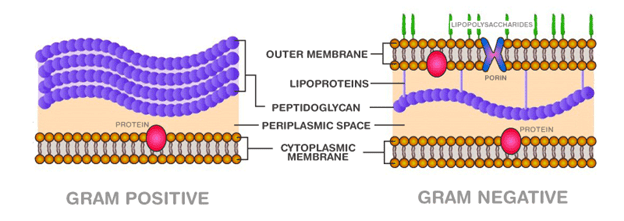
Microorganisms are minute creatures from time immemorial and omnipresent in all kinds of ecosystems on earth. Microorganisms are of different types: Bacteria, Fungi, Protozoa, Algae and Virus. These are not visible through naked eye and require microscope for their structural and functional evaluation.

Industrial microbiology deals with the application of various microorganisms for industrial processes that are beneficial to mankind.

Characteristics of industrially important microorganisms include:

  • Non pathogenic and non-contagious
  • Easy and rapid growth on industrial scale
  • Need of inexpensive medium for culture and growth
  • Production of spores
  • Easy inoculation
  • Desired product should be produced rapidly on large scale
  • capability to genetic manipulation, if required

Major industrial products produced by microorganisms:

  • Pharmaceutical drugs
  • Vaccines
  • Organic acids and solvents
  • Steroids
  • Dairy products
  • Enzymes
  • Beverages
  • Antibiotics
  • Amino acids
  • Vitamins

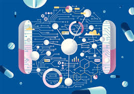
Figure 1: Major applications of microorganisms

Beverages:

Yeast species was widely used from thousands of years to produce beverages like wine, brandy, whiskey etc. Yeasts fall under kingdom Fungi and are eukaryotic, single celled organisms. Yeasts species, Saccharomyces cerevisiae  was allowed to grow on malted cereals and fruit juices to produce ethyl alcohol.

Beyond yeast, bacteria – Acetobacter, Lactobacsp. B. bucheri, etc and fungi – Pichia fermentans, Cyberlindnera mrakii etc are usually used in wine production

Antibiotics:

Discovery of Penicillin, an antibiotic by Alexander Fleming in 1928 is a significant step in medical microbiology history in the 20th century. Antibiotic is a bioactive substance produced by a microorganism that has the ability to either inhibit the growth or kill the other microorganism. Fungi are the main source of antibiotics.

Fungi of Actinomycetes group produce antibiotics like tetracyclin, streptomycin, actinomycin D etc. While, antibiotics like Penicillin, Cephalosporin etc are produced by filamentous fungi.

Organic acids and solvents:

Organic acids and solvents are produced from the microorganisms mainly for the pharmaceutical and other industrial needs. These compounds can be produced either from glucose or in the form of end products from pyruvate or ethanol.

Microorganisms that produce organic acids include:

Aspergillus niger – Citric acid

Acetobacter aceti – Acetic acid

Lactobacillus – Lactic acid

Salmonella – Formic acid

Escherichia coli – Butric acid and malic acid

Acetobacter xylinum – Ascorbic acid

Enzymes:

Enzymes, also called biological catalysts occur naturally in the living system and regulate biochemical reactions. As enzymes have wide applicability in both medical and non-medical fields, microorganisms are genetically modified to produce industrially important enzymes. Amylase was the first industrial enzyme to be produced in the year 1896. Amylase has the ability to regulate indigestion and other digestive system related disorders.

Enzymes are widely used in food preservation, food processing, leather and paper industries, detergents and scientific research and development sectors like molecular biology.

MicroorganismSubstrate for growthEnzyme producedApplications
Aspergillus niger, Penicillium sp.PectinPectinaseAlcohol production, clarification of fruit juices
Saccharomyces diastaticusStarchAmylaseAlcohol production, starch removal glucose syrups production
Bacillus sp.ProteinsProteaseBread, baked foods, waffles production
Aspergillus sp.LipidsLipaseFood and aroma enhancement, biofuel degradation
Cellulomonas sp.CelluloseCellulaseAlcohol and glucose production
Streptomyces sp.XylanXylanasePaper production, biofuel production
Actinomyces, Streptomyces sp.ChitinChitinaseFood additive, therapeutic agent, antifungal and antitumor

Table 1: Enzymatic application of microorganisms

Amino acids:

Amino acids are building blocks of proteins and have high demand as supplements in food and nutraceutical industries. These are also used as supplements in bakery and packed foods. Microorganisms utilize amino acids for their metabolism and growth. Microorganisms are stimulated to produce extra amino acids, such that the extra amino acids are excreted into the surrounding medium.

Lysin and glutamic acid are the prime amino acid supplements in the preparation of bread and other nutritional supplements. Glutamic acid in the form of Monosodium Glutamate is used as flavor enhancing compound in the packed foods.

Glutamic acid is being produced from Corynebacterium gluatmicum. Corynebacterium sp. also used in:

  • Steroid conversion – an important process in the development of pharmaceutical products.
  • Hydrocarbon degradation – breakdown of plastic and oils for environmental protection.

Other amino acid producing bacteria include:

L-alanine – Corynebacterium dismutans, Pseudomonas dacunhae, Escherichia coli

L-arginine – Serratia marcescens, Bacillus subtilis

L-aspartic acid – Escherichia coli

Vitamins:

Vitamins are critical for vital functions and a healthy life. As the human body unable to synthesize these compounds, it is necessary to be supplied in the diet in small amounts. Vegetables and meat are sources of vitamins.

Microorganisms are grown in bulk quantities for the commercial production of vitamins like – thiamine, riboflavin, folic acid, vitamin B12, ascorbic acid, beta-carotene etc.

Some microorganisms that produce vitamins include:

Beta-carotene – Blakeslea trispora, Phycomyces blakesleeanus

Riboflavin – Mycocandida riboflavin, Candida flareri, Clostridium buytilicum

Vitamin B12 – Pseudomonas denitrificans, Bacillus megaterium, Propionibacterium freudenreichii.

Pharmaceutical drugs:

Trichoderma polysporum produces ‘Cyclosporin A’ which is used as immunosuppressive agent during organ transplantation. Moscus purpureus produces ‘Statins’ that has the ability to reduce blood cholesterol levels.

Single cell proteins:

Microorganisms that are rich in protein content can be used as protein supplements for human and domestic applications and are called single cell protein (SCP).

Algal species, Spirulina is the most popular SCP being produced commercially as a protein supplement.

Other SCP producing microorganisms include:

Bacteria – Pseudomonas fluorescens, Lactobacillus, B.megaterium

Algae – Chlorella pyronoidosa, Chondrus crispus

Fungi – Aspergillus fumigates, A. niger, Rhizopus cyclopium

Yeast – S.cerevisae, C.tropicalis, C.utilis

Apart from the above applications, microorganisms have major applications in vaccine production, biofuel production and in treatment of malnutrition.

ARTIFICIAL INTELLIGENCE AND THE HEALTHCARE INDUSTRY TODAY

BY: Ria Fazulbhoy (MSIWM031)

The healthcare industry is going through a massive shift in many ways and using methods. The new world of artificial intelligence and technology has left no stone unturned, including the field of biology. From personal assistants like Siri, Alexa and Google home to smart watches tracking your steps, heart rates and sleep patterns, and mobile applications directing fitness and mental health, this amalgamation of technology and biology has made its way into everyone’s life. This is just the beginning, and we have a long way to go in the exploration of applications of this unique innovation.

Various emerging Industry Applications:

  1. Medical imaging: Many companies today are involving Artificial Intelligence – associated platforms in the medical field for scanning devices in order to improve image clarity and clinical outcomes. This is done by reducing exposure to radiation (For Example: CT scans for liver and kidney lesions).
  1. Management of chronic diseases: Machine learning is being integrated into devices like sensors, to detect, monitor and automate the delivery of treatment. This reduces manpower and increases efficiency.
  1. Drug discovery: A number of applications are there which involve the use of AI with drugs. This includes designing of drugs, polypharmacology, drug repurposing, screening of drugs and chemical synthesis.
  1. Telemedicine: Electronic consultations, medicine management and analyzing medical records and other data of the patients drastically helps the doctors, nurses as well as the patients. Today’s technology has enabled telemedicine to be an emerging field and has helped millions across the globe, especially during the trying times of the Covid-19 pandemic.
  1. Robot assisted surgeries: Data from real surgical processes are collected by cognitive surgical robotics to improve on and improve the already existing approaches to surgery. This greatly helps with minimizing the patient’s treatment time and chance or probability of error.

These are a few upcoming trends amongst various others.

Noteworthy examples of Integration of artificial intelligence in healthcare:

  1. KENSCI

KENSCI is a company targeted towards AI for hospital risk prediction. Its main goal is to combine artificial intelligence and big data to predict financial, operational and clinical risk in hospitals. It takes data from different and existing sources, which can then predict who may get sick and also keeps hospital costs in check.

  • XTALPI (Cloud based digital drug discovery)

This unique technology combines cloud and quantum physics which results in the prediction of small molecules’ chemical and pharmaceutical properties which plays an extremely important role in drug design and development, and thus, drug discovery. This company has a polymorph prediction i.e., crystal structure prediction technology, which is able to predict complex molecular systems and structures within days, as compared to the normal time of weeks and months.

  • ATOMWISE 

Atom wise constitutes a neural network for clinical trials, known as Atom Net. This helps to identify differing biological activity of patients, including their characteristics. The technology can screen anywhere between 10 to 20 million genetic compounds every day and has been found to deliver results 100 times faster than other competitive pharmaceutical companies in the markets. Clinical trials are essential for new discovery of pharmaceuticals and vaccines, and this practice is helping us move toward a cure for serious diseases, including Ebola and multiple sclerosis.

  • DEEP GENOMICS

This is a Canadian based platform which aims at helping researchers find candidates that would be compatible with drugs that are mainly targeted towards neuromuscular and neurodegenerative disorders. Finding the right candidate can improve chances of successful clinical trials. This also aids in the revolutionizing stream of personal medicine.

CORONARY ARTERY DISEASE/ ISCHEMIC HEART DISEASE

BY: Ria Fazulbhoy (MSIWM031)

How does it occur?

This disease is currently the leading cause of death, worldwide. The ischemic heart disease is also known as coronary artery disease (CAD), atherosclerotic heart disease and coronary heart disease (CHD).

The disease occurs due to the blockage of the arteries leading to inadequate blood supply to the heart due to a number of reasons like building of plaque, excess exertion, high levels of cholesterol, etc. While narrowing may be caused by a blood clot or blood vessel constriction as well, it is more often caused by plaque accumulation, called atherosclerosis.

In the blood, cholesterol particles begin to accumulate on the walls of the arteries that supply the heart with blood. Eventually, it can form deposits called plaques. These deposits narrow the arteries and obstruct the flow of blood eventually.

The amount of oxygen supplied to the heart muscle is decreased by this reduction in blood flow. The heart muscle cells die, which is called a heart attack or myocardial infarction, when the blood supply to the heart muscle is completely blocked (MI).

Risk factors that increase chances of the disease:

The risk of developing ischemic heart disease is raised by a variety of variables. Not all individuals with risk factors are going to get ischemic heart disease. There are risk factors for ischemic heart disease include:

  • Diabetes
  • Family history of the condition of the heart
  • High cholesterol in the blood
  • Blood Pressure High
  • Strong triglycerides in the blood
  • Obesity
  • Inactive physique
  • Smoking and other forms of tobacco

Symptoms:

People can experience ischemic heart disease symptoms either regularly or only occasionally, depending on the severity of the case. Some common symptoms experienced by patients include:

  1. Angina pectoris

When the heart muscle is deprived of enough oxygen, the pain endured is called angina pectoris. This is a clinical condition characterized by chest, chin, shoulder, back, or arm pain that is normally exacerbated by exercise or emotional stress and immediately relieved by rest or nitroglycerin use.

  1. Breath shortage: One can experience shortness of breath or severe exhaustion with exercise if the heart can’t pump enough blood to meet the needs of your body.
  1.  Heart attack. A coronary artery that is totally blocked can cause a heart attack. Crushing pressure in one’s chest and pain in your shoulder or arm, sometimes with shortness of breath and sweating, are the classic signs and symptoms of a heart attack.
  1.  Arrhythmia – abnormal heart rhythm: Inadequate blood flow to the heart or heart tissue damage can interfere with the electrical impulses of your heart, causing irregular heart rhythms.

Diagnosis:

  1. Electrocardiogram – ECG or EKG: The electrical activity, rate, and regularity of your heartbeat are calculated by
  2. Echocardiogram: A image of the heart is generated using ultrasound (special sound wave).
  3. Exercise stress test: Tracks your heart rate as you walk on a treadmill. This helps to decide how well the heart functions as more blood needs to be pumped.
  4. Chest X-ray:  In order to create an image of the heart, lungs, and other organs in the chest, x-rays are used.
  5. Coronary angiogram: Monitors blockage and passage of blood into the coronary arteries. In order to detect dye injected through cardiac catheterization, X-rays are used.
  6. Coronary artery calcium scan:  A computed tomography (CT) scan searches for calcium accumulation and plaque in the coronary arteries.

Treatment:

Treatment given to patients suffering from Ischemic heart disease can include drug therapy, regular exercise, quitting of smoking/ tobacco, healthy dietetic.

Medications used to treat the disease include:

  • Angiotensin-converting enzyme (ACE) that relax blood vessels and decrease blood pressure.
  • Angiotensin receptor blockers (ARBs) which help in lowering the blood pressure.
  • Anti-ischemic agents like ranolazine, for example (Ranexa)
  • Antiplatelet drugs that stop blood clots from forming
  • Beta-blockers that decrease the heart rate
  • Calcium channel blockers that lower the heart muscle workload

PARKINSON’S DISEASE

BY: Ria Fazulbhoy (MSIWM031)

Parkinson disease is a disease related to the nervous system and nerve damage. It drastically affects the patient and progressively worsens over time. The progression takes place in 5 stages, which go from low risk symptoms to more drastic ones.  It affects over 100,000 people worldwide each year. At present, approximately more than 10 million people suffer from this disease. However, it is noticed that men have a higher chance of developing the disease, a reason not clear as yet. Other factors like genetics, environmental cues (like exposure to toxins), age, etc can also play a role in the development of PD.

What happens in Parkinson’s disease

  1. Parkinsons affects the central nervous system and the brain.
  2. It mainly affects a region in  the brain, in the basal ganglia, which is known as substantia nigra.
  3. Substantia nigra consists of some cells which produce the chemical hormone and neurotransmitter, dopamine – the feel good hormone.
  4. Dopamine is an extremely important catecholamine in the brain which is responsible for various cues, functions, carrying chemical messages and also contributes to the pleasure pathway i.e how we feel positive emotions like joy, satisfaction and pleasure.
  5. In Parkinson disease, the levels of dopamine in a individual drop due to the death of cells which produce dopamine and are present in substantia nigra.
  6. When these dopamine levels decrease, it causes abnormal activity of the brain, which in turn leads to severe symptoms like impaired movement, depression, sleeping problems, etc.

Symptoms of Parkinson’s disease

The signs and symptoms generally differ from patient to patient. Early signs are almost always undetectable, and the disease is diagnosed most commonly in the later stages. Sometimes, symptoms can be present only on one side of the body (left or right), remain severe on this side and eventually spread to the rest of the body. Parkinson’s disease symptoms include:

  • Tremors: Patients often feel tremors in their limbs such as hands, fingers and legs. Can later also occur in jaws and tongue. It becomes progressively worse and can also cause problems in daily life activities such as eating, bathing, wearing clothes etc.
  • Impaired balance and posture: Posture becomes topped, and patients find it hard to keep balance. External help and support is required in the later stages.
  • Depression and bad dreams: Owing to the decreased levels of dopamine in the brain, and other attributes such as loss of physical control over body, slow movement and psychological effects can lead to depression and other personality disorders. This is accompanied with anxiety, fear and loss of motivation.
  • Changes in talking and speech: In some cases, the speech of the patient can become highly affected. It becomes slurry, soft, sometimes quiet, other times slow, and is expressionless and monotonous.
  • Bradykinesia (slowed movement) : Simple tasks like walking, sitting, eating become time consuming and movement becomes slowed. Steps might get shorter. External help and assistance is needed.

Treatment and prevention

Unfortunately, as of today, there is no complete solution or cure for the complete recovery from Parkinson’s disease Intense research work is being done in labs across the globe to find a solution for the same. Symptoms and side effects can be used by the intake of prescription drugs (like anti depressants), dopaminergics, muscular antagnostics, etc. Physical exercise can also help keep the body moving and fit. Life expectancy can increase with proper care and attention towards the patient.

CELL WALL POLYSACCHARIDES: BACTERIAL PEPTIDOGLYCAN

BY: Ria Fazulbhoy (MSIWM031)

Introduction

Bacteria require a thick, rigid extracellular wall that protects them from osmotic lysis. There is a structure known as the peptidoglycan (also known as muerin) which protects the bacteria and gives the bacterial envelopes their strength and rigidity. Peptidoglycan is a polymer made up of amino acids and sugars which forms a mesh like layer. It is a linear, alternating copolymer of N Acetylglucosamine (GlcNAc) and N-acetylmuramic acid (Mur2Ac), linked by beta 1->4 glycosidic bonds, which is crosslinked by short peptides attached to the Mur2Ac. Both GlcNAc

and Mur2Ac are activated by attachment of a uridine at their anomeric carbons during the assembly of the polymer of this complex macromolecule.

Bacterial peptidoglycan synthesis

The peptidoglycan outside the plasma membrane of bacteria is synthesised in the following manner:

  1. In the first step, GlcNAc 1-phosphate condenses with UTP to form UDP-GlcNAc.
  2. UDP-GlcNAc then reacts with phosphoenolpyruvate to form UDP-Mur2Ac, with NADPH present.
  3. To this, 5 amino acids are added to form Mur2Ac pentapeptide moiety.
  4. The Mur2Ac pentapeptide moiety is made from amino acids L alanine, D glutamate, L lysine, D ala D alanine.
  5. This pentapeptide moiety is then transferred to membrane lipoid dolichol, a long chain isoprenoid alcohol, from the uridine nucleotide.
  6. UDP GlcNAc also donates a GlcNAc.
  7. In a number of bacteria, five glycines are then added in peptide linkage to the amino group of the Lys residue of the pentapeptide.
  8. After this, the disaccharide decapeptide which is formed, is added to the non reducing end of an already existing peptidoglycan molecule.  Dolichol leaves the macromolecule in this step.
  9. A transpeptidation reaction occurs in which there is crosslinking of the adjacent polysaccharide chain. This step is catalysed with the help of the enzyme transpeptidase.
  10. Thus, a huge, strong macromolecule is formed which contributes to the macromolecular wall surrounding the bacterial cell.
  11. Most of the effective antibiotics that are used today against  bacteria, have a mode of action by inhibiting the reactions which are involved in the synthesis of bacterial peptidoglycan.
  12. Numerous other oligosaccharides and polysaccharides are synthesized by similar routes, where sugars are activated for subsequent reactions by attachment to nucleotides

Difference of peptidoglycan in gram positive and gram negative bacteria

Importance of peptidoglycan in bacteria

The peptidoglycan which surrounds the bacteria is very important, and sometimes is essential for their survival. They have 2 main functions:

  1. To counteract and maintain the osmotic pressure of the cell. If the peptidoglycan is absent, the bacteria undergoes very abrupt osmotic lysis. Hence, a lot of antibiotics target the peptidoglycan of the bacteria, since it plays a crucial part in the maintenance of osmotic pressure and protection of the bacterial cell. These antibiotics inhibit the synthesis of peptidoglycan in bacteria and initiate osmotic lysis of it.
  2. Another very important function of the peptidoglycan is the regulation of the molecules entering and leaving the bacterial cell. The peptidoglycan regulates the diffusion of cells which play important roles like division of the cell and anchoring the cell wall (eg : teichoic acid)
  3. Some bacteria also release peptidoglycan fragments, which play an important role in cell to cell communication.

TRANSGENIC ANIMALS AND DISEASE RESISTANCE

BY: Reddy Sailaja M (MSIWM030)

TRANSGENIC ANIMALS

‘Transgenesis’ is a molecular method of introducing a foreign gene (of interest) into the genome of an organism to express the desired trait or characteristic and further pass the trait to the progeny successfully. The gene that is being introduced is called a ‘transgene’.

A transgenic animal or genetically modified animal is the one that is being introduced with a desired foreign gene into its genetic material through recombinant DNA technology, a molecular biology technique.

Transgenesis has been widely applied in most of the domestic animals, aquaculture and agriculture that aids in human welfare and development.

Ralph Brinster and Richard Palmiter were the pioneers in creating first transgenic animal – “Super mouse” in 1982 by introducing human growth hormone in the mouse genome. The offspring produced were larger in size than the parent.

Figure 1: Transgenic super mouse (right) produced by recombinant DNA technology

Pig, goat, sheep, fish, cattle and insects like Drosophila melanogaster (fruit fly) are the most common transgenic animals that are being used in basic and applied research for human welfare.

PRODUCTION OF TRANSGENIC ANIMALS

Two methods are principally followed to generate transgenic animals

  1. Embryonic stem cell method
  2. Pronucleus method by microinjection
  1. Embryonic stem cell method for Transgenesis:
  2. Inner cell mass of mammalian blastocysts contain embryonic stem cells (ESCs). ESCs have the ability to produce all kinds of organisms’ cells, including gametes.
  3. Desired gene is selected from the donor organism.
  4. Vector DNA is chosen that carries the desired DNA to the host cell.
  5. Vector contains promoter and other regulatory sequences that are crucial for transgene transfer, selection and expression in the host organism.
  6. ESCs were cultured along with the vector containing desired DNA
  7. Successfully transformed cells will be selected based on the selection methods like antibiotic resistance.
  8. The transformed cells are injected into inner cell mass of embryonic blastocysts of the mouse for further propagation.
  9. A pseudo pregnant mouse (stimulus of mating results in making mouse uterus receptive for the blastocysts due to hormonal changes) was prepared and the transformed blastocyst stage embryo was introduced into the uterus.
  10. Blastocyst would implant successfully and the mouse gives birth to pups. 10-20% pups will be having the transgene and is heterozygous in nature (only one copy of the gene was transformed and the other was wild).
  11. Heterozygous mice are allowed to mate to get homozygous offspring (1 in 4, Mendelian ratio) was selected and propagated further to generate transgenic trait.

Figure 2: Embryonic stem cell method for transgenic animal generation

  • Microinjection method:
  • Manipulation of the pronucleus is the most common method to create a transgenic animal and is first described by Gordon et al.
  • Superovulating female is induced with specific hormones and the eggs are harvested.
  • The male and female pronuclei are visible under microscope several hours after the sperm is allowed to enter into the oocyte. As male pronucleus is larger in size, the transgene is microinjected easily into it.
  • Pronucleus stage is advantageous as it allows early incorporation of the transgene into the host DNA and the entire host cells could express it.
  • Once the transgene is introduced, male and female pronuclei are allowed to fuse to form a fertilized egg.
  • Once the blastocyst stage is reached, it was implanted into the pseudo pregnant mother and the progeny was checked for the transgene expression as in the ESCs method. 

Figure 3: Generation of transgenic animal by microinjection method

Other techniques followed to generate transgenic animals are listed in table 1.

TRANSGENIC TECHNIQUESINTERPRETATION
Cre-lox techniqueIdeal technique with more control over resulting phenotype; time-consuming
Viral vectorsdifficult; largely restricted to avian species
Cytoplasmic injectionLess efficient than direct pronuclear microinjection
Primordial germ cellsChimeric animals result
Nuclear transferLarge potential for genetically modifying livestock
Spermatogonial manipulationTransplantation into recipient testes

Table 1: Other techniques used to generate transgenic animals

APPLICATION OF TRANSGENESIS

  1. Disease resistant transgenic animals:

Selection and cross breeding of animals is a natural way of producing superior quality livestock animals with respect to disease resistance, more milk production, larger size etc. However, maintaining these qualities to be passed to the generations is unpredictable.

  1. Neurodegenerative disease resistant animals: Spongiform encephalopathy (Scrapie disease) in sheep, bovine spongiform encephalopathy (BSE) (Mad cow disease) in cattle, Creutzfeldt Jacob disease in humans are some of the major neurodegenerative diseases. These diseases occur because of the expression and misfolding of “prion” protein. ‘Gene knock out’ of ‘prion protein through rDNA technology helps in generating prion protein free livestock and resistant to neurodegenerative disorders. RNA interference (RNAi) is the new rDNA technique that helps in knocking down of the desired gene by forming a double stranded DNA construct and suppressing its expression.

RNAi method has extensive applicability, one of which is to generate knock down transgenic animals that can survive RNA based viral infections like foot and mouth disease, classic swine fever and the most resent SARS-CoV-2 disease.

Figure 4: Prion free sheep (Denning et al 2001)

  • Bacterial disease resistant cattle: Mastitis is a bacterial infection of the mammary gland in the cows that affects quality and quantity of the milk being produced. Scientists have developed transgenic cattle that express lysostaphin protein, which kills mastitis causing bacteria by cleaving their cell wall.

Similarly, lysozyme producing transgenic goats are also generated that prevents mastitis causing bacterial lysis and healthier mammary glands.

  • Disease resistance in fishes: Catfish often prone to microbial infection and death. Cecropin B is a small protein expressed in Hyalophora cecropia moth that has anti-microbial protperties. Scientists have generated Cecropin gene expressing transgenic catfish that confers resistance against microbial infections.
  • Disease resistant cattle against Brucellosis: Brucellosis is a deadly zoonotic disease, that can spread across animals without limit and even to humans. A large number of animals in American Bison area have been affected badly and the grazing cattle used to acquire the infection that lead to abortions, low fertility rates, reduced milk production etc. In humans, the disease is called undulant fever and its effects are severe. Recently, it was discovered that bovineNRAMP1 gene is efficient in providing resistance to brucellosis. Transgenic cattle with this gene offer protection against the disease.

Mastitis resistant transgenic cow (Agricultural research service, US)

Identification and integration of genes through transgenesis is the need of the hour that provides disease resistance and improved immune response in livestock and poultry. Scientists are focusing on genetic engineering based disease resistant animal models that would help livestock show resistance to diseases as shown in the table 2.

Table 2: Diverse applications of disease-resistant genetically engineered animals.

  • Medical applications:
  • Disease models: Understanding the disease and its effects are crucial for effective drug development and vaccine creation. Transgenic method is widely applied to generate disease models to understand causes and effects of human diseases. For example, mouse with various cancers or cystic fibrosis were produced through rDNA technology. These models give insights into the disease and further effective drug development and treatment.
  • Understanding gene functions: Mouse/rat genetic composition is closely related to humans. Hence, mice or rat models are chosen to produce genetically modified organism with alteration of gene like, gene knock-out (removal of a particular gene) or gene knock-in (insertion of a gene) or damaging the gene.

This category of transgenic models helps to understand the crucial functions of the gene and its role in human development and disease. This method is widely used to produce transgenic animals with superior quality. For example, transgenic cow – with disease resistance and improved milk production.

  • Production of therapeutic proteins and antibodies: Animals like horse, goat and cows were genetically modified to develop and secrete useful chemical substances like antibodies, and therapeutic proteins that help treat the human diseases efficiently. For example, transgenic cow that secretes egg proteins into its milk. These transgenic animals with therapeutic reagents production are also called ‘walking pharmacies’.
  •  Production of xenotransplants: Scientists have developed transgenic farm animals by ‘knocking out’ the gene that is responsible for eliciting immune response and rejection of an organ when introduced into the human body. For example, knock out transgenic pig organs can be now used for organ transplantation in humans. This method solves the issue of organ donor shortage and saves many critical lives.
  • Transgenic fishes: Transgenic fishes are produced by introducing genes responsible for disease resistant/temperature tolerant/ better growth etc. For example, a company called Aqua Bounty Farms has requested United States Food and Drug Administration (USFDA) to approve its genetically modified salmon that has the ability to grow three times bigger size than normal within a year of its growth.

DISADVANTAGES OF TRANSGENIC ANIMALS

  • Genetically modified animals (like disease laboratory study models) will show negative impact on ecosystem if they escape and released into the environment.
  • May act as human disease reservoirs for critical pathogens like virus, prions etc.
  • May cause severe allergies in humans as it is not a natural product.
  • Genetically modified animals may show alterations in its behavior if the foreign gene undergoes any changes like mutations, leading to any unexpected harm to the mankind.

Even though, the chance of adverse effects is minimal, one can’t rule out completely.

ETHICAL CONCERNS OF TRANSGENIC ANIMALS

It is considered as unethical to produce transgenic animals because it is kind of violating animal rights and disrespect to animals.

Unless there is a balance maintained between need and the production of transgenic animals and effective application in human welfare like medical purpose, agriculture and scientific understanding and application.

CONCLUSION

From its origin, transgenic method has been creating revolutionary output for human well being by producing therapeutic proteins, superior quality breeds of animals and plants and xenografts. A transgenic animal has full potential to play a significant role in biomedical field. However, it is important to maintain ethical standards in effective usage of transgenic method for human welfare. As there is an equal chance of enormous harm that may cause to humans and the environment with the misuse of the technique.

BIOAEROSOLS

BY: K. Sai Manogna (MSIWM014)

Bioaerosols are airborne particles created by biological materials and generate a great deal of energy to distinguish the small particles from the larger particles. Based on their sizes, bioaerosols are graded and often range in diameter from 0.02-100µm. The name of these bioaerosols is given to microorganisms distributed in the atmosphere by the transport and deposition process followed by the launch process.

Launching: 

Launching is the process in which the particles filled by microbes are suspended in the earth’s atmosphere. It is achieved predominantly by aquatic and terrestrial sources. For example, the sneeze exposes the atmosphere to bioaerosols. 

Three considerations are included in this process: 

(a) Air turbulence caused by the human, animal, and machine movement; 

(b) The production, storage, processing, and disposal of waste materials; 

(c) natural mechanical processes, such as the movement of water and wind on solid or liquid surfaces that are contaminated; and 

(d) As a result of regular fungal life cycles, the production of fungal spores. Any other examples may be a passing aircraft releasing a biological warfare agent or a passenger jet releasing unburnt carbon particles’ source as an instantaneous linear source. 

Conveyance of bioaerosols: 

Transport or dispersion is the mechanism by which a viable particle travels at wind speed from one point to another or when it is released by force into the air. The airborne particle’s force depends on its kinetic energy derived from the force at which it is launched into the atmosphere and the speed of the wind. Bio-aerosol transport can be described in terms of time and distance. Inside buildings or other enclosed spaces, this method of transport is standard. 

Deposition of Bio Aerosols: 

The deposition is the last pathway involving the distribution of bioaerosols in the atmosphere. It is then split into the other three forms. 

1. Settling Gravity 

2. Effect on the Surface 

3. Deposition of Rain 

Settling Gravity: 

The action of gravity on particles is the primary mechanism associated with deposition. Strength works more intensely on the particles than air, dragging them down. Larger particles would have higher speeds and settle more rapidly down the aero microbiology pathways However it should be noted that gravitational deposition may be negligible for particles of microbiological interest exposed to winds above 8103 m/hr. 

Impacting the surface: 

It is the mechanism in which the particles of bioaerosols have contact with surfaces such as leaves, trees, walls, with the effect of kinetic energy loss. The potential for impact allows a particle to collide with the surface and encourages its binding to it. However, after a collision, depending on the nature of a particle’s surface, it will bounce. 

Bouncing off a surface allows the particle at a lower rate to re-enter the air current, which can have one of two effects: 

1. It allows for subsequent molecular downward diffusion and gravitational settling, resulting in deposition on or on another nearby surface. 

2. It will cause the particle to escape from the surface and re-enter the air current once more. 

Deposition of Rain: 

The deposition also impacts rainfall and electrostatic charges. It occurs as the condensation reaction between two particles, which combine and produce a massive mass bioaerosol, making it settle faster. The overall efficiency of the deposition of rain also depends on the particle plume’s distribution area. Massive, more diffuse plumes have a substantial impact than smaller, more diffuse plumes. The rainfall rate also influences rain deposition. On the other hand, electrostatic deposition still operates the same way, condenses bioaerosols, but is based on electrovalent particles’ attraction. Both particles appear to have an associated charge of some kind. Usually, microorganisms have an overall negative charge at neutral pH associated with their surfaces. Such negatively charged particles may interact with other airborne particles of positively charged, leading to electrostatic condensation.

Mechanisms for Laboratory Regulation of Bioaerosols: 

Two such indoor conditions are hospitals and microbiology laboratories that fall under intramural aero microbiology, with probably the highest potential for pathogenic microbe aerosolization. The centers for the care of immense numbers of patients with a range of diseases are hospitals. It accounts for a high percentage of individuals being the active carriers of several contagious airborne pathogens or microorganisms, including workers and patient visitors. In this respect, microbiological laboratories are just as important as they also serve as a breeding ground for pathogenic species. 

Physical Bioaerosol Removal through filtration: 

Technologies that tackle the bioaerosol threat fall into two categories: 

(1) capture or physical elimination from the air stream of bioaerosols, and 

(2) inactivation on-line or airborne. 

Technologies that make up the former group have typically not been established explicitly for bioaerosols but aerosols’ general regulation. In the latter case, to make airborne microbes non-infectious and exclusively target bioaerosols, technologies apply external stress such as heat or ultraviolet light. Since bioaerosols are physically identical to non-biological particles of the same aerodynamic size and composition, it is possible to apply standard aerosol control devices (air filters, electrostatic precipitators. that physically extract particles from the airstream for bioaerosol control. Filtration is the most successful method for particle removal, both viable and nonviable. For example, high-performance particulate air (HEPA) filters have a 99.97 percent removal efficiency of 0.3-μm sized particles by definition. 

Mechanically, filters extract particles by integrating four simple filtration components. 

Mechanisms: inertial effect, gravitational settling, interception, and diffusion. Impact occurs with larger aerosols that do not adjust to changes in a flow streamline induced by a collector (fiber, granule.) due to their inertia. Gravity, especially when the flow velocity is insufficient, may also cause larger particles to contact a collector. For particles in the submicrometric scale, the two dominant mechanical collection mechanisms are diffusion and interception. As they deviate from a flow streamline by Brownian motion, aerosols are collected by diffusion and eventually deposited on a collector. Aerosols follow a streamline during interception and contact a collector when the streamline distance from the collector is equal to the particle’s radius. 

Disinfection by Air Filter: 

Due to the risks associated with bioaerosols sustained viability, many technologies for disinfecting filter media have been developed. These include photocatalytic oxidation (PCO), UV illumination, and other technologies, as well as anti-microbial filters. A brief overview of several unique technologies for filter disinfection are described below:  

UV light: Irradiation with UV light of bioaerosols (without the presence of UV light, photocatalyst) may cause inactivation. This procedure, known as ultraviolet germicidal irradiation (UVGI), creates thymine dimmers in DNA and inhibits replicating the targeted microbe. 

Anti-microbial filters: Bioaerosols have also been tested against air filters, which have been treated with biocidal chemicals such as iodine. For iodine treated filters, inactivation is hypothesized through the penetration of iodine molecules through the cell wall of microbe and subsequent damage to the capsid protein. In addition to killing microbes obtained from the filter, it is speculated that microbes passing through the filter can be inactivated by iodine species, leading to a decrease in viable bioaerosols’ penetration. A benefit of anti-microbial filters is that additional equipment (e.g., UV light) is not required and can therefore be readily integrated into respirators. 

Technologies for Airborne-Inactivation: 

Besides the physical elimination by filtration of bioaerosols from an airstream, air 

With airborne-inactivation technologies, it can be disinfected. Technologies may be mounted in an on-line system, e.g., ventilation and cooling of heating systems) 

System) to process air that is polluted. Descriptions of several airborne-inactivation technologies are given below: 

UVGI: UVGI lamps may be placed before or after an air filter by direct irradiation of the suspended microbes to minimize the amount of penetrating infectious bioaerosols. The implementation of UVGI is relatively complicated because several factors must be taken into account in the engineering design: airflow patterns, residence time (dose) of the microbe, relative humidity, different resistance of bioaerosols to UV light, ray-tracing optics, power consumption, lamp dust, shielding effect of the material surrounding the bioaerosol, and ozone production from UV lamps. 

Microwave irradiation: By direct irradiation, bioaerosols may be inactivated. Microwave radiation at a frequency of 2.45-GHz decreases the concentration of laboratory-generated and atmospheric bioaerosols. Electron microscopy of irradiated cells revealed that cell death could be responsible for structural damage. 

Cold plasma: Inactivation of plasma has been used in surface disinfection and disinfection. 

Sterilization, however, on-line bioaerosol inactivation has recently been implemented. 

Dielectric barrier discharge (DBD) – a non-thermal technique that uses electrical discharge between electrodes separated by a dielectric material – can generate plasma for disinfection purposes. DNA and cell membrane damage likely cause microbe death. 

Toxic vapours: It has been shown that chemicals such as chlorine dioxide (ClO2) reduce the concentration of culturable airborne bacteria and fungi effectively and thus decontaminate buildings. ClO2 is an oxidizing agent suspected of causing microbes’ death through membrane damage or protein synthesis destruction. Unlike the systems discussed above, due to the vapor’s toxicity, harmful vapours cannot be incorporated in an on-line environment and cannot be used with human occupants. 

Ultra-high temperature (UHT) treatment: UHT methods have historically been used to sterilize or disinfect liquids (e.g., milk) in order to destroy resistant bacterial spores (applying temperatures of > 125 °C for several seconds). However, recent studies have shown their effectiveness against bioaerosols. Airflow was heated to temperatures greater than 1,000 °C for less than a second for inactivation in UHT bioaerosol tests. 

LOOP MEDIATED ISOTHERMAL AMPLIFICATION

BY: Reddy Sailaja M (MSIWM030)

INTRODUCTION

Infectious diseases affect living forms on earth that include but not limited to plants, animals including human beings. Pathogenic micro organisms i.e., bacteria, virus, parasites etc attack, hinder the growth and development of an organisms and sometimes lead to death.

Infectious diseases that turn out to be pandemic have had bad effect on human beings in the history like bubonic plague, influenza, Spanish flu, avian flu and the most recent on COVID-19 (SARD-CoV-2). When the diseases spread rapidly and cross the country’s border, it is called as a pandemic.

Rapid diagnosis and treatment are the only mode of spreading the disease and to save lives. Standard traditional methods of microbial detection include – microbial culture (aerobic and anaerobic), Gram staining, colony morphology and other biochemical analysis. However, these traditional microbial methods take 5-7 days to give result, by then patients would be severely affected and sometimes might die.

Henceforth, rapid diagnosis with in a day or in hours of time is critical for effective treatment and patient management. Recently, Nucleic acid amplification technique (NAAT), a major molecular biology application has gained interest in the diagnostic field for its rapid and sensitive pathogen detection in short time. Polymerase chain reaction (PCR), a NAAT method gave hope for the early infection detection as it is fast and sensitive method in pathogen detection. PCR uses thermoresistant DNA polymerase enzyme (Taq polymerase), sequence specific primers and under specific cyclic conditions amplify the pathogenic DNA isolated from the sample to billions of copies in few hours. Due to sensitivity and speed, PCR became the choice of pathogen detection in medical microbiology field.

However, PCR has certain draw backs as follows:

  • Need of a thermocycler (high cost)
  • Need of carcinogenic material like Ethidium bromide for DNA band visualization
  • Need of a trained technician
  • Sophisticated molecular biology lab setup with at least three isolated rooms
  • Can’t be setup at point of care centres like rural areas

Loop-mediated Isothermal amplification (LAMP) is a revolutionary NAAT method, discovered by Notomi et al in 2000. It has the potential to rapidly detect the pathogenic DNA more sensitively and specifically in comparison with PCR at a constant temperature (isothermal). LAMP method is based on the auto cycling of the DNA using strand displacement reaction and utilizes DNA polymerase like Bst, Bsm, Gsp etc

Due to its high sensitivity, speed and efficiency, it has varied applications in medical microbiology field.

CHARACTERISTICSLAMPPCR
Type of reactionIsothermalCyclic
Reaction time30-60 minutes2-4 hours
TemperatureIsothermal Temperature (60-65⁰C range).Variable Temperature. Denaturation (95⁰C); Annealing (50-60⁰C); Extension (72⁰C)
Need of thermocyclerNo. simple dry bath is enoughYes
SensitivityLimit of detection is higher.Limit of detection is lower.
SpecificityHigh as it uses 4-6 primersLower than LAMP
Sample kindDetection is good even with crude samplesPure DNA is required
Primers characteristics4-6 primers. Loop primers increase reaction speed2 primers
Mode of detectionFluorescence detection in real time. Visual detection with naked yes, gel electrophoresis or turbidity.  Fluorescence detection in real time. Visual detection is only through gel electrophoresis

Table 1: Differences between LAMP and PCR techniques

LAMP PRIMERS

LAMP makes use of 4 primers that are designed specifically to recognise 6 different regions on the target gene.

  1. Forward inner primer (FIP) – FIP comprises of two primers namely, F2 region at the 3’ end and F1C region at 5’ end (F1C of the primer is similar to F1C portion of the target DNA).
  2. Backward inner primer (BIP) – BIP comprises of two primers namely, B2 region at the 3’ end and B1C region at 5’ end (B1C of the primer is similar to B1C portion of the target DNA).
  3. Forward outer primer (F3) – F3 is the outer primers and is short in its length. F3 is complementary to F3C region of the target DNA.
  4. Backward outer primer (B3) – B3 is one of the outer primers and is complementary to B3C region on target DNA.
  5. Loop primers – Loop forward and loop backward (LF and LB) are the two additional primers utilized in the LAMP reaction to increase the speed.

Figure 1: LAMP primers

STAGES IN THE LAMP REACTION

1. F2 portion of FIP primer binds to F2C region on the target DNA and initiates new strand synthesis and amplification.

2 F3 outer primer then binds to the F3c region on the target DNA and extends the strand by displacing the FIP associated complementary strand. This displaced strand forms a loop lie structure at the 5′ end.

3. Th synthesized ssDNA with a loop at the 5′ end assists as a template for BIP. B2 binds to B2c region on the target DNA and synthesizes new complementary strand by opening of the 5′ end loop.

4. Now, the B3 primer binds to B3c region on the target DNA and extends by displacing the BIP connected complementary strand. This results in dumbbell shaped DNA formation.

5. Then the F1 primer gets extended by opening up the loop at the 5′ end with the help of Bst DNA polymerase. At this stage, dumbbell shaped DNA become a stem loop structure and initiates the LAMP reaction. This stage is called the LAMP reaction’s second stage.

6. Further in LAMP cycling, the FIP binds to the loop region of the stem-loop DNA and initiates strand synthesis by displacing F1 primer and formation of a new loop at the 3′ end.

7. Extension happens at the 3′ end of B1 by displacing the FIP strand, forming a dumbbell shaped DNA. Self-primed DNA synthesis by strand displacement gives out one complementary strand of the original stem loop DNA along with one more stem loop DNA with a gap repair.

8. Both the new stem loop DNAs act as template for a BIP primed strand displacement reaction in the succeeding cycles. Consequently, for every half LAMP cycle, 13 fold amplification of the target DNA occurs.

The ultimate amplification LAMP products contain combination of stem loop DNA with varied lengths looking like a cauliflower like structure with multiple loops.

LAMP REACTION SET UP

To setup LAMP assay, target DNA, an isothermal DNA polymerase with strand displacement activity, primers and buffer are sufficient. LAMP assay can be setup in a simple water bath or a heat block at a constant temperature (ideally at 65°C).

Figure 2: Over view of LAMP reaction

LAMP DETECTION

LAMP reaction can be detected as follows:

  1. Fluorescence based detection: using florescent dyes like SYBR green, Pico green, Eva green

Figure 3: LAMP detection using SYBR green fluorescent dye

  1. Visual detection: Based on turbidity and precipitation in the positively reacted tubes. Leuco crystal violet is a dye that detects positive reaction based on turbidity.

Figure 4: LAMP detection based on turbidometry

  1. Colorimetric dyes are also used as they react with the free Mg2+ being produced in the reaction. Colour change happens based on the pH change. For example, phenol red show pink colour before the reaction and turns yellow if the sample is positive for the pathogen (as the pathogenic DNA multiplies, more Mg2+ will be released into the reaction tube).

Figure 5: Colorimetric detection of LAMP reaction (Source: NEB)

ADVANTAGES OF LAMP TECHNIQUE

  • Require simple water bath or heat block (no costly thermocycler).
  • Amplification at isothermal conditions.
  • More specificity and sensitivity as it utilizes 4-6 primers.
  • Cost effective
  • Easy deployment at point of care centres at rural areas.
  • No trained technician is required. Setup is quite simple.

APPLICATIONS OF LAMP TECHNIQUE

  • Rapid diagnosis of bacterial, viral, fungal and parasitic organisms.
  • Helps to detect pathogens at both genus level and species level. In case of viruses, various strains can be easily differentiated.

MICROBIOLOGY OF WATER INDICATOR ORGANISMS

BY: K. Sai Manogna (MSIWM014)

For life, water is essential. There must be sufficient, stable, and usable supplies available to everyone. Improving access to clean drinking water will lead to substantial health benefits.  Most individuals fail to gain access to clean water. The supply of clean and filtered water to each house may be the standard in Europe and North America, but access to clean water and sanitation is not the rule in developing countries, and water-borne infections are common. There is no access to better sanitation for two and a half billion people, and more than 1.5 million children die from diarrheal diseases each year. According to the WHO, the mortality of water-related illnesses exceeds 5 million individuals per year; more than 50 percent of these are microbial intestinal diseases, with cholera standing out first and foremost.

A prominent public health concern in developing countries is acute microbial diarrheal diseases. Those with the lowest financial resources and the worst hygiene services are those affected by diarrheal diseases. Children under five, especially in Asian and African countries, are the most affected by water-borne microbial diseases. Often affecting developing countries are microbial water-borne diseases—the most severe water-borne bacterial illnesses.

Table 1: Some bacterial diseases transmitted through drinking water.

DiseaseBacterial agent
CholeraVibrio cholerae, serovarieties O1 and O139
Typhoid fever and other serious salmonellosisSalmonella enterica subsp. enterica serovar Paratyphi Salmonella enterica subsp. enterica serovar Typhi Salmonella enterica subsp. enterica serovar Typhimurium
Bacillary dysentery or shigellosisShigella dysenteriae Shigella flexneri Shigella boydii Shigella sonnei
Gastroenteritis – vibrioMainly Vibrio parahaemolyticus

Indicator Organisms:

1. Microorganisms such as viruses and bacteria in water bodies, which are used as a proxy to determine the existence of pathogens in that area, are indicator organisms.

2. It is preferred that these microorganisms are non-pathogenic, have no or limited water growth, and are consistently detectable at low concentrations.

3. In larger populations than the related pathogen, the indicator species should be present and preferably have comparable survival rates instead of the pathogen.

4. In the monitoring of water quality, different indicator species can be used, and the efficacy of predicting pathogens depends on their detection limit, their tolerance to environmental stresses and other pollutants.

Cholera:

Tiny, curved-shaped Gram-negative rods with a single polar flagellum are Vibrio. Vibrios are optional anaerobes capable of metabolism that are both fermentative and respiratory. For all animals, sodium promotes the development and is an absolute prerequisite for most.

1. The majority of plants are oxidase-positive, and nitrate is reduced to nitrite.

2. Pili cells of some microbes, such as V. cholerae, V. parahaemolyticus, and V. vulnificus, have structures consisting of TcpA protein.

3. The production of TcpA is co-regulated with cholera toxins expression and is a primary determinant of in vivo colonization.

4. Several species of Vibrio can infect humans. The most significant of these species is, by far, V. cholerae.

5. Several forms of soft tissue infections have been isolated from V. alginolyticus.

6. The cells of Vibrio cholerae will expand at 40°C at pH 9-10.

7. The presence of sodium chloride stimulates growth. Vibrio cholerae is a bacterial genus that is very diverse.

8. It is split into 200 serovarieties, distinguished by the lipopolysaccharide (LPS) structure (O antigens). Only O1 and O139 serovarieties are involved in true cholera.

9. Gastroenteritis may be caused by many other serovarieties, but not cholera.

10. Biochemical and virological features are the basis for the differentiation between Classical and El Tor biotypes.

Disease characterization:

1. The incubation period for cholera is 1-3 days.

2. Acute and severe diarrhea, which can reach one liter per hour, characterises the disease.

3. Patients with cholera feel thirsty, have muscle pain and general fatigue, and display anuria symptoms accompanied by oliguria, hypovolemia, and hemoconcentration.

4. In the blood, potassium decreases to deficient levels. With cyanosis, there is circulatory collapse and dehydration.

Several factors depend on the seriousness of the illness:

(a) personal immunity: both previous infections and vaccines can confer this immunity;

(b) inoculum: disease arises only after the absorption of a minimum quantity of cells, approx. 108.

(c) Gastric barrier: V. cholera cells like simple media and the stomach is also an adverse medium for bacterial survival, usually very acidic. Patients that take anti-acid drugs are more vulnerable than healthy patients to infection.

(d) Blood group: persons with O-group blood are more vulnerable than others for still unexplained causes.

5. In the absence of treatment, the cholera-patient mortality rate is approx—fifty percent.

6. The lost water and the lost salts, mostly potassium, must be replaced.

7. Water and salts can be administered orally during light dehydration, but rapid and intravenous administration is mandatory under extreme conditions.

8. Presently, doxycycline is the most effective antibiotic. In some instances, if no antibiotic is available for treatment, the administration of salt and sugar water will save the patient and help with recovery.

9. Two significant determinants of infection exist:

(a) the adhesion of bacterial cells to the mucous membrane of the intestine. It depends on the presence on the cell surface of pili and adhesins;

(b) development of a toxin from cholera.

Salmonellosis:

1. Gram-negative motile straight rods include the genus Salmonella, a member of the family Enterobacteriaceae.

2. Cells are oxidase-negative and positive for catalase, contain D-glucose gas, and use citrate as a sole source of carbon. There are many endotoxins in Salmonellae: O, H, and Vi antigens.

3. S. Subsp enterica. enterica serovar Enteritidis is the most widely isolated serovariety worldwide from humans. Other serovarieties can, however, be prevalent locally.

4. A fermented juice historically extracted from the palm-tree was the source of insulation.

Disease Characterization:

1. Two forms of salmonellosis can be pathogenic to humans:

(a) typhoid and paratyphoid fever (not to be confused with rickettsia-induced typhus disease);

(b) Gastroenteritis.

2. Low infection doses are sufficient to cause clinical symptoms (less than 1,000 cells).

3. There are different clinical signs of salmonellosis in newborns and children, from a severe typhoid-like disease with septicemia of a range to a mild or asymptomatic infection.

4. The infection is commonly spread through the hands of staff in pediatric wards.

5. Ubiquitous Salmonella serovars, such as Typhimurium, are often caused by food-borne Salmonella gastroenteritis.

6. Symptoms such as diarrhea, vomiting, and fever occur about 12 hours after consuming infected food and lasts 2 to 5 days.

7. Spontaneous healing typically happens. All kinds of food can be associated with Salmonella.

8. The prevention of food-borne Salmonella infection is focused on the prevention of contamination, the prevention of food-borne Salmonella multiplication (persistent storage of food at 4oC), and where possible, the use of pasteurization (milk) or sterilization (other foods).

9. When infected with fertilizers of the fecal origin or washed with polluted water, vegetables and fruits can carry Salmonella.

10. The incidence of typhoid fever decreases as a country’s development level grows, such as pasteurization of milk, dairy products, and controlled water sewage systems.

11. The risk of fecal contamination of water and food remains high where these hygienic conditions are absent, and so is the occurrence of typhoid fever.

Bacillary Dysentery or Shigellosis:

Shigella are members of the Enterobacteriaceae family that are Gram-negative, non-spore-forming, non-motile, straight-rod-like. Without gas production, cells ferment sugars. There is no fermentation of salicin, adonitol, and myo-inositol. Cells do not use citrate, malonate, and acetate as the primary source of carbon and do not create H2S. It is not decarboxylated with lysine. Cells are oxidase-negative and positive for catalase. Members of the genus Shigella have a complex antigenic sequence, and their somatic O antigens are the basis of taxonomy.

Disease Characterization:

1. The incubation time for shigellosis is 1-4 days.

2. Typically, the illness starts with fever, anorexia, tiredness, and malaise. Patients exhibit irregular, low-volume, sometimes grossly purulent, bloody stools, and abdominal cramps.

3. Diarrhea progresses to dysentery after 12 to 36 hours, with blood, mucus, and pus appearing in feces that decrease in volume (no more than 30 mL of fluid per kg per day).

4. Even though the molecular basis of shigellosis is involved, the colonic mucosa’s penetration is the initial phase in pathogenesis.

5. Degeneration of the epithelium and acute inflammatory colitis in the lamina propria define Shigella infection’s resulting concentration.

6. Desquamation and ulceration of the mucosa eventually contribute to leakage into the intestinal lumen of blood, infectious elements, and mucus.

7. The colon’s water absorption is hindered under some circumstances, and the amount of stool depends on the flow of ileocecal blood.

8. As a consequence, normal, scanty, dysenteric stools can move through the patient.

9. The bacterium must first adhere to its target cell in order for Shigella to penetrate an epithelial cell.

10. The bacterium is usually internalized into an endosome, which is then lysed to obtain entry to the cytoplasm where replication occurs.

AERO MICROBIOLOGY

BY: K. Sai Manogna (MSIWM014)

Introduction:

The study of living microbes that are suspended in the air is known as Aero microbiology. Such microbes are known as bioaerosols. There are significantly fewer microorganisms in the atmosphere than in the oceans and in the soil; there are still many microorganisms that can impact the atmosphere. With the help of wind and precipitation, these microbes have a chance to migrate long distances and increase the rate of infectious diseases caused by these microbes. In humans, animals, and plants, these aerosols are ecologically important because they can be associated with the disease. Microbes can suspend themselves in the atmosphere, where they can communicate and precipitate with the clouds and create specific shifts in the clouds. 

The air has two microbial ecosystems. 

A. Atmosphere 

B. Clouds 

A. Atmosphere: 

1. High light intensities, extreme temperature fluctuations, low amount of organic matter, and a lack of water availability; characterize the atmosphere as a habitat, making it a non-hospitable environment for microorganisms and a generally inadequate habitat development. 

2. In the lower regions of the atmosphere, however, large numbers of microbes are contained. 

B. Clouds: 

1. In the atmosphere, an apparent mass of concentrated watery vapor floating, usually well above the general ground level. 

2. Clouds, with a pH ranging from 3 to 7, are also an acidic environment. 

Sources of airborne microorganisms: 

1. Air is not a favourable microbial growth environment because it does not provide adequate moisture and nutrients to sustain growth and reproduction, and there is also no indigenous flora growth in the air. 

2. Quite a range of sources responsible for introducing microbes into the air have been identified and researched. 

3. The most popular of these is dirt. Microbes are suspended in the air with wind flow and remain there and often accumulate. 

4. Microbes are often released into the air by human activities such as digging, sloughing, and running. 

5. Microbes are often released into the air through air currents and splashes of water. 

6. Besides, air currents strip plant and animal pathogens from their surfaces and disperse them across the atmosphere. 

7. In contrast to animal pathogens, plant pathogens can spread more quickly. For example, a gamine flies over a thousand kilometers with Puccini spores.

Examples of airborne plant pathogens:

Examples of airborne animal pathogens:

8. Human beings are the primary cause of the introduction of bacteria into the air.

Examples of airborne human pathogens:

9. The most comprehensive source is human activity. The pathogenic bacteria in the human respiratory tract and the mouth’s microbes are continuously released into the air, but they cough, sneeze, and laugh. 

Depending on the size and moisture content, the microbes released into the air come in three forms. Those are the three forms: 

1. Droplets 

2. Nuclei Droplets 

3. Dust that is contagious 

Droplets: As we sneeze, millions of droplets are released, and mucus is expelled from about 200 miles away. Such droplets are water droplets that hold microorganisms if a diseased individual releases them. Saliva and mucus comprise these droplets. Most of the microorganisms they transport are from the respiratory tract. The droplet size determines how long microorganisms live on the droplet. Large-sized droplets settle quickly in the air. The source of these droplets carrying the microorganism can be a source of infectious disease. 

Nuclei Droplets: 

Water particles emitted 1 to 5 micrograms in diameter during sneezing and coughing. For respiratory disorders, droplet nuclei are known to be the raw material. On its surface, it contains saliva and mucus. They are stuck in the air for a more extended period because of their small scale. Droplet nuclei, if the bacteria are the constant source of bacterial infections, are known to be 

The present remains viable on its surface. The viability of bacteria depends on physical conditions, such as humidity, sunlight, moisture, and droplet size. 

Dust Particles: 

By bed making, holding a handkerchief, working with a patient with dried secretion, digging and ploughing, these dust particles are released into the air. Microorganisms adhere to these droplets’ surface and are then suspended by the above techniques to dry them. There is a more significant size of dust particles laden with bacteria and settle down in the air. Two forms of droplets cause airborne diseases. 

a. Droplet infection due to droplets with a diameter greater than 100-micron meters. 

b. Any dried droplet residues cause airborne infections. 

Infection with droplets remains localized and concentrated, while airborne infection may be long-distance. Microorganism can grow on dust particles for a more extended period. It is proven harmful in hospitals and laboratories when closed bottles of dried specimens are opened, and cotton plugs are removed from the bottles. 

Factors influencing airborne microbes:

Factors that influence microbial survival in the atmosphere are 

a. Temperature

b. Moisture/Humidity

c. Content of Nutrients 

d. pH and Acidity 

1. The main factor in regulating the growth of microbes in the air is temperature. 

2. High temperatures hinder the production of microbes and often denature the microbes’ structural conformation. 

3. Very few microbes can live and withstand high temperatures, i.e., extremophiles. 

4. Likewise, when ice crystal formation occurs, shallow temperatures are also not ideal for microbial growth. 

5. Humidity has a role in preserving the development of airborne microbes. 

6. Gram-Bacteria associated with aerosols tend to live in low humidity for a more extended period. 

7. The abundance of nutrients in the atmosphere is lower, so it does not help microbial growth.